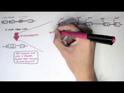
0:00 Armando Hausudugan, Biology and Medicine videos, please make sure to subscribe, 0:05 join 0:06 the forming group. 0:07 For the latest videos, please visit Facebook, Armando Hausudugan, and here, 0:10 make sure to 0:11 like, leave, and you can also ask questions, answer questions, and please post 0:14 some interesting 0:15 things, such as artworks. 0:17 And you can also change the quality settings, which I highly suggest, to the 0:21 highest one, 0:21 such as HD or 720, for better graphics. 0:25 And in this video, we're going to talk about the diversity of antibodies and T 0:28 cell receptors, 0:29 and actually how they become diverse, and how they become highly specific for a 0:34 particular 0:35 antigen, you can say. 0:37 So we begin our journey in the bone marrow, which is within the bone. 0:43 And in the bone marrow, we have a progenitor lymphoid cell, which comes from a 0:47 stem cell 0:47 in the bone marrow. 0:48 Now, this progenitor lymphoid cell can become two things. 0:54 It can become a progenitor T cell, or it can become a progenitor B cell. 0:59 Now, let's follow this life of a progenitor T cell first. 1:04 This progenitor T cell will then migrate from the bone marrow into the thymus, 1:10 where T cell 1:11 development occurs. 1:12 So T cell development occurs in the thymus. 1:14 The progenitor T cell will become a naive T cell here. 1:19 And it becomes a naive T cell through a process called somatic recombination, 1:25 or V-D-G recombination, 1:27 where it will obtain a unique T cell receptor, which will bind to a specific 1:34 antigen. 1:35 And so this increases the diversity and specificity of the T cell receptors. 1:41 Now, after the progenitor T cell has become the naive T cell, and developed 1:45 into the naive 1:46 T cell through cytokines or whatnot, it will then migrate into the lymph nodes. 1:51 Now, let's go back to the progenitor B cell. 1:55 The progenitor B cell will also undergo in the bone marrow somatic recomb 2:00 ination, also 2:01 known as V-D-J recombination. 2:05 And through this recombination, it will become an immature B cell with a unique 2:11 antibody, 2:12 which will bind to a specific antigen. 2:15 So through somatic recombination, it will obtain a unique type of antibody, 2:20 usually immunoglobulin 2:21 M. 2:25 And once this immature B cell has developed with an immunoglobulin M, it will 2:28 then migrate 2:29 also into the lymph nodes. 2:31 So both the naive T cell and the immature B cell are in the lymph nodes. 2:35 So let's have a look inside the lymph nodes. 2:38 So here we have the immature B cell, which has matured, just become a mature B 2:43 cell once 2:44 it's entered the lymph nodes. 2:45 And then we have the naive T cell. 2:47 The naive T cell will become activated when an antigen presenting cell presents 2:52 an antigen 2:53 to the naive T cell. 2:55 Once the antigen presenting cell, such as a macrophage or dendritic cell, 2:58 presents an 2:59 antigen to the naive T cell, the naive T cell will begin to proliferate and 3:04 differentiate. 3:05 It will differentiate into a cytotoxic T cell or a T helper cell. 3:10 The T helper cell can then activate the mature B cell, or alternatively, the 3:14 mature B cell 3:14 can become activated when it binds onto a pathogen, the antigen of a pathogen. 3:20 Or both, the T helper cell or the antigen of the pathogen can both activate the 3:24 mature 3:25 B cell. 3:26 Now within the lymph node, there is an area called a germinal center. 3:32 And the germinal center consists of a dark zone and a light zone. 3:36 When the mature B cell becomes activated either from the T helper cell or the 3:40 antigen pathogen 3:41 or both, the mature B cell will move into the germinal center, into the dark 3:48 zone first, 3:49 where it will undergo somatic hypermutation and then become a central blast. 3:55 So it will undergo somatic hypermutation. 3:58 And what somatic hypermutation does, it causes mutations within the gene, which 4:02 further increases 4:04 the affinity actually, and diversity, specificity of the antibodies of the B 4:10 cell. 4:11 So the central blast from the dark zone will then move into the light zone 4:15 where it becomes 4:16 a central site and then here in the light zone, it can undergo class switching 4:21 and 4:22 clonal expansion, where they will differentiate and proliferate into either 4:29 memory B cells 4:30 or plasma cells, the antibodies to creating cells. 4:35 Point to make is that the central blast and central sites are still the B cells 4:38 . 4:39 It's just that they're called different names. 4:41 And also class switching basically involves when the immunoglobulin, which I 4:46 just mentioned, 4:47 is usually IgM, can become IgE, IgA, et cetera. 4:51 So this occurs in the germinal center. 4:54 Now let's take a look and go back to the progenitor B cell and learn a bit more 4:59 about 5:00 the diversity of these antibodies. 5:03 So we begin with the progenitor B cell in the bone marrow, right? 5:06 And the progenitor B cell through VDJ recombination or somatic recombination 5:11 becomes immature 5:12 B cells, each with a unique antibody which will bind to a specific type of 5:18 antigen. 5:19 And all of these immature B cells, they all have unique antibodies, right? 5:24 So let's look at the antibody structure to appreciate the diversity of these 5:29 molecules. 5:30 So an antibody consists of a light chain, an orange here, and a heavy chain, 5:35 the rest 5:36 of it. 5:37 And they're both connected through disulfide bonds. 5:40 Another way of looking at the antibody, the same antibody in this particular 5:45 immature 5:45 B cell, is that it contains a variable region, there's blue areas, also known 5:50 as V regions, 5:51 and a constant region. 5:53 So here we have CCC, Constance and V. And also the other diagram we have CCC 5:58 and VVV. 5:59 As you can see, there are more constant regions on the heavy chain. 6:04 Now what's important about immunoglobulin structure, such as this one, is that 6:07 the variable region 6:09 has the unique binding site, and has a unique binding site for a specific 6:14 antigen. 6:15 So the variable region is basically what determines what type of antigen it 6:19 binds to. 6:20 The constant region is essentially determines what class this immature B cell 6:26 is. 6:26 And usually, as I mentioned, immature B cells is usually immunoglobulin M. And 6:32 so this constant 6:32 region is an immunoglobulin M constant region. 6:37 So let's have a closer look at what I mean by antibody diversity, and how each 6:41 of these 6:42 immature B cells possesses a unique antibody, which we'll buy into a specific 6:45 antigen. 6:46 Let's have a look at this particular immature B cell, let's call this immature 6:49 B cell B 6:49 cell 682. 6:50 Now B cell 682 cannot bind to this particular antigen, because the V region of 6:57 the antibody 6:58 does not match that of the pathogen's antigen, because as you can see, this 7:01 pathogen number 7:02 2 has circular looking antigens, whereas the binding site of the B cell has a 7:10 triangular 7:11 looking V region, the binding site. 7:15 The B cell 682 can also bind to this particular pathogen, because the antigen 7:20 is square looking, 7:22 and so it does not fit into the variable region, the binding site. 7:26 However, the B cell 682 can bind to this particular pathogen, because this 7:31 particular 7:32 pathogen has triangular looking antigens, which are complementary to the 7:39 antibody's 7:40 variable region, the antibody's binding site. 7:44 And so as you can see, the antibodies are highly specific, and so binds to a 7:49 specific 7:50 type of antigen. 7:53 So let's look at this connection, the contact between antibody and antigen, in 7:58 a bigger 7:59 picture. 8:00 So here we have a pathogen with its specific type of antigen on it. 8:05 And here we have the antibody, the antibody consisting of a light chain and 8:12 heavy chain. 8:13 The top portions are the variable regions and the rest of the constant regions. 8:17 So here is the antibody, and the V region contains the binding site, which 8:21 binds and 8:22 latches makes contact with the antigen of a pathogen. 8:25 So let's have a closer look at this heavy chain of the antibody binding to the 8:31 antigen. 8:32 So here we have the protein structure of the antigen of the pathogen, and here 8:37 we have 8:37 the heavy chain variable region of the antibody. 8:41 And the site of which the antigen binds or makes contact with the heavy chain, 8:48 binding 8:49 site is known as the epitope. 8:51 So the epitope binds or makes contact with the binding site of the variable 8:57 region, essentially. 8:59 And that concludes the video on the diversity of antibodies and the T cell 9:03 receptors. 9:04 I hope you enjoyed that. 9:07 But if you click on the links provided, and I know I always said that in my 9:11 last video 9:12 on antigen recognition, but if you click on the links, you can look at the som 9:16 atic recombination. 9:17 The progenitor T cell to the naive T cell, and how this increases the 9:22 specificity and 9:22 diversity of the T cell receptors, and also you can click on the link of the 9:26 somatic recombination 9:27 from the progenitor B cell to the immature B cell, and how this increases the 9:32 diversity 9:32 and specificity of the antibodies, or also known as the VDJ recombination. 9:38 Also in the link notes, you can click on the link on somatic hybrid mutation 9:41 and how this 9:41 increases the affinity and diversity of the antibodies once again, so that it 9:46 binds to 9:46 a specific type of antigen, and also on class switching and clonal expansion, 9:51 how the B cells 9:51 differentiate into the B memory cells or the plasma cells. 9:55 Hope you enjoyed this video. 9:56 Please like, comment, and share. 9:57 Thank you.