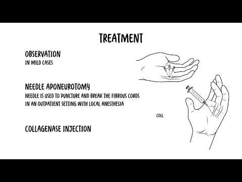
0:00 Hello, in this video, we're going to look at the clinical anatomy of the hand 0:08 and also 0:08 the fingers. 0:09 So we'll actually focus on the wrist joint mainly. 0:13 Now we'll begin by focusing on the right hand and here's the right hand. 0:19 We're looking at it from a palm of you, so the palms of your hands and actually 0:23 the surface 0:24 anatomy here, you can actually feel your scaphoid bone, the tubercle of your sc 0:31 aphoid 0:32 bone, and there's also the piriformis bone you can feel, as well as above it, 0:36 the hook 0:36 of hand-made somewhere. 0:39 Let's look at the right hand again and introduce some of the bones now. 0:43 Here are the forearm bones, the ulna medially, and the radius laterally. 0:50 You can remember the radius is lateral because lateral is where you can feel 0:54 the radial archery. 0:56 The wrist bones consists of many small bones. 1:01 To put it simply, there are eight, four sitting on the top and four below. 1:08 They are collectively known as the carpal bones. 1:12 You can remember the eight carpal bones by starting at the thumb and moving 1:17 around in 1:18 a clockwise motion and remember this saying, so long to pinky here comes the 1:24 thumb. 1:25 So S is for scaphoid, L for lunate, T for triquentrum, P for pysiform, moving 1:36 up, H is 1:38 for hamate, C for capitate, and then trapezoid and then trapezium. 1:51 So again, so long to pinky, here comes the thumb. 1:57 The bones of the hands are made up of metacouples, then you have the phalanges, 2:03 and then you 2:04 have the, of course, the distal, middle, and proximal phalanges, which are the 2:08 fingers 2:08 essentially. 2:09 But remember the thumb has a metacouple, but it has proximal and then a distal 2:17 phalanges, 2:18 so there's no middle phalanges. 2:22 Drawing the same right hand diagram again, looking at it from a palma view, and 2:28 imagine 2:29 cutting a slice through these two sections at the carpal bones. 2:34 So again, we are basically looking at the four carpal bones on the top, or the 2:39 distal 2:39 four, and then the other proximal four carpal bones. 2:43 So focusing on the distal four carpal bones, from the pinky, here comes the 2:49 thumb. 2:50 So H is for hamate, C for capitate, T for trapezoid, and then trapezium. 2:57 Trapezoid and trapezium might be confusing because they sound similar, but to 3:02 remember 3:02 which one comes first, think of the last letter of each word. 3:06 So the D is before the M, and so trapezoid is first, and then its trapezium, 3:12 which is 3:12 closest to the thumb. 3:15 The hamate has a hook, and traveling across the hamate and the trapezium is the 3:20 flexor 3:20 retinaculum. 3:22 The flexor retinaculum is a very important structure to remember. 3:27 But first, let's look at the other proximal four carpal bones. 3:33 So long to pinky, S is for skafoid, L for lunate, T triquetrum, and P-pisiform. 3:42 The skafoid has a tubercle that can be felt, and here again is the flexor retin 3:49 aculum. 3:50 The flexor retinaculum is an important part of the hands anatomy, the wrist 3:53 anatomy, because 3:54 things run over it, and things run under it. 3:58 So let's have a closer look, and let's stick with the right hand for now. 4:03 But before going on, I want to introduce to you an attendant, a tendon that 4:08 runs over 4:08 your flexor retinaculum, called a tendon of palmaris longus. 4:13 This tendon is a big tendon you can see in the middle of your wrist, and this 4:20 tendon, 4:21 it originates essentially from the medial epicondyle, and is responsible for 4:26 flexing 4:26 the wrist. 4:27 It is innervated by the median nerve. 4:31 So here I'm drawing the cobble bones of the right hand. 4:34 Let us now look at things that run superficial to the flexor retinaculum, and 4:39 things that 4:40 run deep to the flexor retinaculum. 4:43 Let's begin by looking at the deep structures. 4:47 And there are many structures that run deep to the flexor retinaculum. 4:51 It's easy to divide it into four things, one, the median nerve, two, the tend 5:00 ons of flexor 5:01 digitorium superficialis, three, the tendons of flexor digitorium profundis, 5:09 and the fourth 5:10 is the tendon of flexor palisis longus, which goes to the thumb. 5:18 And the flexor digitorium superficialis and flexor digitorium profundis has 5:24 four actual 5:25 tendons, each. 5:32 I actually drew this wrong, the tendon of flexor palisis longus should be at 5:37 the right 5:38 hand side, because that's the side of the thumb, sorry. 5:42 Anyways, let's now look and talk about the structures which run superficial to 5:47 the flexor 5:48 retinaculum. 5:51 I just want to add the hook of hamet here and the trapezium here. 5:54 Again, these are the distal cobble bones. 5:57 Okay, so the structures that run superficial to the flexor retinaculum include 6:03 the tendon 6:03 of palmaris longus, which I talked about. 6:09 Because we are looking at the right hand, the ulna structures are on the left 6:15 side, medially. 6:17 So here is the ulna artery and the ulna nerve. 6:23 Palma cutaneous branch of the ulna nerve and the palma cutaneous branch of the 6:28 median 6:28 nerve can also be found on the other side. 6:31 Okay, some clinical relevance of the flexor retinaculum and the median nerve. 6:38 So here is the right hand again. 6:40 Here is the flexor retinaculum. 6:43 It's also sometimes referred to as the carpal ligament, which makes sense 6:47 because it literally 6:48 goes over a carpal bones. 6:51 Anyway, remember the four main groups of structures that run under it, one of 6:57 which is the median 6:58 nerve. 6:59 The median nerve is sensory for the middle, index and thumb region, and also 7:05 motor for 7:06 these regions as well. 7:09 Carpal tunnel syndrome is where you get compression of the median nerve at the 7:13 flexor retinaculum, 7:14 causing numbness, parasthesia, and pain in the median nerve distribution. 7:22 Carpal tunnel syndrome affects females more than men, and there are many causes 7:27 that actually 7:28 can cause carpal tunnel and irritate the median nerve. 7:33 Going back to the diagram we drew earlier, do you remember the tendon of palmar 7:37 is longus? 7:38 The tendon that goes over your flexor retinaculum, the tendon you can see on 7:43 your wrist. 7:44 Well, this tendon, it has some branches that come off it and form what's called 7:48 the aponeurosis, 7:49 the palma aponeurosis, and this has some clinical relevance. 7:53 Another clinical relevance is Jupiter's contracture, which is localized thick 7:58 ening and contracture 8:00 of the palmar aponeurosis. 8:03 It causes the proximal and middle phalanx, or phalanx, to flex. 8:10 The distal phalanx is unaffected. 8:15 Let's look in more detail at the anatomy of the finger, the phalanx. 8:20 So here we are mainly looking at either the middle, index, or pinky, or ring 8:26 finger, but 8:26 we're not looking at the thumb, because the thumb does not have a middle phalan 8:31 x. 8:32 And these other fingers, they consist of the distal phalanx, the middle phalanx 8:36 , and the 8:37 proximal phalanx, and then you have your metacapel. 8:41 Now remember the tendons that run under the flexor retinaculum? 8:46 Well this is where we talk about some of those tendons and where they attach on 8:50 the finger. 8:52 See the flexor digitorium superficialis tendons run towards the proximal phal 8:58 anges, then bifurcates, 9:00 and attaches to the middle phalanx. 9:05 And so we can say that the flexor digitorium superficialis tendon flexes the 9:10 proximal interflangeal 9:11 joint. 9:17 The flexor digitorium profundis tendon run below, deep to the flexor digitorium 9:24 superficialis, 9:26 and attaches to the distal phalanx. 9:29 It does not bifurcate. 9:32 Thus, we can say the flexor digitorium profundis tendon flexes the distal inter 9:38 flangeal joint. 9:41 Alright, some clinical anatomy. 9:45 Your finger is a condition where there is localized thickening of long flexor 9:51 tendons, 9:52 which then prevent movement of the tendon within the fibrous sheath. 9:57 So just think about it as part of the flexor tendon thickening, creating a lump 10:02 which prevents 10:02 movement of the finger. 10:04 So let's just draw it out. 10:06 Here is a finger like your index finger. 10:09 Here are your phalanges, and approximately is your metacapal bones. 10:15 Sorry, I know I wrote metatarsal. 10:19 Tassels are in your foot. 10:20 This is meant to be metacapal. 10:22 Now in this diagram, it's also important to know that we have these fibrous she 10:27 ath sort 10:28 of running on your palm surface of your fingers. 10:33 And these fibrous sheath, they sort of act as a pulley. 10:36 And under it is the flexor tendon. 10:40 So the flexor tendon runs under the fibrous sheath. 10:45 A trigger finger is essentially where you have sort of a nodule or thickening 10:51 of the 10:52 flexor tendon that runs under the fibrous sheath. 10:56 And this causes a sort of restrictive movement of that finger. 11:02 In this case, the flexor tendon thickening or nodule is occurring in between 11:06 the metacapal 11:06 bone and the proximal phalanx. 11:10 This inhibits the flexion between the metacapal and proximal interflangeal 11:15 joint.