

Members have early access to this video
Learn about membershipFoot Anatomy: Bones, Joints, Arches, Muscles, Vessels & Nerves
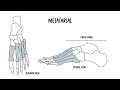
Discover the bones of the foot—tarsals, metatarsals, and phalanges—and learn practical clinical anatomy for diagnosing fractures and common pathologies. This video breaks down foot anatomy with clear explanations of the tarsal group (calcaneus, talus, navicular, cuneiforms, cuboid), the five metatarsals, and the toe phalanges, plus the functional division into hindfoot, midfoot, and forefoot. You’ll understand how the calcaneus supports weight and anchors the Achilles, how the talus links foot to leg at the ankle, and how the navicular and cuneiforms serve as tendon attachment points. Key clinical insights cover calcaneal and talar fractures from high-energy trauma, the mechanics and consequences of tarsal coalition, and metatarsal injuries: stress fractures common in athletes, Jones fractures with poor blood supply and nonunion risk, avulsion fractures of the fifth metatarsal, shaft fractures from direct blows, and proximal fractures from crush injuries. The video highlights where injuries occur, why healing can be complicated, and how bone anatomy relates to tendon attachments and load transmission. Ideal for medical students, clinicians, and allied health professionals, this concise anatomy-and-clinical primer equips you to recognize patterns of injury, link symptoms to underlying structures, and appreciate functional implications for movement and weight-bearing. Watch to sharpen your diagnostic reasoning and apply anatomical knowledge to clinical scenarios—subscribe for more focused musculoskeletal lessons.