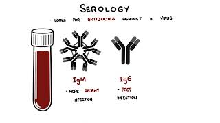
0:00 In this video, we're going to talk about diagnostic tests for viruses. 0:10 So when we test for viruses, we are usually looking for one of three things, 0:15 the virus 0:15 itself, the parts of the virus, or the body's immune response to the virus. 0:20 In practice, the main tests are PCR, antigen tests, file cultures, and serology 0:27 . 0:27 Let's look at each of these. 0:31 So PCR stands for polymerase chain reaction. 0:34 What each word means, polymerase is an enzyme that builds new strands of DNA, a 0:39 chain is 0:40 the reaction that happens in a repeated series of steps, and a reaction is 0:45 obviously the 0:46 chemical process taking place in the test tube or machine. 0:50 So overall, PCR or polymerase chain reaction is a lab technique that makes many 0:55 copies 0:56 of a piece of DNA so it can be detected more easily. 1:01 So PCR is one of the most sensitive tests for diagnosing a current viral 1:05 infection. 1:06 PCR looks for the virus's genetic material, which may be DNA or RNA depending 1:11 on the virus. 1:12 A sample is collected from the site where the virus is most likely to be found, 1:16 such as 1:17 the nasal or throat swab, for SARS virus or influenza, and a vesicle swab, so a 1:24 blister 1:25 swab, for example, for varicella zoster virus or a blood and other bodily 1:31 fluids in selected 1:32 infections. 1:34 Now in the laboratory, the viral genetic material is extracted from the sample. 1:40 If the virus is an RNA virus, such as the SARS-COVID-2 or influenza, the RNA is 1:47 converted 1:48 into complementary DNA by reverse transcriptase, which is why the test is 1:54 sometimes called 1:55 RT PCR, reverse transcriptase PCR. 2:01 Anyway, the machine then uses repeated temperature cycles to denature the DNA, 2:08 allows short primers 2:10 to bind to viral-specific sequences and extend new copies using DNA polymerase. 2:16 This repeated amplification makes millions of copies of the target sequence, 2:22 allowing 2:22 even very small amounts of viruses to be detected. 2:27 Because PCR amplifies the signal, it is highly sensitive and is very useful 2:32 early in infection 2:34 when the virus is present in the sampled site. 2:37 So for example, with COVID-19, a nasal swab can be tested for reverse 2:43 transcriptase PCR 2:45 to detect SARS-COVID-2 RNA, with shingles or chickenpox, PCR is performed on 2:53 fluid from 2:54 a skin lesion as this is one of the most useful ways to confirm varicella z 3:00 oster viral infections, 3:02 which is a DNA virus, just a standard PCR. 3:10 Next we have antigen tests. 3:13 antigen tests look for viral proteins rather than genetic materials. 3:18 They are usually faster and simpler than PCR, but they are generally less 3:22 sensitive so 3:23 they can miss infections if the viral amount is low. 3:27 To perform the test, really what happens is a sample is collected from the 3:31 nasal or throat 3:32 swab depending on the virus. 3:34 The sample is mixed with a reagent and placed on a test strip or cartridge 3:39 containing antibodies 3:41 that are designed to bind to specific viral proteins. 3:45 This is an immuno assay. 3:48 So here you can see the strip and you can see already antibodies that are the 3:54 cartridge 3:54 already containing the antibodies. 3:57 And so if the viral antigen is present, it binds to these antibodies and 4:01 produces a visible 4:02 signal, often a colored line or a machine red result. 4:07 Obviously, if it's negative only the control line will light up. 4:12 Now this is the principle used in many rapid antigen tests for SARS-CoV-2, 4:19 Influenza and 4:20 RSV. 4:21 These tests are useful when a quick answer is needed, but a negative result may 4:25 need 4:25 confirmation if clinical suspicion remains high. 4:29 So again, for example, a rapid COVID antigen test uses a nasal swab and checks 4:34 for SARS-CoV-2 4:35 proteins. 4:36 A rapid influenza diagnostic test works in a similar way by detecting influenza 4:42 viral 4:42 antigens from a respiratory sample. 4:50 Next are viral cultures, which really involves trying to grow the virus in the 4:55 lab. 4:55 This is slower and more specialized, so it is not used routinely for many viral 5:01 infections. 5:01 Now that PCI is widely available. 5:04 To perform viral culture as a clinical sample such as a swab, blood or lesion 5:09 fluid is inoculated 5:10 onto living cells in a lab. 5:13 Viruses cannot grow on their own, so they need living host cells. 5:17 If the virus is present, it infects the cells and may produce visible changes 5:22 called cytopathic 5:24 effect. 5:25 The virus can then be identified by its growth pattern or by additional tests. 5:31 Viral cultures is rarely used now, but in some situations it may be useful. 5:36 For example, proving live infectious virus, investigating of an unknown or 5:42 emerging virus, 5:44 doing antiviral susceptibility testing, selecting reference lab confirmation, 5:49 or for research 5:50 purposes. 5:56 Next we have serology, serology looks for the antibodies against the virus 6:02 rather than the 6:03 virus itself. 6:05 This usually involves taking a blood sample. 6:08 In the laboratory, the patient's blood or serum is then exposed to viral antig 6:13 ens, to 6:14 see whether antibodies such as IgM or IgG bind to them. 6:19 So it's kind of like the reverse, we're actually giving or exposing the person 6:24 's blood to the 6:26 viral antigen to see if they have a response. 6:29 Now many serology assays use techniques such as a lysor or other immuno assays. 6:35 In general, IgM may suggest more recent infection and IgG may suggest a past 6:41 infection, immunity 6:43 or previous exposure, although interpretation depends on the specific virus and 6:48 the clinical 6:49 context. 6:52 Because antibodies take time to develop, serology is usually more useful later 6:56 in the course 6:56 of the illness or when asking whether the patient has had a previous infection. 7:01 So you can see in this graph, IgM initially spikes up after the infection but 7:06 then tapers 7:07 down and goes away whereas IgG persists. 7:11 Let's look at an example, let's just say you want to look at cytomegalovirus 7:16 infection. 7:17 Well, cytomegalovirus of serology can help determine whether a person has 7:21 evidence of 7:22 recent or past cytomegalovirus exposure. 7:27 If an adult, patient has a positive CMV IgG and a negative CMV IgM, this 7:35 suggests a past 7:36 CMV infection rather than a new acute infection. 7:45 More complicating example is around the EBV virus. 7:48 In a young adult with a fever, sore throat, lympharinopathy and fatigue, EBV 7:53 serology 7:53 is useful because it helps determine whether this is a current primary EBV 7:59 infection or 8:01 evidence of past exposure. 8:04 In acute infection, the EBV viral capsin antigen IgM is usually positive early 8:12 whereas the 8:13 EBV viral capsid antigen IgG also appears early and then persists for life 8:22 while the 8:23 EB nuclei antigen antibodies are usually absent early and appear later, very 8:32 complicated. 8:34 Then you can add PCR to this so that EBV PCR can also be used but it answers a 8:40 different 8:41 question. 8:42 Serology is usually more helpful for stage in typical infectious mononucleosis 8:46 which is 8:47 caused by EBV whereas PCR is more useful when you want to detect or quantify EB 8:53 V DNA directly 8:54 such as in immunocompromised patients or when reactivation, high viral load or 9:00 EBV associated 9:02 lymphoprolifative diseases is a concern. 9:07 Complicated stuff 9:09 So in summary, a very important principle is that timing matters. 9:14 Early infection, PCR and sometimes antigen testing are more useful because they 9:19 detect 9:20 the virus directly. 9:21 Later in the infection when the immune system has had time to respond, serology 9:26 becomes 9:26 more informative. 9:28 The type of sample also matters. 9:30 Respiratory viruses are often tested from nasal or throat swabs, skin viruses 9:36 from lesion 9:37 swabs and some systemic viruses from blood or other bodily fluids. 9:44 So in simple terms, PCR looks for the viral genetic material by amplifying it. 9:50 Antigen testing looks for the viral protein using an antibody based immuno 9:55 assay. 9:56 Viral culture tries to grow the virus in living cells and serology looks for 10:00 antibodies 10:01 in the blood, IgM and IgG. 10:03 Thank you for watching. 10:07 �