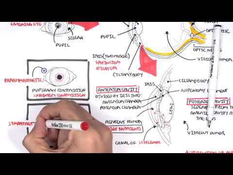
0:00 So in this video, we're going to learn about the orbit of the eye. 0:06 So here we can see the bones which surround and contain the eyeball. 0:13 The bones of the orbit have boundaries. 0:17 The roof is made up of the frontal and your sphenoid bone. 0:23 The medial boundary, which is closest to your nose, is made up of mainly the 0:28 maxilla, 0:29 macromal bone, ethmoid, and a small part of the sphenoid, which is here 0:35 actually. 0:37 The inferior border of the orbit is mainly, is actually the roof of your max 0:42 illary sinus. 0:43 It's made up mainly of the bones, maxilla, zygomatic, and palentine. 0:48 So here's your maxilla, zygomatic, and your palentine bones. 0:57 The lateral border is made up of the bones, the zygomatic bone, and the greater 1:01 wing of 1:02 the sphenoid bone. 1:04 Now these boundaries are significant because an orbital fracture can have some 1:08 serious 1:08 consequences. 1:10 For example, the orbital fracture usually occurs in the floor. 1:15 So fractures usually occur on the floor of the orbit or the medial walls 1:19 because they 1:20 are the most weakest. 1:23 The lateral wall is the strongest wall. 1:26 Members of the floor, which is the inferior wall, may involve the maxillary sin 1:33 us, causing 1:34 sinus sinus. 1:35 Because remember, the floor of the orbit is also the roof of the maxillary sin 1:41 us. 1:42 And the roof of the maxillary sinus is not very strong. 1:45 Okay, now let's zoom into the stuff inside the orbit here. 1:52 And there are some important holes. 1:55 Now we learned about the bones around the area. 1:59 Well, there are three important holes here. 2:03 The superior orbital fissure, the inferior orbital fissure, and the optic for 2:09 amen. 2:10 Let us learn more about these holes, these three holes, and what structures 2:14 pass through 2:15 them. 2:16 So here again are the three holes. 2:19 This is the optic foramen, and two important structures pass through here. 2:25 The optic nerve cranial nerve number two, and the ophthalmic artery, which 2:31 supplies the retina. 2:33 The other hole is the superior orbital fissure. 2:38 There are many structures that pass through here. 2:41 Let us begin with the nerves first. 2:44 There are branches of the ophthalmic nerve, which is a branch of cranial nerve 2:49 five, the 2:50 V1 branch. 2:53 You also have the trochlear nerve, which is cranial nerve number four. 2:58 There is also the superior branch of the oculomotor nerve, cranial nerve three. 3:03 There is also the inferior branch of the oculomotor nerve, cranial nerve number 3:08 three. 3:09 Then you also find the abducens nerve, cranial nerve number six. 3:14 These nerves are very important because they pass through the superior orbital 3:19 fissure, 3:19 and they innervate the muscles responsible for moving your eyeball. 3:24 Then you have an important vein called the superior ophthalmic vein. 3:30 The third hole is the inferior orbital fissure, and two important structures 3:35 pass through here. 3:36 The inferior ophthalmic vein, and the maxillary nerve, which is a branch of the 3:43 trigeminal 3:44 nerve. 3:45 The maxillary nerve is the V2. 3:48 Again, this here represents where the muscle, the tendons of the eyeball origin 3:55 ates. 3:56 And again, cranial nerve three, the oculomotor nerve, cranial nerve four, the 4:00 trochlear nerve, 4:01 the cranial nerve six, the abducens nerve, supplies these muscles. 4:07 They are motor nerves. 4:10 Now let us look at the eyelid when we blink, the eyelid. 4:14 The eyelid is surrounded by muscles, it's composed of muscles. 4:19 These muscles are innervated by nerves. 4:21 So the trigeminal nerve, cranial nerve five, gives off the maxillary nerve 4:27 branch, which 4:28 is the V2, which has another branch called the infororbital nerve. 4:35 The infororbital nerve goes through the infororbital foramen. 4:40 So if you remember the inferior orbital fissure here, we have an auxiliary 4:47 nerve off the trigeminal, 4:49 which passes the lower margin of the orbit and exits through the infororbital 4:56 foramen, and 4:57 becomes the infororbital nerve. 5:01 The infororbital nerve supplies the inferior tarsus muscles, which are the 5:06 muscles of the 5:06 eyelid. 5:09 The superior tarsus is supplied by branches of the supra trochlear nerve. 5:20 Because we have an infororbital nerve, which we talked about, we also have a 5:24 supraorbital 5:25 nerve, which is a branch of the ophthalmic nerve, V1, which is a branch of the 5:31 trigeminal nerve, 5:34 cranial nerve number five, not the maxillary nerve, that is a mistake. 5:38 So again, the ophthalmic nerve, V1, is a branch of the trigeminal nerve, which 5:43 is cranial nerve 5:44 number five. 5:47 And funny story, a maxillary nerve, which is V2, is a branch of the trigeminal 5:52 nerve 5:52 as well. 5:55 Anyway, another nerve is the lacrimal nerve, which supplies muscles of the 6:00 lateral aspect 6:01 of the eyelid. 6:03 An important muscle to introduce now is the tendon of levator. 6:08 This is of important clinical significance because a problem in this tendon can 6:13 result 6:13 in ptosis, which is drooping of the eyelid. 6:18 So again, drawing the muscles of the eyelid out, we have tendon of levator, pap 6:23 abrae superioris, 6:24 which is the proper long name. 6:26 So tendon levator, papabrae superioris, and we are looking at the right eye in 6:33 this diagram. 6:35 So we have the lateral papabrae muscles, the medial papabrae, and the superior 6:40 and inferior 6:41 ptarsis. 6:42 So these are the muscles that make up the eyelid. 6:46 Now looking at some clinical things that are important, Horner syndrome is 6:51 where we have 6:52 a lesion, which leads to a loss of sympathetic function in the head. 6:57 And this lesion can be due to cancer, for example. 7:00 Remember, it is a problem with this sympathetic innervation. 7:04 And as a result, we have three clinical signs of Horner's. 7:08 And these three are meiosis, ptosis, and anhydrosis. 7:13 Tosis is where there is paralysis of the dilator pupillary muscles, so you 7:19 cannot dilate your 7:20 pupils. 7:23 Tosis is paralysis of the superior tarsal muscles, which results in the dro 7:27 oping of your eyelid. 7:29 A problem with levator, papabrae superioris can also cause partial ptosis. 7:35 But this tendon is not sympathetic innervation, but oculomotor nerve, cranial 7:41 nerve number 7:41 three innervated. 7:44 I hope that makes sense. 7:46 Anhydrosis is loss of innervation to sweat glands, which means no sweating on 7:50 the affected 7:51 side. 7:54 And in Horner's, you can also be flushed warm with dry skin. 7:59 So I hope you enjoyed this video on the eyelid and the orbit. 8:05 Thank you for watching.