

Petechial, Purpuric Rash
OVERVIEW
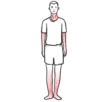
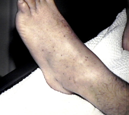
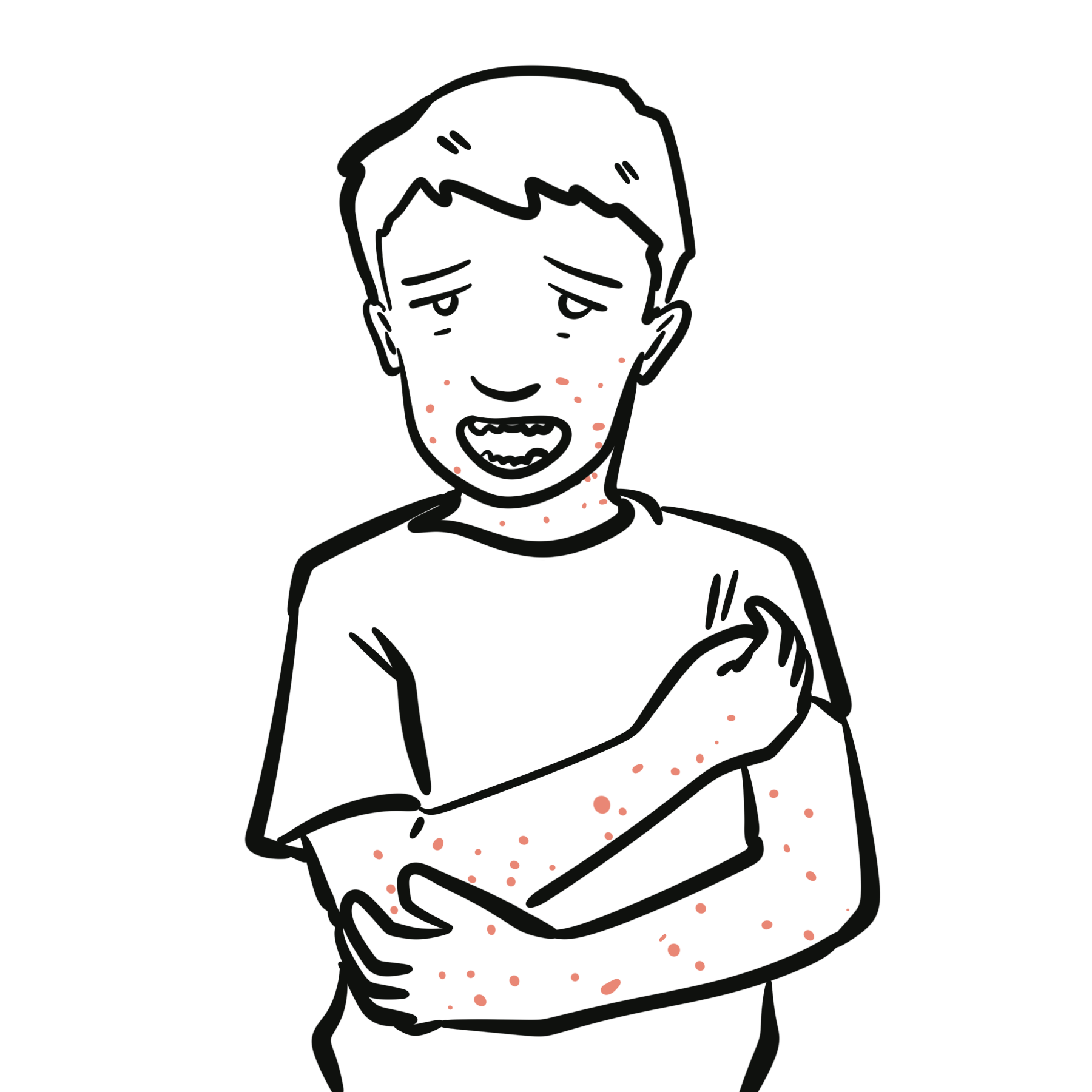
Petechiae and purpura are areas of bleeding into the skin or mucous membranes that do not blanch with pressure.
- Petechiae: Pinpoint lesions, generally less than 2 mm.
- Purpura: Larger lesions, approximately 2–10 mm.
- Ecchymoses: Broader areas of cutaneous bleeding, usually greater than 10 mm.
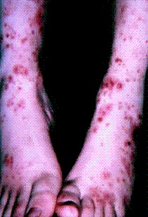
- Palpable purpura: Raised, non-blanching lesions caused by inflammation and damage of small blood vessels, classically seen in cutaneous small-vessel vasculitis.
- Purpura fulminans: Rapidly progressive, painful or necrotic purpura associated with severe infection, disseminated intravascular coagulation and tissue ischaemia.
A petechial or purpuric rash may result from thrombocytopenia, platelet dysfunction, coagulation abnormalities, vascular inflammation, infection, trauma or benign capillary fragility. The priority is to distinguish a stable patient with a localised or chronic eruption from a patient with meningococcal disease, sepsis, disseminated intravascular coagulation, thrombotic thrombocytopenic purpura or another life-threatening disorder.
Use the “Glass Test” (diascopy) immediately at triage: firm pressure applied with a transparent glass over the lesions. If the lesions do NOT fade or blanch, red blood cells have extravasated into tissue. In a febrile child, treat this as an immediate life-threatening emergency.
Mechanistic Clues:
- Petechiae with mucosal bleeding: Suggest thrombocytopenia or platelet dysfunction.
- Large ecchymoses, haemarthroses or deep-muscle bleeding: Suggest a coagulation-factor deficiency or anticoagulant effect.
- Palpable purpura over dependent areas: Suggest cutaneous small-vessel vasculitis.
- Fever with a rapidly spreading non-blanching rash: Treat as invasive infection until proven otherwise.
- Thrombocytopenia with anaemia, schistocytes and organ dysfunction: Consider thrombotic microangiopathy or disseminated intravascular coagulation.
- Localised petechiae above the nipple line after coughing or vomiting: May reflect transient pressure-related capillary rupture.
A non-blanching rash is a physical sign, not a diagnosis. Always assess the patient’s physiological state, platelet count, coagulation profile, organ function and associated clinical features before attributing the eruption to a benign cause.
TRIAGE & ALGORITHM
Step 1: Emergency Triage & Red Flag Screening (The “Unwell Child”)
- Red Flags for Septic Shock / Invasive Meningococcal Disease (IMD):
- Physiology: Tachycardia out of proportion to fever, prolonged capillary refill time (> 2 seconds), cold peripheries, mottled skin, hypotension (decompensated shock), tachypnea, altered mental status / extreme lethargy.
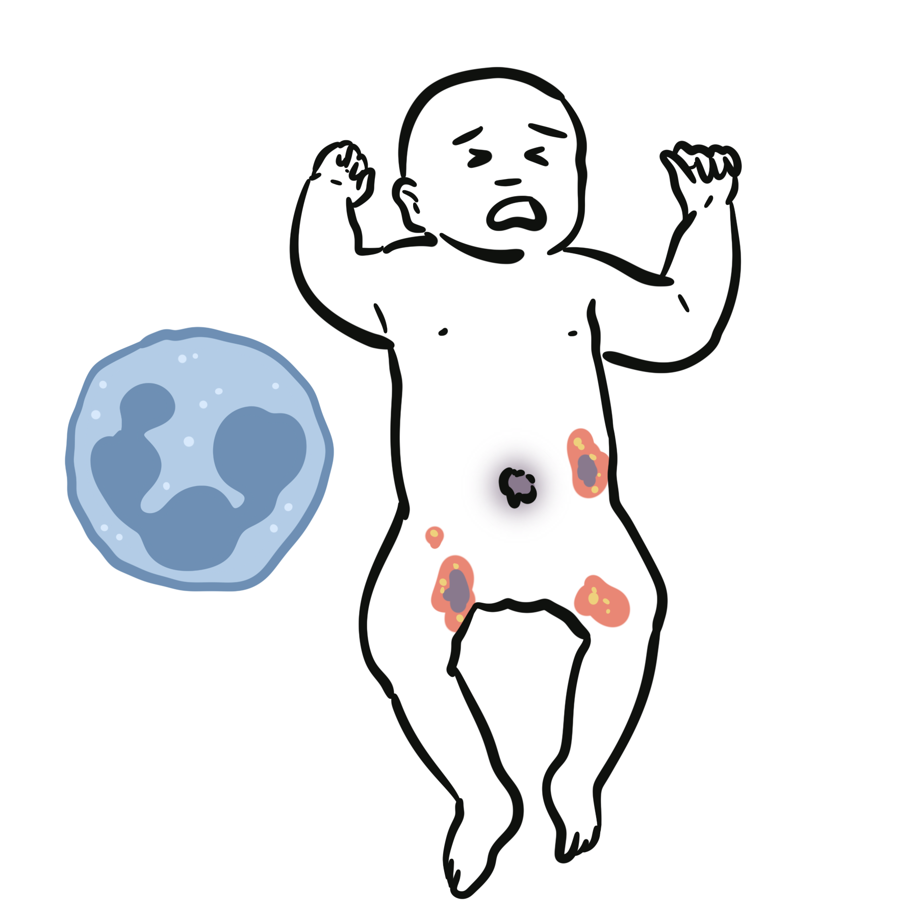
- Rash Progression: Rapidly evolving petechiae that coalesce into dark purple, necrotic, irregular plaques (Purpura Fulminans).
- Immediate Action: High-flow oxygen, 2x large-bore IV/IO access, fluid resuscitation (10–20 mL/kg balanced crystalloid bolus), and immediate parenteral antibiotics (Ceftriaxone / Cefotaxime).
- Other Critical Red Flags:
- Non-Accidental Injury (NAI): Bruising/purpura in non-mobile infants, bruises on soft tissues (ears, neck, abdomen, buttocks), delayed presentation, inconsistent story.
- Thrombotic Microangiopathy (TMA / HUS): Pallor, petechiae, oliguria following a bloody diarrheal illness.
Step 2: Differentiate Palpable vs. Non-Palpable (Macular) Purpura
- Palpable Purpura (Elevated / Infiltrated Lesions):
- -> Points directly to Cutaneous Vasculitis (e.g., IgA Vasculitis / Henoch-Schönlein Purpura) or infectious embolization.
- Flat / Macular Purpura & Petechiae:
- -> Points to Platelet Disorders (Thrombocytopenia, platelet dysfunction), Coagulopathy, or Mechanical / Capillary Fragility.
Step 3: Branching Logic Guided by Platelet Count & Well-being
- Child Unwell / Febrile: -> Sepsis / Meningococcemia, Disseminated Intravascular Coagulation (DIC), Hemolytic Uremic Syndrome (HUS), or Acute Leukemia.
- Child Well + Isolated Profound Thrombocytopenia (Platelets < 20–30 x 10^9/L): -> Immune Thrombocytopenia (ITP).
- Child Well + Normal Platelet Count + Palpable Purpura on Lower Limbs: -> IgA Vasculitis (Henoch-Schönlein Purpura).
- Child Well + Normal Platelets + Petechiae strictly above the Nipple Line: -> Mechanical / Pressure-Induced (Post-coughing, vomiting, screaming).
Rapidly progressive purpura with fever, hypotension, altered consciousness or severe limb pain is a medical emergency. Do not wait for the platelet count, coagulation results or rash progression before commencing resuscitation and empirical treatment for suspected invasive infection.
DIFFERENTIAL DIAGNOSIS
Infectious Causes
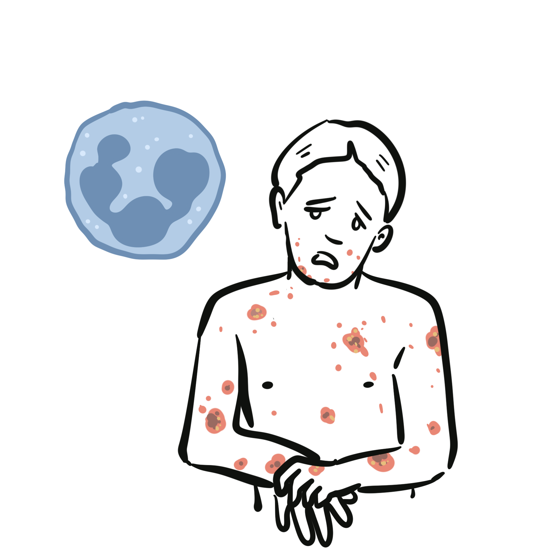
- Meningococcal disease: Fever, severe systemic illness and a petechial or purpuric rash that may spread rapidly. Headache, neck stiffness, altered mental state, hypotension, limb pain, cold extremities or multiorgan failure may occur. Rash may be absent early and meningism is not required.
- Sepsis with disseminated intravascular coagulation: Bleeding from puncture sites, widespread petechiae, ecchymoses, purpura, acral ischaemia or necrosis with shock and organ dysfunction.
- Purpura fulminans: Painful, rapidly progressive retiform purpura and skin necrosis caused by severe coagulation disturbance, commonly associated with overwhelming infection.
- Infective endocarditis: Fever, cardiac murmur, embolic phenomena, splinter haemorrhages, petechiae or Janeway lesions.
- Viral infection: May cause transient thrombocytopenia or a petechial eruption. Consider HIV, hepatitis viruses, Epstein–Barr virus, cytomegalovirus and other clinically relevant infections.
- Rickettsial or other invasive infection: Consider according to geography, travel, tick exposure and systemic features.
Platelet Disorders
- Immune thrombocytopenia: Isolated thrombocytopenia with petechiae, bruising or mucosal bleeding. The patient is often otherwise clinically well and the blood film lacks schistocytes. It remains a diagnosis of exclusion.
- Drug-induced immune thrombocytopenia: Abrupt, sometimes profound thrombocytopenia following exposure to a causative medicine.
- Bone-marrow failure or infiltration: Thrombocytopenia accompanied by anaemia, neutropenia, constitutional symptoms, lymphadenopathy, organomegaly or an abnormal blood film.
- Hypersplenism: Thrombocytopenia with splenomegaly, often in the context of portal hypertension or systemic disease.
- Platelet dysfunction: Mucocutaneous bleeding despite a normal or mildly reduced platelet count. Consider antiplatelet therapy, uraemia, myeloproliferative disease or an inherited disorder.
Thrombotic Microangiopathies
- Thrombotic thrombocytopenic purpura: Thrombocytopenia and microangiopathic haemolytic anaemia, often with neurological, cardiac, gastrointestinal or renal involvement. Fever and the complete historical pentad are frequently absent.
- Haemolytic uraemic syndrome: Microangiopathic haemolysis, thrombocytopenia and acute kidney injury. Shiga-toxin-associated disease often follows bloody diarrhoea, although other forms occur.
- Pregnancy-associated microangiopathy: Consider pre-eclampsia, HELLP syndrome, acute fatty liver of pregnancy and pregnancy-triggered TTP or atypical haemolytic uraemic syndrome.
Coagulation Disorders
- Disseminated intravascular coagulation: Thrombocytopenia, prolonged clotting times, increased D-dimer, falling fibrinogen and fragmented red cells in an appropriate clinical setting.
- Liver disease: Reduced coagulation-factor synthesis, thrombocytopenia from portal hypertension and qualitative platelet dysfunction may coexist.
- Vitamin K deficiency or warfarin excess: Prolonged prothrombin time with bruising or bleeding.
- Haemophilia or acquired factor inhibitor: Deep bleeding and extensive ecchymoses are more typical than isolated petechiae.
- Direct oral anticoagulant effect: Routine coagulation tests may not reliably quantify the anticoagulant effect.
Vasculitic and Inflammatory Causes
- Cutaneous small-vessel vasculitis: Palpable purpura, usually symmetric and concentrated over dependent lower limbs. It may be idiopathic or associated with a medicine, infection, autoimmune disease or malignancy.
- IgA vasculitis: Palpable purpura with arthralgia or arthritis, abdominal pain and renal involvement. Adults may have more severe renal disease than children.
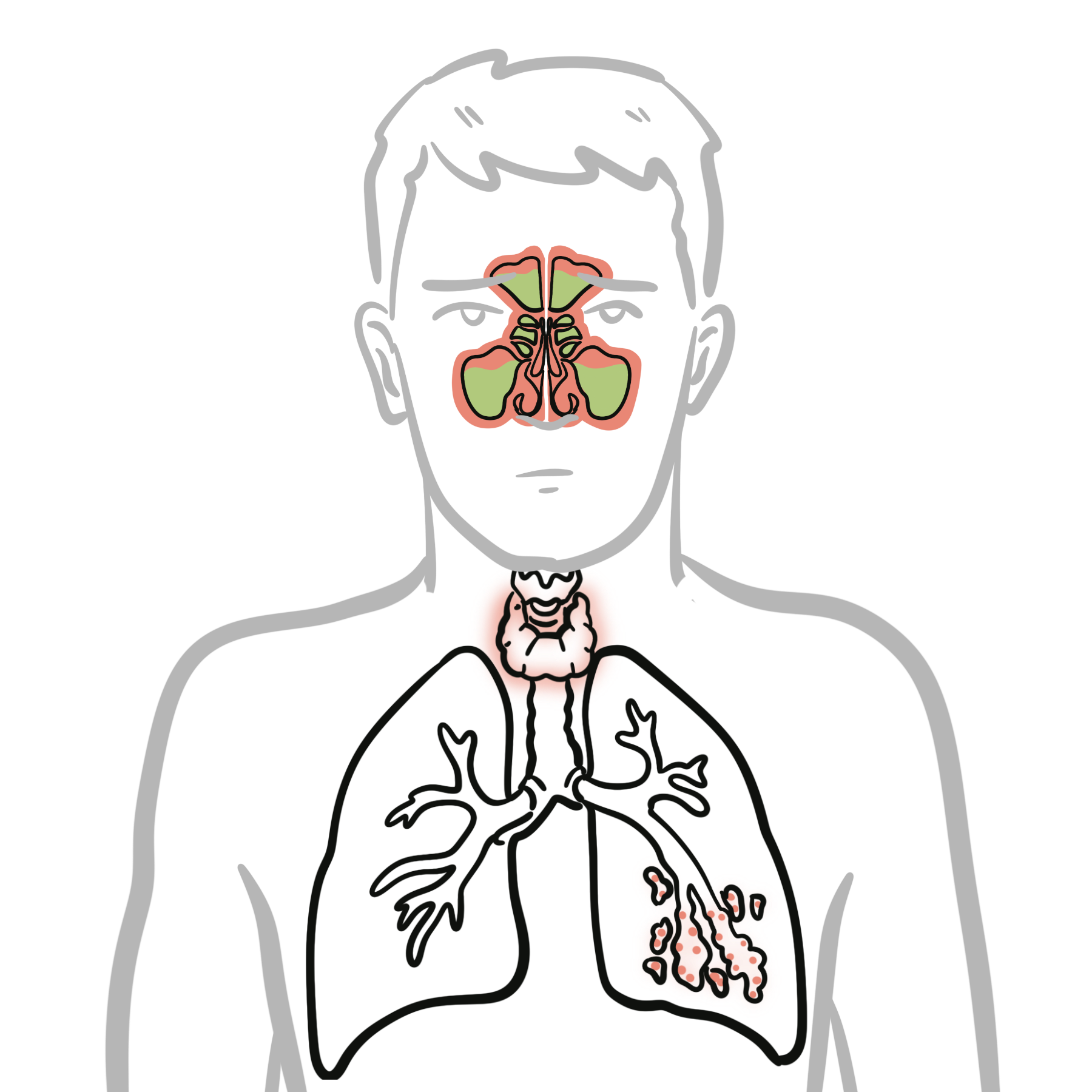
- ANCA-associated vasculitis: Purpura accompanied by glomerulonephritis, pulmonary disease, ENT involvement, neuropathy or constitutional symptoms.
- Cryoglobulinaemic vasculitis: Purpura, arthralgia, weakness, neuropathy or renal disease; assess for hepatitis C, autoimmune disease and lymphoproliferative disorders.
- Connective-tissue disease-associated vasculitis: Consider systemic lupus erythematosus, rheumatoid vasculitis and Sjögren disease where supported by the phenotype.
Vascular-Occlusive Disorders
- Antiphospholipid syndrome: Thrombosis, pregnancy morbidity, livedo or retiform purpura.
- Calciphylaxis: Extremely painful retiform plaques progressing to necrosis, usually in advanced kidney disease but occasionally without it.
- Cholesterol embolisation: Livedo reticularis, blue toes, purpura, renal impairment and eosinophilia, often following a vascular procedure.
- Septic emboli: Painful or necrotic peripheral lesions in a patient with bacteraemia or endocarditis.
- Warfarin-induced skin necrosis: Painful plaques and necrosis developing after warfarin initiation, particularly in areas rich in subcutaneous fat.
Benign, Mechanical and Dermatological Causes
- Pressure-related petechiae: Localised lesions following vomiting, coughing, seizures, strenuous activity or tourniquet pressure.
- Trauma or suction injury: Distribution corresponds to the mechanical exposure.
- Actinic or senile purpura: Recurrent ecchymoses on sun-damaged extensor surfaces in older adults.
- Corticosteroid-associated purpura: Thin, fragile skin with easy bruising after systemic or potent topical corticosteroid exposure.
- Pigmented purpuric dermatosis: Chronic, non-palpable red-brown or “cayenne pepper” lesions, commonly affecting the lower legs.
- Scurvy: Perifollicular haemorrhage, corkscrew hairs, gingival disease, poor wound healing and nutritional risk.
- Non-accidental injury: Consider when the lesion pattern, developmental stage or history is inconsistent with accidental trauma.
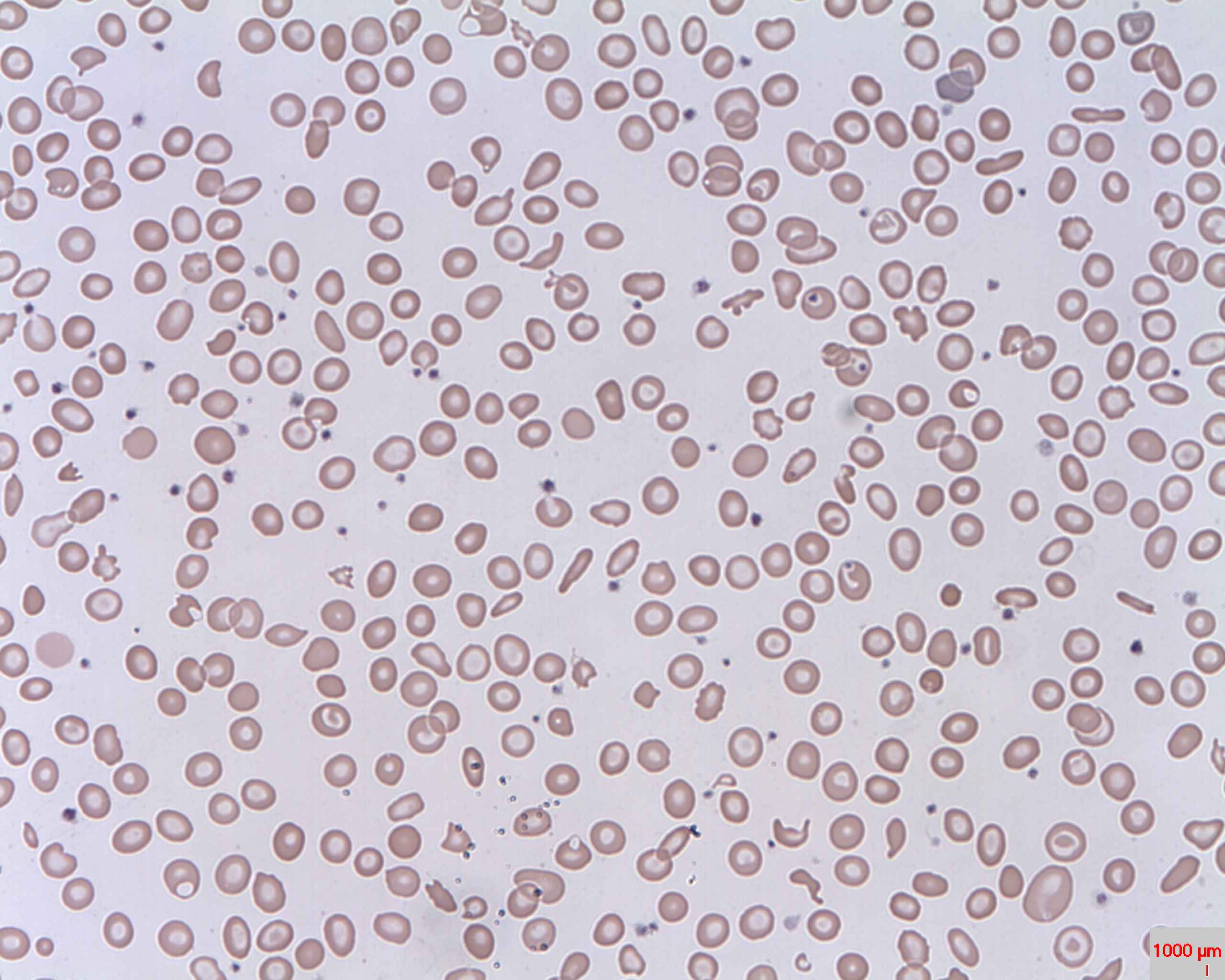
Thrombocytopenia with schistocytes is not immune thrombocytopenia until thrombotic microangiopathy and disseminated intravascular coagulation have been excluded. TTP may present without fever, renal failure or the complete classical pentad.
INVESTIGATIONS
Immediate Bedside Assessment
- Record temperature, heart rate, respiratory rate, blood pressure, oxygen saturation, capillary refill and mental state.
- Assess for shock, active bleeding, meningism and focal neurological abnormalities.
- Examine the complete skin surface and document progression; mark the margins of rapidly evolving purpura where appropriate.
- Perform urinalysis for blood and protein.
- Measure capillary glucose in an acutely unwell or neurologically impaired patient.
- In children, assess age-appropriate behaviour, feeding, interaction and hydration.
First-Line Laboratory Tests
- Full blood count: Determine platelet count and assess for anaemia, neutropenia, leucocytosis, lymphocytosis or pancytopenia.
- Peripheral blood film: Exclude platelet clumping and look for schistocytes, blasts, dysplasia, abnormal lymphocytes or another haematological clue.
- Coagulation profile: Prothrombin time/INR, activated partial thromboplastin time and fibrinogen.
- D-dimer: Supports activation of coagulation and fibrinolysis but is non-specific.
- Renal function and electrolytes: Identify acute kidney injury and complications of haemolysis or sepsis.
- Liver function tests: Assess hepatic dysfunction, haemolysis and systemic illness.
- Inflammatory markers: CRP and, where appropriate, ESR.
- Urinalysis and urine protein quantification: Detect glomerular haematuria and proteinuria in systemic vasculitis.
- Pregnancy testing: When pregnancy is possible and would alter the differential or management.
Investigation for Suspected Infection or Sepsis
- Obtain blood cultures before antibiotics when this does not delay treatment.
- Measure venous or arterial blood gas, lactate, glucose and acid–base status.
- Obtain meningococcal PCR or other pathogen-specific testing according to local protocols.
- Perform lumbar puncture only when meningitis is suspected and it is safe to proceed.
- Do not delay antibiotics to obtain lumbar puncture or imaging.
- Evaluate for another infection source with targeted urine, respiratory, wound or imaging investigations.
- Notify public-health authorities promptly when meningococcal disease is suspected or confirmed.
Investigation for Haemolysis or Thrombotic Microangiopathy
- Blood film for schistocytes, reticulocyte count, lactate dehydrogenase, bilirubin and haptoglobin.
- Direct antiglobulin test, creatinine and urinalysis.
- Troponin and ECG where TTP is suspected.
- ADAMTS13 activity and inhibitor testing before plasma treatment where possible.
- Stool Shiga-toxin testing when diarrhoea-associated haemolytic uraemic syndrome is suspected.
Use a validated pre-test tool such as the PLASMIC score to support assessment for severe ADAMTS13 deficiency, but do not allow scoring or ADAMTS13 results to delay treatment when clinical suspicion for TTP is high.
Investigation for Suspected Vasculitis
- Urine microscopy and urine protein-to-creatinine or albumin-to-creatinine ratio.
- Creatinine and estimated glomerular filtration rate.
- ANA, extractable nuclear antigens, double-stranded DNA antibodies and complement where connective-tissue disease is suspected.
- ANCA when the clinical phenotype suggests ANCA-associated vasculitis.
- Serum immunoglobulins and electrophoresis.
- Cryoglobulins, collected and transported under appropriate temperature-controlled conditions.
- Hepatitis B, hepatitis C and HIV testing where clinically indicated.
- Blood cultures and echocardiography when endocarditis is possible.
- Chest imaging when pulmonary vasculitis or pulmonary haemorrhage is suspected.
Skin Biopsy
- Biopsy a fresh, representative lesion when cutaneous vasculitis, vascular occlusion or another inflammatory dermatosis is suspected.
- For suspected small-vessel vasculitis, obtain a punch biopsy from a recently developed purpuric papule for routine histopathology.
- Obtain a separate specimen from a fresh lesion for direct immunofluorescence when IgA vasculitis or immune-complex vasculitis is possible.
- Avoid sampling only necrotic tissue because it may not demonstrate the primary pathological process.
- Do not delay emergency treatment to obtain a biopsy.
Additional Haematological Testing
- HIV, hepatitis B and hepatitis C testing.
- Vitamin B12, folate and iron studies.
- Coagulation-factor assays or inhibitor testing.
- Platelet-function testing after specialist discussion.
- HIT assessment using the 4Ts score followed by appropriate laboratory testing.
- Bone-marrow examination for unexplained cytopenias, abnormal circulating cells or suspected marrow failure or malignancy.
- Abdominal imaging when splenomegaly, liver disease or occult malignancy is suspected.
Always review the blood film personally or obtain expert laboratory review when severe thrombocytopenia, haemolysis or an unexplained systemic illness is present. Platelet clumping can produce pseudothrombocytopenia, whereas schistocytes may signal a time-critical thrombotic microangiopathy.
CRITICAL MANAGEMENT
Suspected Meningococcal Disease or Sepsis
- Initiate immediate ABC assessment and continuous physiological monitoring.
- Obtain intravenous or intraosseous access and support oxygenation and ventilation as required.
- Obtain blood cultures and relevant samples promptly, but do not delay antibiotics.
- Administer immediate age-appropriate parenteral antimicrobial therapy according to local meningococcal and sepsis guidelines, commonly ceftriaxone or cefotaxime.
- Give intravenous fluids judiciously and reassess frequently.
- Initiate vasopressor support when shock persists despite appropriate initial fluid resuscitation.
- Monitor lactate, urine output, glucose, electrolytes, coagulation and organ function.
- Seek urgent critical-care and infectious-diseases input.
- Apply appropriate infection-control precautions.
- Notify public health and arrange chemoprophylaxis for eligible close contacts according to local guidance.
Disseminated Intravascular Coagulation and Purpura Fulminans
- Treat the underlying cause immediately, particularly sepsis.
- Involve critical care, haematology and relevant surgical teams.
- Monitor platelet count, prothrombin time, activated partial thromboplastin time, fibrinogen and D-dimer serially.
- Provide platelets, fibrinogen replacement or plasma when clinically significant bleeding, an urgent procedure or severe haemostatic depletion warrants it.
- Do not transfuse solely to normalise laboratory results in a stable, non-bleeding patient.
- Assess urgently for limb ischaemia, compartment syndrome and evolving skin necrosis.
Suspected Thrombotic Thrombocytopenic Purpura
- Contact haematology immediately.
- Collect ADAMTS13 testing before plasma treatment if this causes no delay.
- Commence urgent therapeutic plasma exchange and corticosteroid therapy when immune TTP is strongly suspected.
- Consider early caplacizumab and rituximab according to specialist guidance and availability.
- Monitor neurological, cardiac and renal complications.
- Avoid prophylactic platelet transfusion unless there is life-threatening bleeding or an essential invasive procedure, because transfusion may worsen microvascular thrombosis.
Severe Immune Thrombocytopenia
- Base treatment on bleeding severity, platelet count, comorbidities, anticoagulant use and the need for procedures.
- Use corticosteroids as first-line therapy when treatment is indicated.
- Use intravenous immunoglobulin when a rapid platelet increase is required or significant bleeding is present.
- Life-threatening bleeding requires urgent haematology-directed combined treatment, generally including corticosteroids, intravenous immunoglobulin and platelet transfusion.
- Avoid aspirin, NSAIDs, intramuscular injections and unnecessary invasive procedures.
- Do not diagnose immune thrombocytopenia until alternative causes of thrombocytopenia have been assessed.
Heparin-Induced Thrombocytopenia
- Calculate the 4Ts score.
- If the clinical probability is intermediate or high, stop all heparin products, including flushes.
- Send appropriate HIT testing and commence a non-heparin anticoagulant when safe and indicated.
- Investigate for thrombosis.
- Do not wait for confirmatory testing before acting when the probability is high.
Suspected Systemic Vasculitis
- Assess urgently for glomerulonephritis, pulmonary haemorrhage, mononeuritis multiplex, gastrointestinal ischaemia and other organ-threatening disease.
- Obtain appropriate cultures and exclude important infectious mimics.
- Seek rheumatology, renal, respiratory or other specialist input according to the organs involved.
- Commence immunosuppression promptly when organ- or life-threatening vasculitis is established or strongly suspected, while balancing the risk of occult infection.
- Skin-limited disease without systemic involvement may be managed less urgently according to its cause and severity.
Supportive Measures
- Stop or reverse causative medications when appropriate.
- Apply direct pressure to active external bleeding.
- Avoid intramuscular injections and unnecessary invasive procedures in severe thrombocytopenia or coagulopathy.
- Use red-cell transfusion according to clinical bleeding, haemodynamic status and haemoglobin.
- Treat pain while avoiding medicines that worsen platelet dysfunction where possible.
- Provide clear safety-net advice for patients managed outside hospital.
Fever with a rapidly progressive petechial or purpuric rash is a treatment-before-confirmation emergency. Likewise, suspected TTP requires plasma-exchange-based treatment before ADAMTS13 results return. In both conditions, waiting for diagnostic certainty can be fatal.
REFERENCES
- National Institute for Health and Care Excellence. Meningitis (bacterial) and meningococcal disease: recognition, diagnosis and management. NICE guideline NG240 [Internet]. London: NICE; 2024. Available from: https://www.nice.org.uk/guidance/ng240
- Centers for Disease Control and Prevention. Chapter 8: meningococcal disease. Manual for the Surveillance of Vaccine-Preventable Diseases [Internet]. Atlanta: CDC; 2024. Available from: https://www.cdc.gov/surv-manual/php/table-of-contents/chapter-8-meningococcal-disease.html
- Neunert C, Terrell DR, Arnold DM, et al. American Society of Hematology 2019 guidelines for immune thrombocytopenia. Blood Adv. 2019;3(23):3829–3866. doi:10.1182/bloodadvances.2019000966.
- Cuker A, Arepally GM, Chong BH, et al. American Society of Hematology 2018 guidelines for management of venous thromboembolism: heparin-induced thrombocytopenia. Blood Adv. 2018;2(22):3360–3392. doi:10.1182/bloodadvances.2018024489.
- Zheng XL, Vesely SK, Cataland SR, et al. ISTH guidelines for the diagnosis of thrombotic thrombocytopenic purpura. J Thromb Haemost. 2020;18(10):2486–2495. doi:10.1111/jth.15006.
- Zheng XL, Vesely SK, Cataland SR, et al. ISTH guidelines for treatment of thrombotic thrombocytopenic purpura. J Thromb Haemost. 2020;18(10):2496–2502. doi:10.1111/jth.15010.
- Franchini M, Lippi G, Manzato F. Recent acquisitions in the pathophysiology, diagnosis and treatment of disseminated intravascular coagulation. Thromb J. 2006;4:4. doi:10.1186/1477-9560-4-4.
- Goeser MR, Laniosz V, Wetter DA. A practical approach to the diagnosis, evaluation, and management of cutaneous small-vessel vasculitis. Am J Clin Dermatol. 2014;15(4):299–306. doi:10.1007/s40257-014-0076-6.
- Carlson JA. The histological assessment of cutaneous vasculitis. Histopathology. 2010;56(1):3–23. doi:10.1111/j.1365-2559.2009.03443.x.
- World Health Organization. Micronutrient deficiencies and nutrition guidance [Internet]. Geneva: WHO. Available from: https://www.who.int/health-topics/micronutrients











Members only discussions coming soon…