

Temporal Arteritis (Giant Cell Arteritis)
Overview
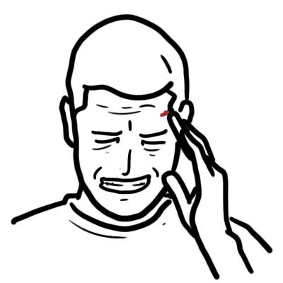
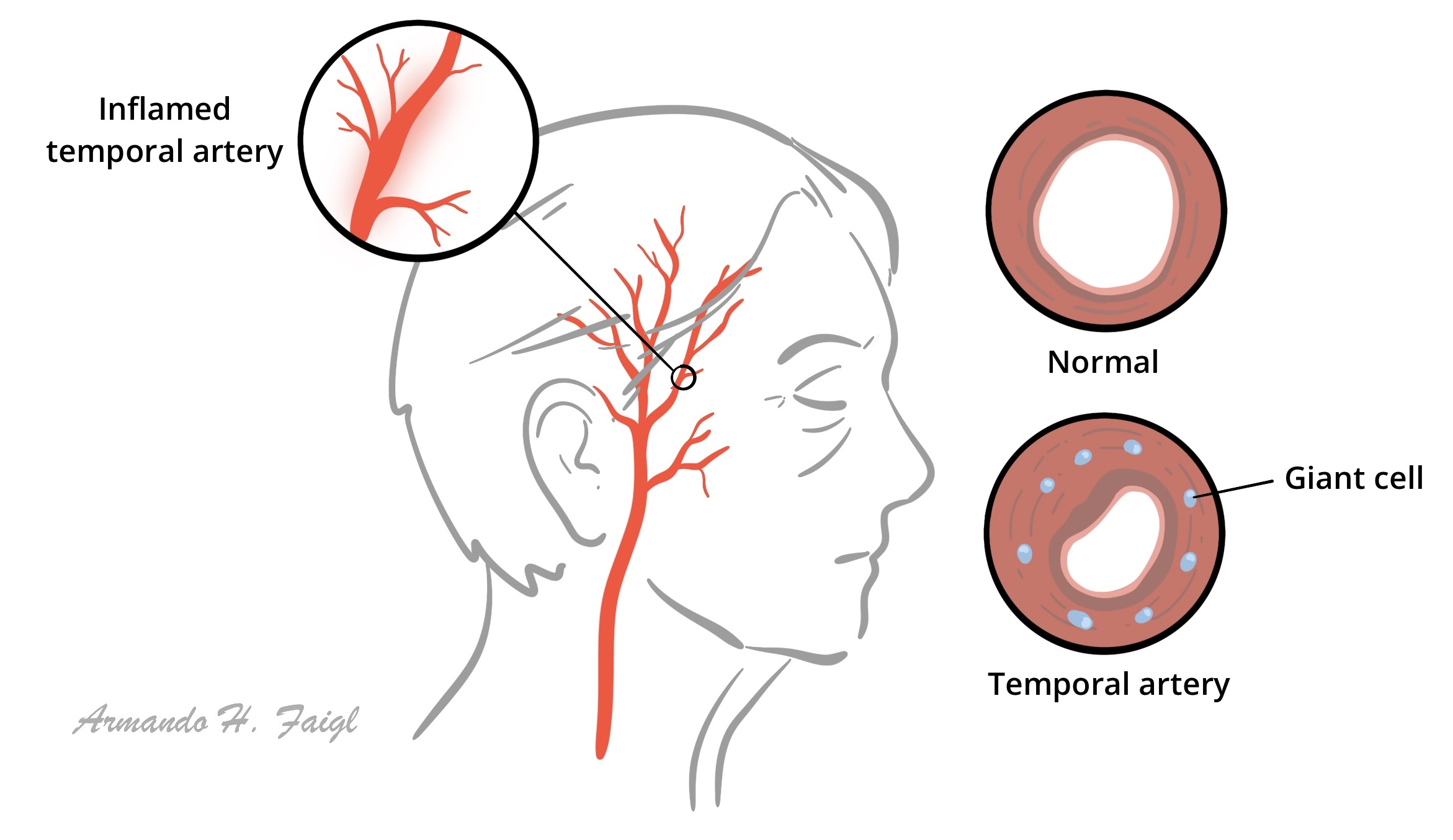
Giant cell arteritis (GCA), also known as temporal arteritis or cranial arteritis, is the most common systemic vasculitis in adults. It is a granulomatous panarteritis that selectively targets medium and large-sized arteries, displaying a strong tropism for the branches of the external carotid artery (particularly the superficial temporal artery) and the aorta.
The disease occurs almost exclusively in individuals older than 50 years of age and is closely associated with Polymyalgia Rheumatica (PMR). GCA is considered a medical emergency: transmural inflammation and luminal stenosis of ophthalmic arteries can lead to irreversible anterior ischemic optic neuropathy (AION) and sudden, permanent blindness. Prompt recognition and immediate empiric corticosteroid therapy are critical to prevent catastrophic visual loss.
Giant cell arteritis, the most common type of vasculitis in adults, affects medium and large arteries and can result in blindness.
Giant cell arteritis does not always involve the temporal artery and can affect other medium and large arteries including the aorta.
Definition
Giant cell arteritis: Systemic inflammatory vasculitis of unknown aetiology that occurs in older persons
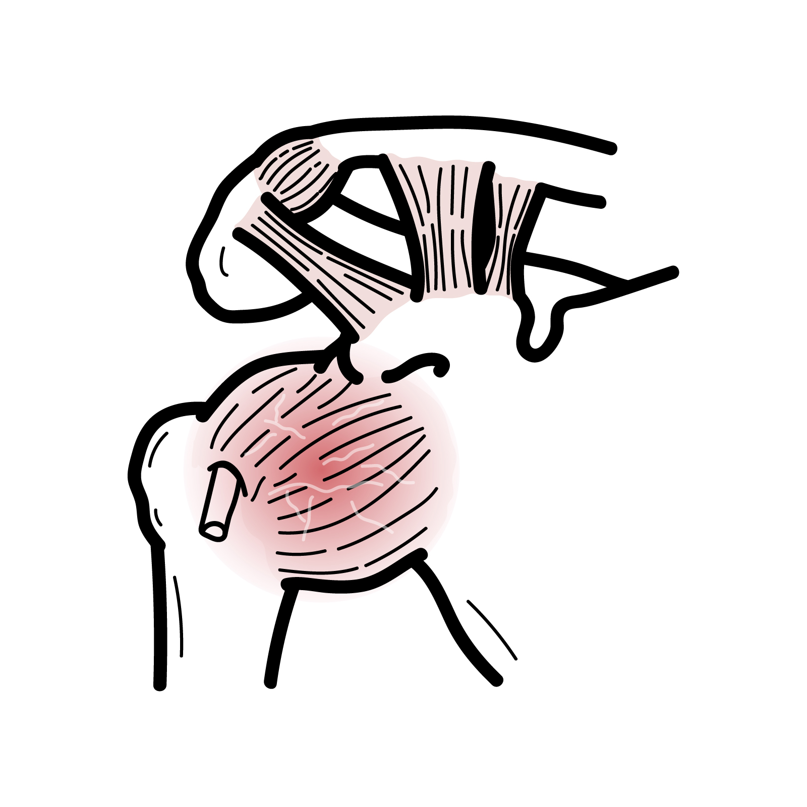
Polymyalgia rheumatica: Commonest inflammatory rheumatic disorder affecting older people. Patients typically present with bilateral shoulder pain, morning stiffness, raised inflammatory markers, and have a rapid response to low-dose corticosteroids
Takayasu’s arteritis: chronic granulomatous vasculitis affecting large arteries: primarily the aorta and its main branches.
Classification
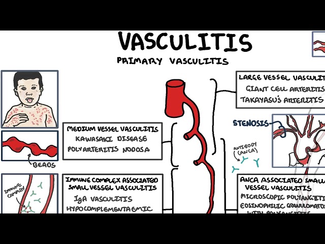
Under the Chapel Hill Consensus Conference criteria, GCA is classified as a Large Vessel Vasculitis (LVV) alongside Takayasu Arteritis.
| Feature | Giant Cell Arteritis (GCA) | Takayasu Arteritis |
| Age of Onset | > 50 years (peak 70–80 years) | < 50 years (peak 20–30 years) |
| Geographic / Ethnicity | Northern European descent | Asian descent |
| Primary Vessels | Cranial branches of external carotid, aorta | Aorta and its major primary branches |
| PMR Association | Strongly associated (~50% of cases) | Not associated |
The precursor protein determines the disease subtype, but age is the fundamental clinical discriminator between the two major large-vessel vasculitides: GCA occurs in patients > 50 years, whereas Takayasu occurs in patients < 50 years.
Pathophysiology
- Immune Activation in Adventitia: Resident dendritic cells in the arterial adventitia become activated, releasing chemokines that recruit circulating CD4+ T-lymphocytes (differentiating into Th1 and Th17 subsets).
- Granulomatous Panarteritis: Activated T-cells secrete Interferon-gamma (IFN-gamma), which transforms macrophages into multinucleated giant cells within the media and intima, forming characteristic granulomas.
- Arterial Wall Destruction: Macrophages release Matrix Metalloproteinases (MMPs) and Reactive Oxygen Species (ROS), causing fragmentation of the internal elastic lamina.
- Intimal Hyperplasia & Luminal Occlusion: Vascular Endothelial Growth Factor (VEGF) and Platelet-Derived Growth Factor (PDGF) drive smooth muscle proliferation and intimal thickening, culminating in arterial lumen stenosis, thrombosis, and distal tissue ischemia.
Granulomatous inflammation in GCA centers on the internal elastic lamina, and its degradation is a structural hallmark on biopsy.
Clinical Manifestation
Jaw claudication is the single most specific clinical feature of GCA. Amaurosis fugax is a medical emergency requiring immediate high-dose steroids to prevent permanent blindness.
Polymyalgia rheumatica Commonest inflammatory rheumatic disorder affecting older people. Patients typically present with bilateral shoulder pain, morning stiffness, raised inflammatory markers, and have a rapid response to low-dose corticosteroids.
Diagnosis
- Elevated ESR/CRP
- Temporal artery biopsy (Gold Standard)
- Histopathology: Shows transmural inflammation, multinucleated giant cells, histiocytes, T-cell infiltration, intimal hyperplasia, and fragmentation of the internal elastic lamina.
- Biopsy Length: Specimen length must be at least 1.5–2.0 cm to account for “skip lesions”.
- Biopsy should be performed within 1–2 weeks of starting steroids; do NOT delay steroid administration while awaiting biopsy.
- Imaging Studies (support diagnosis and can be used second line for diagnosis)
- Temporal Artery Ultrasound: Non-invasive first-line alternative showing the “Halo Sign” (hypoechoic circumferential wall edema around the arterial lumen).
- PET-CT / CT Angiography / MR Angiography: Used to evaluate extracranial large-vessel involvement (aortitis, subclavian, or axillary artery involvement).
A normal ESR/CRP makes GCA highly unlikely (high negative predictive value), but a biopsy MUST still be obtained to confirm the diagnosis. Never delay steroids while waiting for a biopsy!
Imperative to obtain adequate temporal artery biopsy (>2cm) because GCA presents as skip lesions.
ESR maybe normal initially.
Treatment
Management centers on rapid suppression of arterial inflammation using systemic corticosteroids and steroid-sparing biologic agents.
Corticosteroid Therapy (Immediate Initiation)
- Uncomplicated GCA (No visual loss or amaurosis fugax):
- Start Oral Prednisone 40–60 mg daily immediately upon clinical suspicion.
- Complicated GCA (Active visual loss or amaurosis fugax):
- Start IV Pulse Methylprednisolone 500–1000 mg daily for 3 consecutive days before transitioning to high-dose oral Prednisone.
- Tapering Schedule: Maintain high dose for 2–4 weeks until symptoms resolve and inflammatory markers normalize, then slowly taper over 12–24 months.
Steroid-Sparing Biologic Agents
- Tocilizumab (Anti-IL-6 Receptor Monoclonal Antibody)
- IL17 and JAKi have also been used (clinical trials show good results)
Adjuvant & Preventative Care
- Low-dose Aspirin (75–100 mg daily) to reduce cranial ischemic events.
- Bisphosphonates, Calcium, and Vitamin D co-prescription for all patients on long-term steroid therapy.
- PPI
If you suspect GCA do ESR and start prednisolone immediately without waiting biopsy results!
If you suspect GCA in a patient with visual symptoms, administer IV Pulse Methylprednisolone immediately. Every hour of delay increases the risk of permanent blindness in the affected or fellow eye
Complications & Prognosis
Complications
- Permanent Blindness: Bilateral in up to 20–50% of untreated cases due to irreversible AION or central retinal artery occlusion (CRAO).
- Thoracic aortic aneurysms occur 17 times more frequently in GCA patients than age-matched controls, often presenting late (years after initial diagnosis).
- Ischemic stroke secondary to vertebrobasilar or carotid artery occlusion.
- Iatrogenic Corticosteroid Toxicity
- Osteoporosis/fractures
- Diabetes mellitus
- Hypertension, cataracts
- Secondary infections
- Myopathy.
Prognosis
- Vision lost prior to initiating steroid therapy rarely recovers, but immediate treatment protects the contralateral eye in > 95% of cases.
- Relapses occur in 30–50% of patients during steroid tapering, requiring prolonged treatment (average duration 2 years).
- Periodic imaging (e.g., chest X-ray, CT, or MRI every 2–5 years) is recommended to screen for late thoracic aortic aneurysms.
GCA and PMR are among the most common inflammatory rheumatic diseases in the elderly; the prevalence of these diseases is expected to increase due to ageing of the population.
GCA patients require long-term surveillance even after successful steroid tapering due to the late risk of silent thoracic aortic aneurysm formation.
References
- Weyand CM, Goronzy JJ. Clinical practice. Giant-cell arteritis and polymyalgia rheumatica. N Engl J Med. 2014;371(1):50-57. doi:10.1056/NEJMcp1214825
- Dejaco C, Brouwer E, Mason JC, et al. To scan or not to scan in giant cell arteritis: imaging first strategy. Ann Rheum Dis. 2023;82(2):167-174. doi:10.1136/ard-2022-223326
- Stone JH, Tuckwell K, Dimonaco S, et al. Trial of Tocilizumab in Giant-Cell Arteritis. N Engl J Med. 2017;377(4):317-328. doi:10.1056/NEJMoa1613849









Members only discussions coming soon…