

Overview
A cervical stinger or burner is a transient peripheral nerve injury caused by traction, compression or direct trauma to a cervical nerve root or the upper brachial plexus. It typically occurs during collision or contact sport and produces sudden unilateral burning, stinging or electric-shock pain radiating from the neck or shoulder into the arm.
The C5 and C6 nerve roots or upper trunk of the brachial plexus are most commonly affected. Associated paraesthesia and weakness may occur, particularly involving shoulder abduction and elbow flexion. Symptoms usually resolve within seconds to minutes but may persist for hours, days or occasionally longer when axonal injury has occurred.
A stinger is a diagnosis of exclusion following trauma. Bilateral symptoms, lower-limb involvement, persistent neck pain, cervical tenderness, altered consciousness or bladder/bowel disturbance are not typical and must prompt immediate management as a possible cervical spinal cord or unstable spinal injury.
Definition
- Stinger/burner
- Transient unilateral neurological symptoms caused by injury to a cervical nerve root or the brachial plexus.
- Brachial plexus
- A network formed predominantly by the C5–T1 nerve roots that supplies motor and sensory innervation to the upper limb.
- Neurapraxia
- Temporary conduction block without axonal disruption; recovery is usually rapid and complete.
- Axonotmesis
- Disruption of axons with preservation of some supporting connective tissue; recovery requires axonal regeneration and is slower.
- Cervical cord neurapraxia
- Transient spinal cord dysfunction causing bilateral limb symptoms, sometimes including all four limbs; it is distinct from a unilateral stinger and requires urgent assessment.
- Return to play
- Medical clearance to resume sport after symptoms and neurological abnormalities have completely resolved.
Anatomy & Physiology
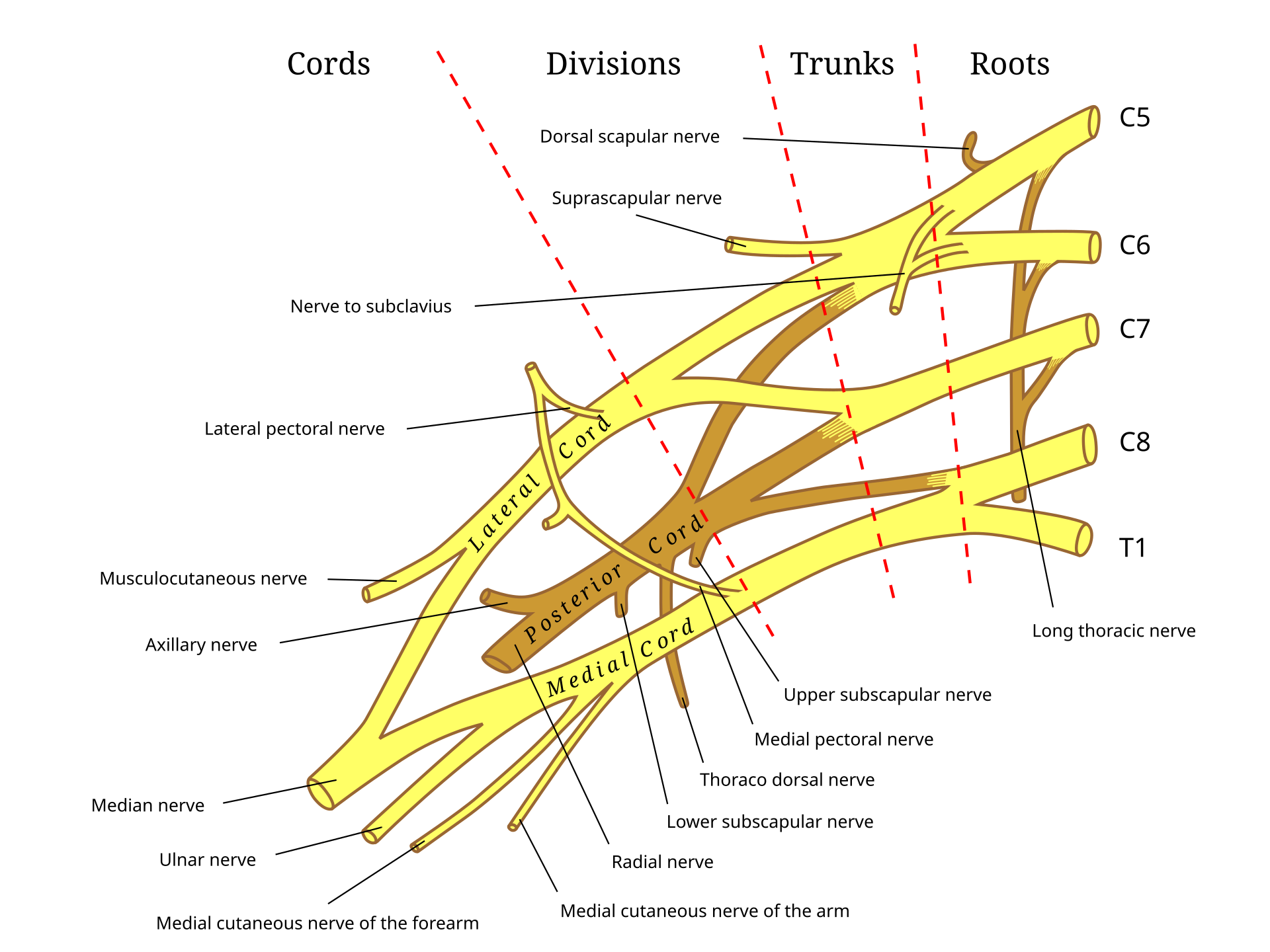
The brachial plexus is formed by the anterior rami of the C5–T1 spinal nerve roots. These roots combine into trunks, divisions, cords and terminal peripheral nerves.
The upper trunk, formed primarily by C5 and C6, supplies muscles including:
- Deltoid: shoulder abduction.
- Supraspinatus: initiation of shoulder abduction.
- Infraspinatus: external shoulder rotation.
- Biceps: elbow flexion and forearm supination.
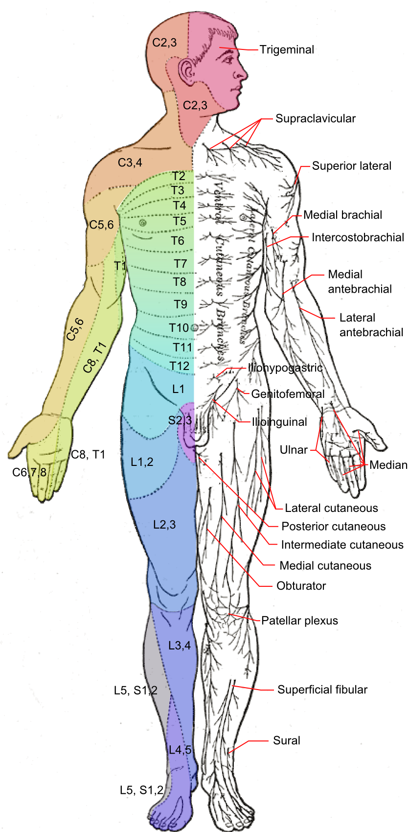
The C5–C6 sensory distribution includes the lateral shoulder, upper arm, lateral forearm and thumb region. Considerable overlap exists between adjacent roots and peripheral nerves, so clinical patterns may be incomplete.
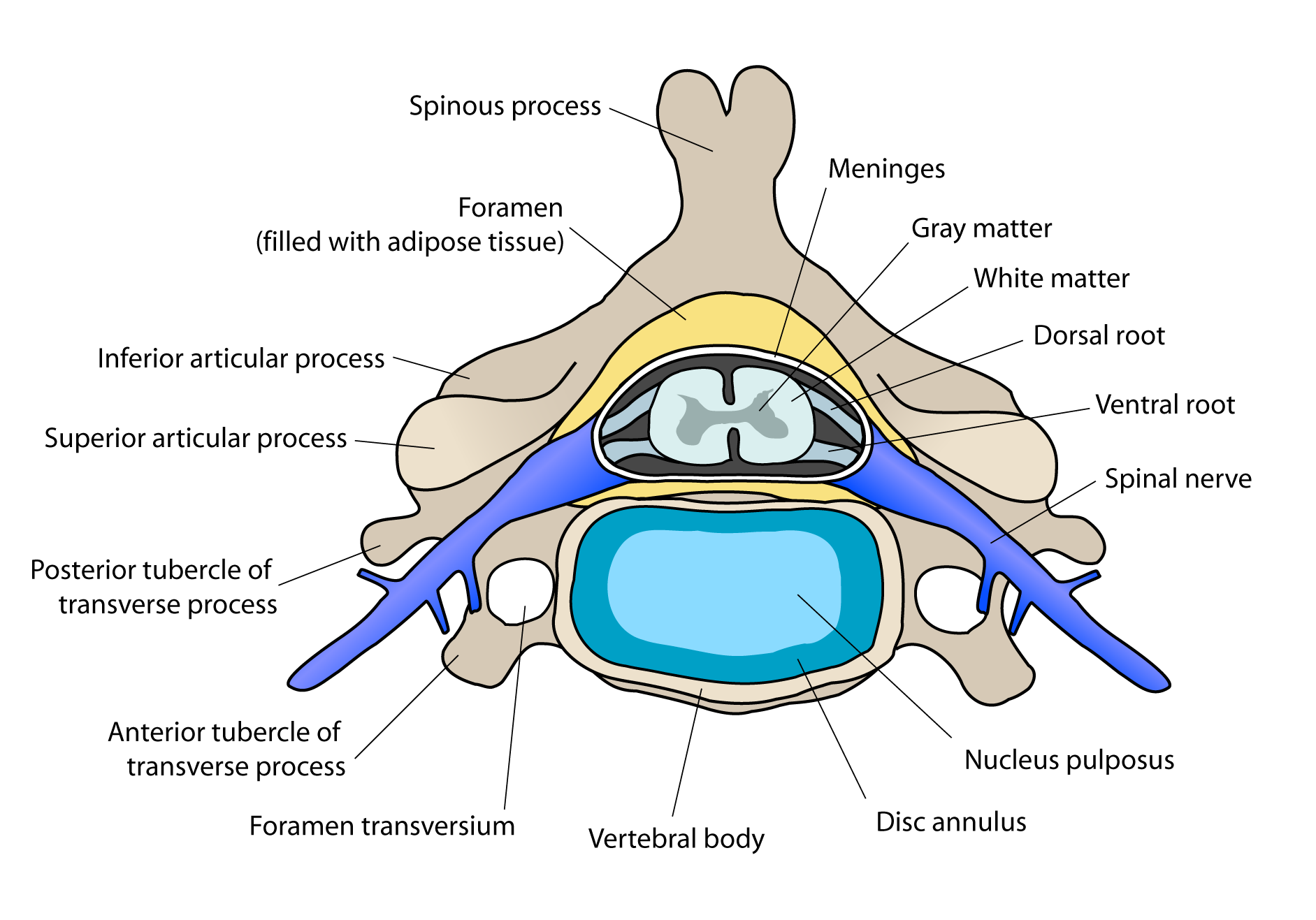
Cervical extension, ipsilateral rotation and lateral flexion narrow the intervertebral foramina. Conversely, depression of the shoulder while the head bends away stretches the cervical roots and upper brachial plexus.
A positive Spurling manoeuvre and neck pain favour a cervical nerve-root mechanism. A painless neck with symptoms produced by shoulder depression and contralateral neck flexion may favour an upper-trunk brachial plexus injury.
Aetiology & Risk Factors
Mechanisms of Injury
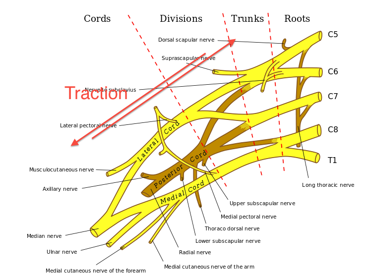
- Traction: the shoulder is forced downward while the neck bends away from the affected side, stretching the upper brachial plexus.
- Compression: the neck is forced into extension, rotation or lateral flexion toward the affected side, narrowing the neural foramen and compressing an exiting cervical root.
- Direct trauma: a blow to the supraclavicular region or lower lateral neck directly compresses the brachial plexus.
- Combined mechanism: collision injuries frequently involve simultaneous traction, compression and direct impact.2,3
Risk Factors
- Collision and contact sports, particularly rugby, American football, wrestling, ice hockey and lacrosse.
- Previous stinger or burner injury.
- Incorrect tackling or blocking technique.
- Leading with the head or “spear” tackling.
- Weak cervical, shoulder-girdle or scapular stabilising muscles.
- Poorly fitted or inadequate protective equipment.
- Cervical foraminal or central canal stenosis.
- Degenerative cervical disc disease or osteophyte formation.
- Congenital cervical canal narrowing.
- Playing positions involving frequent high-energy collisions.
Pathophysiology
A stinger represents a spectrum of peripheral nerve injury:
- Mechanical stretch, compression or direct impact disrupts normal nerve conduction.
- Mild injury produces focal demyelination and neurapraxia without axonal disruption.
- The resulting conduction block causes immediate burning pain, paraesthesia and transient weakness.
- Symptoms resolve as membrane function and conduction recover.
- More severe trauma may disrupt axons, producing axonotmesis, denervation and prolonged weakness.
- Recurrent injury before complete recovery can result in cumulative neurological damage, chronic pain, muscle atrophy and persistent weakness.
Compression injuries more commonly involve the cervical nerve root, whereas traction and direct-impact injuries more commonly affect the upper brachial plexus. This distinction is not always clinically clear.
Clinical Manifestations
Typical Features
- Immediate unilateral burning, stinging or electric-shock pain.
- Pain radiating from the neck or shoulder down one arm.
- Warmth, tingling or numbness in the affected arm.
- Transient weakness, heaviness or a “dead arm” sensation.
- Symptoms commonly affecting a C5–C6 or upper-trunk distribution.
- Reduced shoulder abduction, external rotation or elbow flexion strength.
- Symptoms lasting seconds to minutes in uncomplicated neurapraxia.
Pain commonly resolves before weakness. Persistent weakness may therefore remain even after the athlete reports feeling better.
Clinical Examination
- Unilateral sensory disturbance
- Weakness of the deltoid, biceps, supraspinatus or infraspinatus.
- Reduced biceps or brachioradialis reflex, although reflexes may remain normal.
- Positive Spurling manoeuvre when cervical foraminal compression is responsible.
- The athlete may support the affected arm against the body or with the opposite hand.
- Serial examinations may demonstrate progressive recovery of sensation and strength.
A typical stinger is unilateral. Bilateral symptoms or lower-limb involvement suggest possible spinal cord injury and require immediate cervical-spine precautions and emergency assessment.
Diagnosis
Diagnosis is usually clinical, based on the traumatic mechanism, immediate unilateral upper-limb symptoms and subsequent neurological recovery. The first priority is excluding cervical fracture, instability, spinal cord injury and significant shoulder trauma.=
Imaging
Imaging is not usually required after a first unilateral episode that resolves rapidly and is associated with a benign cervical and neurological examination.
Imaging should be considered when there is:
- Recurrent injury.
- Bilateral neurological symptoms.
- Neck pain, midline tenderness or restricted movement.
- Symptoms lasting longer than 24 hours.
- Persistent weakness or sensory loss.
- Concern about cervical fracture, instability, disc herniation or stenosis.
- An atypical mechanism or clinical presentation.
Plain cervical radiographs may identify fracture, malalignment or degenerative changes.
CT provides detailed assessment of fracture and bony anatomy.
MRI is preferred when spinal cord injury, disc herniation, ligamentous injury, nerve-root compression, foraminal stenosis or central canal stenosis is suspected.
Flexion–extension radiographs should not be performed acutely on the field and should only be considered in an appropriate supervised clinical setting.
Electrodiagnostic Testing
Nerve-conduction studies and electromyography may be useful when:
- Weakness or sensory symptoms persist.
- The lesion site is uncertain.
- Cervical radiculopathy must be distinguished from brachial plexopathy.
- Axonal injury is suspected.
- Recovery needs to be monitored.
Abnormal sensory nerve responses favour a postganglionic brachial plexus lesion. Denervation of cervical paraspinal muscles supports a nerve-root lesion. Fibrillation potentials may take several weeks to appear, so an early normal study does not exclude axonal injury.3
Differential Diagnosis
- Cervical fracture or instability.
- Cervical spinal cord injury or cord neurapraxia.
- Cervical disc herniation with radiculopathy.
- Traumatic brachial plexus rupture or avulsion.
- Shoulder dislocation or fracture.
- Clavicle fracture.
- Rotator-cuff injury.
- Peripheral mononeuropathy.
- Vascular injury to the upper limb.
- Concussion or head injury.
Treatment
Immediate Management
- Remove the athlete from play.
- Do not allow continued participation while pain, paraesthesia or weakness is present.
- Assess for cervical spinal cord injury and other traumatic injuries.
- Initiate cervical-spine precautions when red flags or diagnostic uncertainty exist.
- Arrange emergency transport when bilateral symptoms, lower-limb involvement, persistent cervical pain or other signs of serious injury are present.
- Perform and document serial neurological examinations.
Conservative Management
Most uncomplicated stingers are managed non-operatively:
- Relative rest from contact activity.
- Ice and simple analgesia or an NSAID when appropriate.
- Restoration of pain-free cervical and shoulder movement.
- Progressive strengthening of the cervical, scapular and shoulder muscles.
- Correction of posture, muscular imbalance and sporting technique.
- Review of tackling or collision mechanics.
- Inspection and refitting of protective equipment.
A soft sling may briefly improve comfort but should not be used for prolonged immobilisation. Routine corticosteroid treatment is not supported for an uncomplicated stinger.
Persistent or Recurrent Injury
- Arrange formal sports medicine, rehabilitation, neurology or spinal assessment.
- Obtain cervical imaging where indicated.
- Consider electrodiagnostic testing.
- Continue targeted rehabilitation until strength and function return.
- Investigate cervical stenosis, disc disease or another structural predisposition.
- Consider avoiding further collision sport when persistent neurological deficits, significant instability or recurrent severe injuries are present.
Surgery is rarely required for the stinger itself. It may be considered when a surgically correctable cervical lesion causes persistent nerve-root compression or progressive neurological dysfunction.
Return to Play
Return-to-play recommendations vary, but there is broad agreement that an athlete must have all of the following:3,5,6
- Complete resolution of pain, burning, numbness and paraesthesia.
- Full, pain-free cervical range of motion.
- Normal cervical and upper-limb neurological examination.
- Full, symmetric upper-limb strength.
- No cervical tenderness or evidence of instability.
- Ability to complete sport-specific activity without recurrent symptoms.
An athlete with a first uncomplicated stinger that resolves rapidly may be considered for same-day return after complete reassessment. However, the athlete must not return while any weakness, neck pain or neurological symptom persists.
A second stinger during the same game should exclude the athlete from further participation that day. Recurrent episodes, prolonged symptoms or three or more episodes in a season warrant specialist assessment and cervical imaging before further collision sport.3,5
Return-to-play decisions should not be based solely on MRI or electromyography. Clinical recovery, normal strength, pain-free movement, neurological examination, recurrence history and underlying anatomy must be considered together.
Symptom resolution alone is insufficient. Strength may remain impaired after pain has resolved, so bilateral strength and neurological function must be reassessed before return to play.
Complications & Prognosis
Complications
- Recurrent stinger or burner injury.
- Persistent neuropathic pain or paraesthesia.
- Chronic shoulder or arm weakness.
- Deltoid, biceps or shoulder-girdle muscle atrophy.
- Axonal loss with delayed or incomplete recovery.
- Missed cervical fracture, instability or spinal cord injury.
- Chronic cervical radiculopathy.
- Time lost from training or competition.
Prognosis
Most first-time stingers are neurapraxic injuries and resolve completely within minutes or hours. The prognosis is favourable when symptoms resolve rapidly and the neurological examination returns to normal.1–3
Persistent weakness, muscle atrophy, electrodiagnostic evidence of axonal loss, structural cervical disease and recurrent injury are associated with slower recovery. Neurological recovery may take weeks or months when axonotmesis has occurred.
Previous stinger injury substantially increases the risk of recurrence. Nevertheless, recurrence alone does not necessarily imply permanent neurological injury; each episode requires reassessment for cumulative weakness and underlying cervical pathology.
References
- American Academy of Orthopaedic Surgeons. Burners and stingers [Internet]. Rosemont: AAOS; 2026 [cited 2026 Sep 1]. Available from: https://www.orthoinfo.org/diseases–conditions/burners-and-stingers/.
- Ahearn BM, Starr HM, Seiler JG. Traumatic brachial plexopathy in athletes: current concepts for diagnosis and management of stingers. J Am Acad Orthop Surg. 2019;27(18):677–684. doi:10.5435/JAAOS-D-17-00746.
- Bowles DR, Canseco JA, Alexander TD, Schroeder GD, Hecht AC, Vaccaro AR. The prevalence and management of stingers in college and professional collision athletes. Curr Rev Musculoskelet Med. 2020;13(6):651–662. doi:10.1007/s12178-020-09665-5.
- Swartz EE, Boden BP, Courson RW, Decoster LC, Horodyski M, Norkus SA, et al. National Athletic Trainers’ Association position statement: acute management of the cervical spine-injured athlete. J Athl Train. 2009;44(3):306–331. doi:10.4085/1062-6050-44.3.306.
- Huang P, Anissipour A, McGee W, Lemak L. Return-to-play recommendations after cervical, thoracic, and lumbar spine injuries: a comprehensive review. Sports Health. 2016;8(1):19–25. doi:10.1177/1941738115610753.
- Schroeder GD, Canseco JA, Patel PD, Hilibrand AS, Kepler CK, Mirkovic S, et al. Updated return-to-play recommendations for collision athletes after cervical spine injury: a modified Delphi consensus study with the Cervical Spine Research Society. Neurosurgery. 2020;87(4):647–654. doi:10.1093/neuros/nyaa308.