

Overview
Lung cancer is a malignant tumour arising from cells within the lungs or bronchi. Two main groups:
- Non-small cell lung cancer (NSCLC): approximately 85%
- Adenocarcinoma
- Squamous cell carcinoma
- Large-cell carcinoma
- Small cell lung cancer (SCLC): approximately 15%
- More aggressive, with early nodal and distant metastasis.
Lung cancer is the leading cause of cancer death worldwide. Globally in 2022:
- Approximately 2.5 million new cases
- Approximately 1.8 million deaths
Incidence rises markedly with age. Tobacco smoking is the major risk factor, but lung cancer can occur in people who have never smoked.
Definition
Massive haemoptysis: More than 500 mL of blood loss that is coughed up within a 24-hour period
Horner Syndrome: Symptoms are ptosis, loss of pupillary dilation (miosis), and loss of sweating on the ipsilateral side (anhidrosis) caused by compression of the superior cervical ganglion and resultant loss of sympathetic innervation.
Superior Vena Cava syndrome: Obstruction of venous drainage, usually by external compression of the SVC, leading to edema of the face, neck, and upper part of the torso often with formation of collateral veins on the upper chest.
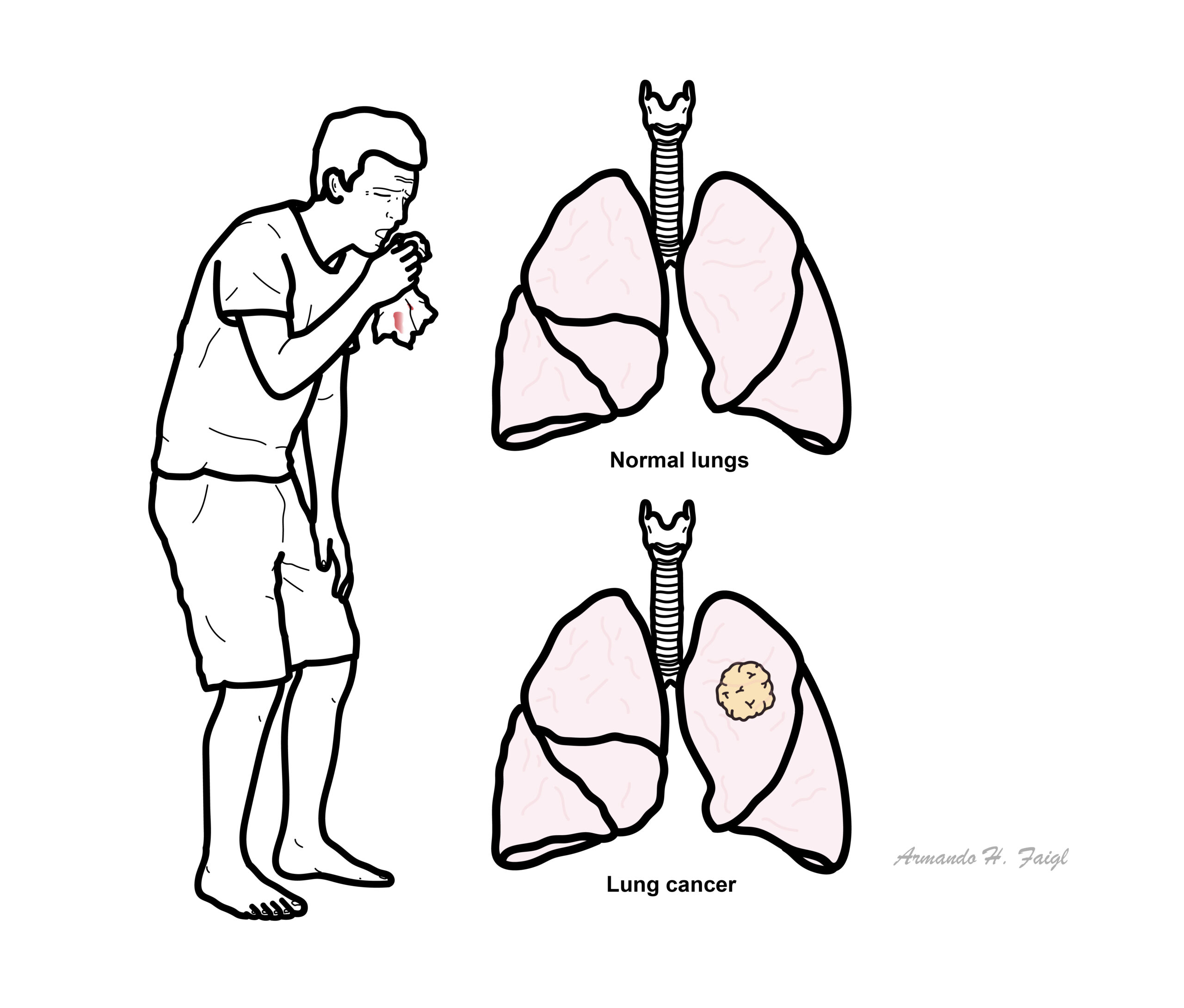
What to notice
Detailed image description
Aetiology & Risk Factors
There are many risk factors for lung cancer. Smoking is the most common.
Other risk factors:
- Passive smoking
- Radon
- Asbestos, silica and diesel exhaust
- Air pollution
- Previous thoracic radiotherapy
- Chronic lung disease
- Genetic susceptibility and family history.
Lung Carcinoma Classification
Small cell lung carcinoma (SCLC – 20%)
Non-small cell lung carcinoma ( NSCLC – 80%)
- Adenocarcinoma
- Squamous cell carcinoma
- Large cell
Staging of SCLC is limited or extensive. NSCLC uses the TNM classification for staging.
| Lung cancer characteristics | ||||
| SCLC | Adenocarcinoma | Squamous cell | Large cell | |
| Location | Central | Peripheral | Central | Peripheral |
| Cavitation | Never | Most likely | ||
| Metastases | Early | Early | Late | Late |
| Neoplastic Syndrome and Extrapulmonary manifestation | ADH, ACTH | Thrombophlebitis | PTH | Superior vena cava syndromes + hoarseness |
Clinical Manifestations
Respiratory symptoms
- Persistent new or changing cough
- Dyspnoea
- Haemoptysis
- Chest or shoulder pain
- Wheeze
- Recurrent or non-resolving pneumonia
- Hoarse voice
- Reduced exercise tolerance
Constitutional symptoms
- Unintentional weight loss
- Anorexia
- Fatigue and weakness
- Fever or night sweats
Early lung cancer is frequently asymptomatic and may be detected incidentally on imaging.
Local invasion or compression
- Recurrent laryngeal nerve involvement: hoarseness
- Phrenic nerve involvement: diaphragmatic paralysis
- Oesophageal compression: dysphagia
- Superior vena cava obstruction: facial/neck swelling, distended chest veins and dyspnoea
- Pancoast tumour: shoulder or arm pain, brachial plexopathy
- Horner syndrome: ptosis, miosis and anhidrosis
- Pleural or pericardial effusion
- Airway obstruction causing collapse or post-obstructive pneumonia.
Metastatic manifestations
Common metastatic sites include the brain, bone, liver, adrenal glands and contralateral lung.
- Brain: headache, seizures, focal neurological deficits or personality change
- Bone: pain, pathological fracture or spinal cord compression
- Liver: hepatomegaly, abdominal discomfort or jaundice
- Adrenal metastases are often asymptomatic.
Paraneoplastic syndromes
- Small cell lung cancer:
- SIADH → hyponatraemia
- Ectopic ACTH → Cushing syndrome
- Lambert–Eaton myasthenic syndrome
- Paraneoplastic cerebellar degeneration
- Squamous cell carcinoma:
- PTH-related peptide secretion → hypercalcaemia
- Digital clubbing and hypertrophic pulmonary osteoarthropathy may occur, particularly with NSCLC.
Most patients with hemoptysis require evaluation with bronchoscopy. Massive hemoptysis may result in death by asphyxiation.
Diagnosis
Investigations
General
- Chest X-Ray – opacity
- Spirometry – if thinking of obstructive or restrictive lung disease
- CT
Investigations for staging
- CT- Biopsy
- PET scan
- Pleural fluid aspiration
- Lung biopsy with bronchoscopy
- Endobronchial ultrasound
- Sputum MCS
Treatment
Once a patient presents with symptoms or radiographic findings suggestive of lung cancer, the next steps are as follows:
- Tissue diagnosis to establish malignant diagnosis and histologic type
- Staging to determine resectability or curative potential
- Cancer treatment: surgery, radiotherapy, or chemotherapy
Non-small cell lung cancer
Early-stage disease: stages I–II
- Surgical resection:
- Lobectomy
- Segmentectomy or wedge resection
- Stereotactic body radiotherapy—SBRT/SABR if medically inoperable
- +/- Neoadjuvant or adjuvant platinum-based chemotherapy
- Selected patients may receive perioperative or adjuvant:
- Immunotherapy
- EGFR-targeted therapy
- ALK-targeted therapy
depending on stage and tumour biomarkers.
Locally advanced disease: stage III
- Requires multidisciplinary assessment.
- Resectable disease: neoadjuvant chemotherapy ± immunotherapy followed by surgery in selected patients.
- Unresectable disease: concurrent platinum-based chemoradiotherapy.
- Consolidation immunotherapy, commonly durvalumab, may follow chemoradiotherapy when there has been no progression and the patient is eligible.
Metastatic disease: stage IV
- Usually treated with systemic therapy rather than curative surgery.
- Perform broad molecular profiling and PD-L1 testing.
- Actionable alterations may include:
- EGFR
- ALK
- ROS1
- BRAF V600E
- KRAS G12C
- MET exon 14
- RET
- NTRK
- HER2
- Treatment options:
- Targeted tyrosine-kinase therapy for an actionable driver
- Immune-checkpoint inhibitor alone in selected patients
- Immunotherapy plus platinum-based chemotherapy
- Chemotherapy when immunotherapy or targeted treatment is unsuitable
- Palliative radiotherapy may treat painful bone disease, brain metastases, haemoptysis, airway obstruction or superior vena cava syndrome.
- Early supportive and palliative care should accompany anticancer treatment.
Small cell lung cancer
Limited-stage SCLC
- Concurrent platinum–etoposide chemotherapy and thoracic radiotherapy
- Surgery is rarely used and is generally limited to very early, node-negative disease.
- Selected responders may receive:
- Consolidation immunotherapy according to local approval
- Prophylactic cranial irradiation or MRI brain surveillance.
Extensive-stage SCLC
- Platinum–etoposide chemotherapy plus:
- Atezolizumab, or
- Durvalumab
- Radiotherapy for selected thoracic disease, brain metastases, bone pain or other symptoms.
- Recurrent disease options include topotecan, lurbinectedin, tarlatamab or other systemic therapies, depending on availability and previous response.
Complications & Prognosis
Complication
NSCLC
- Post-obstructive pneumonia/hypoxia
- Superior vena vaca syndrome
- Paraneoplastic syndromes
SCLC
- Post-obstructive pneumonia/hypoxia
- Superior vena vaca syndrome
- Paraneoplastic syndromes
- Chemothreapy induced hematological toxicity
- Radiation induced esophageal/lung injury
Prognosis
- Early-stage NSCLC may be potentially cured with surgery or definitive radiotherapy.
- SCLC often responds initially to chemotherapy but has a high rate of relapse.
- Prognosis is considerably poorer once distant metastases are present.
- Australian overall five-year relative survival was 27.3% for patients diagnosed during 2017–2021, improving from 9.5% during 1987–1991.
- US population data report approximate five-year survival of:
- Localised: 65.5%
- Regional: 38.2%
- Distant metastatic: 10.5%
References
- World Health Organization. Lung cancer [Internet]. Geneva: World Health Organization; 2026.
- Cancer Australia. Lung cancer in Australia statistics [Internet]. Canberra: Australian Government; 2026.
- Cancer Australia. Types, risk factors and symptoms of lung cancer [Internet]. Canberra: Australian Government; 2026.
- National Cancer Institute. Non-small cell lung cancer treatment (PDQ): health professional version [Internet]. Bethesda: National Cancer Institute; 2025.
- National Cancer Institute. Small cell lung cancer treatment (PDQ): health professional version [Internet]. Bethesda: National Cancer Institute; 2025.