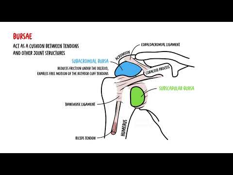
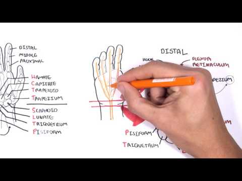
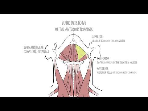
0:00 In this video we're going to look at hydrocephalus. 0:10 This is an overview and introduction. 0:12 Hydrocephalus literally means hydro, water, phthalus head, so water in the head 0:20 . 0:20 It is actually the excessive accumulation of cerebrospinal fluid, CSF, in the 0:26 brain that 0:27 can be due to an increased production of CSF, a CSF flow obstruction, or a 0:37 decreased absorption 0:38 of CSF. 0:42 Hydrocephalus can occur in both adults and newborns. 0:46 In the newborns the signs are more obvious such as the increased head 0:56 circumference. 0:58 The normal brain have ventricles which produce cerebrospinal fluid, CSF. 1:04 The CSF flows and surrounds the brain and serves as a form of protection. 1:10 The CSF is then absorbed or reabsorbed in the cardiovascular system from the 1:16 subarachnoid 1:17 space. 1:19 It is important to know the layers of the head. 1:25 The most outer part is the scalp which is the skin, the subcutaneous tissue and 1:30 the connective 1:31 tissue essentially. 1:34 Below the scalp we have the bone and under the bone is arjura mata. 1:41 Then the arachnoid mata and under the arachnoid mata is a subarachnoid space 1:52 where we find 1:53 the cerebrospinal fluid. 1:56 Surrounding the brain tissue itself is the piamata and then below the piamata 2:01 we have 2:02 the brain tissue. 2:07 In hydrocephalus there is accumulation of CSF and this can lead to ventricular 2:15 enlargement 2:16 and if we take another cross section here enlargement of the subarachnoid space 2:24 . 2:24 The enlargement of the head as mentioned is mainly seen in infants who have 2:31 hydrocephalus. 2:32 In infants this is called congenital hydrocephalus but hydrocephalus can also 2:38 occur in adults. 2:40 This is termed acquired hydrocephalus. 2:44 The signs and symptoms of congenital hydrocephalus include seizures, vomiting, 2:51 irritability, rapid 2:53 increase in head circumference and sutures being open. 2:58 In acquired hydrocephalus the signs and symptoms are that of an increased intr 3:03 acranial pressure 3:04 and can include headaches, nausea, cognitive impairment, blurring of vision, 3:12 double vision 3:13 but the sutures are closed here and the reason sutures are closed is because in 3:19 adults our 3:20 skulls have hopefully already tightened up and closed. 3:27 And another sign of adult hydrocephalus, acquired hydrocephalus can be signs of 3:34 Parkinson's 3:35 and Alzheimer's disease which makes diagnosing acquired hydrocephalus difficult 3:41 because it 3:42 is often missed and so hydrocephalus is often misdiagnosed as either Parkinson 3:47 's disease 3:49 or Alzheimer's disease which both are neurodegenerative diseases. 3:57 To understand the pathophysiology of hydrocephalus is important to look at the 4:02 overview of the 4:03 flow of cerebrospinal fluid in the brain and how and where it comes from. 4:10 So the lateral ventricles is involved in cerebrospinal fluid production. 4:16 The fluid flows into the third ventricle via the intraventricular foramen. 4:24 The fluid from the third ventricle then flows into the fourth ventricle which 4:29 is located 4:30 in front of the cerebellum. 4:34 The fourth ventricle also produces cerebrospinal fluid. 4:39 The cerebrospinal fluid then flows two ways, one the cerebrospinal fluid flows 4:45 down the 4:46 central canal which is in the center of the spinal cord essentially. 4:51 It goes down to the spinal cord or the cerebrospinal fluid actually drains into 4:57 the subarachnoid 4:58 space via the medial and lateral apertures. 5:03 The fluid from the central canal then also moves into the subarachnoid space 5:08 after going 5:08 around. 5:10 The fluid in the subarachnoid space will be reabsorbed by the venous system 5:16 through the 5:17 subarachnoid villi. 5:20 The venous system around the brain are the venous sinus. 5:24 The cerebrospinal fluid then continues on with the vascular system around the 5:31 body. 5:31 The accumulation of CSF, cerebrospinal fluid, can occur in any compartment, the 5:37 lateral ventricle 5:38 the third ventricle, the fourth ventricle or the subarachnoid space. 5:44 Depending on where the accumulation is occurring signifies what the pathology 5:52 may be. 5:53 So the accumulation of CSF in general can be attributed to three things. 5:59 One, overproduction of CSF, two, obstruction of CSF flow around the area, or 6:06 three, decrease 6:07 in CSF absorption by the venous sinus. 6:13 So investigations that can be performed are mainly imaging studies such as CT 6:18 or MRI scans. 6:20 And these imaging techniques are very effective in identifying potential causes 6:25 of hydrocephalus, 6:26 which can be tumours, growths, hemorrhage, as well as congenital malformation 6:32 of the brain 6:33 itself. 6:36 The management of hydrocephalus include controlling cardiovascular risks, and 6:39 this is particularly 6:40 in adults. 6:42 The increase in cerebrospinal fluid may need to be drained, which can be done 6:46 with a ventricular 6:47 peritoneal shunt or a ventricular atrial shunt. 6:53 The concept behind this is essentially a valve and catheter is inserted into 6:58 the ventricles 6:59 where the accumulation of CSF is. 7:02 It catheters and fed under the skin towards the peritoneum. 7:08 And thus the cerebrospinal fluid will drain from the brain into the peritoneal 7:15 cavity. 7:15 Alternatively, the catheter can be fed into the left atrium, where it will 7:21 continue with 7:21 the cardiovascular flow. 7:27 Another option is performing a endoscopic third ventriculostomy. 7:31 This is where a hole is created between the third ventricle and the subrachnosp 7:33 ice to 7:36 redirect CSF flow in cases of obstruction, for example. 7:45 Complications of hydrocephalus are mainly due to operations performed, and this 7:52 can include 7:53 post-operative stroke, shunt infection typically by staphylococcus aureus, 7:59 bleeding because 8:00 of the catheter, subdural hematoma, vascular disease, and cognitive impairment 8:07 because 8:07 of the increase in intracranial pressure, or because of the complications that 8:13 we talked 8:14 about such a stroke.