Discover the anterior triangle of the neck: clear, clinical anatomy for students and clinicians. Anterior triangle anatomy, boundaries, and key contents are explained with practical surgical and examination relevance.
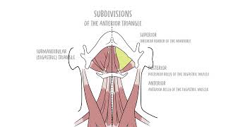
This video breaks down the anterior triangle—bounded by the midline, the anterior border of the sternocleidomastoid, and the mandible—covering its apex, roof, and muscular floor. Learn how the digastric and omohyoid muscles subdivide the area into four clinically vital regions: the submental, submandibular (digastric), carotid, and muscular triangles. For each subdivision you’ll identify precise borders, essential contents, and relevant neurovascular structures: submental lymph nodes and anterior jugular vein in the submental triangle; the submandibular gland, facial vessels, hypoglossal (CN XII) and mylohyoid nerves in the submandibular triangle; the carotid sheath with common carotid bifurcation, internal jugular, vagus (CN X), accessory (CN XI), hypoglossal, and branches of the external carotid in the carotid triangle; and the thyroid, parathyroids, larynx, trachea, esophagus and infrahyoid muscles in the muscular triangle. Clear visuals and stepwise narration make it easy to localize glands, arteries, veins, lymph nodes and cranial nerves relevant to ENT, head & neck surgery, and physical exam. Watch to improve your clinical localization, surgical planning, and anatomy recall with a concise, high-yield walkthrough of one of the neck’s most important surgical maps. Subscribe for more anatomy breakdowns and apply this framework to your next clinical assessment or dissection.