

Members have early access to this video
Learn about membershipMicrobiology Stains Explained Simply | Gram Stain, Acid-Fast, Capsule & Spore
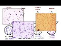
Learn how staining transforms invisible microbes into diagnostic clues with gram stain and acid-fast techniques — essential for identifying bacteria and guiding treatment. This video breaks down why staining is vital, how the gram stain differentiates gram-positive (purple) from gram-negative (pink) bacteria based on cell wall structure, and why that distinction directly impacts antibiotic selection. You’ll see the four steps of the gram stain—crystal violet, iodine mordant, alcohol decolorizer, and safranin counterstain—explained clearly with real-world examples like Staphylococcus aureus and E. coli.
Next, explore the acid-fast (Ziehl–Neelsen) stain used for mycobacteria such as Mycobacterium tuberculosis. Learn how waxy, mycolic-acid–rich cell walls resist conventional stains, why heat and carbol fuchsin are required, and how acid-alcohol decolorization plus a methylene blue counterstain reveal red acid-fast organisms against blue or green non–acid-fast cells. The video also surveys specialty stains—endospore stain for Bacillus and Clostridium, capsule stains for Streptococcus pneumoniae, and silver stains for delicate pathogens—highlighting practical diagnostic applications.
Watch to gain a concise, practical understanding of staining theory, step-by-step procedures, and the clinical benefits: faster identification, informed antibiotic choices, and rapid detection of slow-growing pathogens. Whether you’re a student, lab tech, or clinician, this clear, visual guide sharpens microbiology skills and improves diagnostic confidence. Watch now to see how color and contrast unlock microbial identity and drive better clinical decisions.