

Members have early access to this video
Learn about membershipMicroscopy Explained | Light, Phase Contrast, Fluorescence & Electron Microscopy
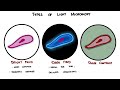
Discover how microscopes reveal the hidden microbial world—light, fluorescent, and electron microscopy explained with practical applications and diagnostic value. In this clear, engaging video you’ll learn how magnification and resolution work, why staining and contrast techniques make bacteria visible, and which microscopy methods suit specific tasks: bright-field for basic morphology and gram stains; dark-field for delicate, transparent organisms like Borrelia; phase-contrast for live, unstained cells; fluorescence for targeted detection of microbes and proteins; and electron microscopy for nanometer-scale views of viruses and organelles.
Watch concise demonstrations of how each technique transforms invisible specks into diagnostic clues—see examples of gram-positive cocci and gram-negative bacilli, fluorescently labeled eukaryotic cell components, and dramatic SEM images of parasites. Gain practical insights into clinical uses: rapid TB detection with fluorescent dyes, identifying infection-causing shapes and arrangements under light microscopes, and when to escalate to electron microscopy for research-grade detail.
This video emphasizes benefits you can use: faster diagnostics, higher specificity with fluorescent labeling, non-destructive live-cell imaging with phase contrast, and unparalleled ultrastructural resolution with TEM and SEM. Ideal for students, lab technicians, clinicians, and curious learners, the presentation balances technical terms with accessible explanations so you leave with a functional understanding of which microscope to choose and why. Dive in to sharpen your microscopy knowledge and improve your lab decision-making—watch now to see the microbial world come into focus.