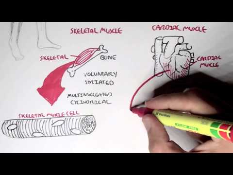
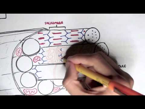
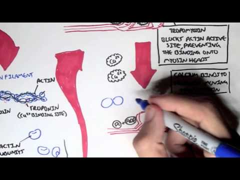
0:00 Armana Hasirunan, Biology and Medicine videos, please make sure to subscribe, 0:05 join the forum 0:06 and group for the latest videos, please visit Facebook, Armana Hasirunan, 0:10 please like, and 0:11 you can also ask questions, answer questions, and post interesting things, 0:15 including art 0:15 works. 0:16 It would be greatly appreciated. 0:17 And you can change your quality settings to the highest one for better graphics 0:20 . 0:20 In this video, we're going to talk about skeletal muscle, just an overview sort 0:25 of thing. 0:26 And so a skeletal muscle cell, as I mentioned in previous, in the introduction 0:32 video, is 0:33 cylindrical shaped and multinucleated. 0:37 And a skeletal muscle cell is known as a muscle fiber. 0:42 And interestingly enough, skeletal muscle cell, the fibers, are striated by a 0:48 highly organized 0:49 internal arrangement. 0:50 So this muscle fiber has an internal arrangement. 0:54 So if we pull some things out from this muscle fiber, we can find myofibrils. 1:02 So muscle fiber contains many myofibrils. 1:05 And if we take a portion of this myofibril, we can see that it's made out of 1:10 some interesting 1:11 structures known as the thin and thick filaments. 1:17 So the myofibrils contain thick and thin filaments. 1:21 The red is a thick and the blue is a thin filaments. 1:24 But of course, the muscle fiber, the muscle cell, is actually part of a bigger 1:32 organized 1:33 structure. 1:35 So if we look at this bone and a skeletal muscle, which attaches on it, and we 1:39 cut across section 1:40 and skeletal muscle, we have the muscle, the organ. 1:47 So this is what the muscle looks like as an organ. 1:50 And the muscle contains, if we look at the cross section, different portions of 1:54 the muscles. 1:55 And we'll look into this soon. 1:56 But the muscle contains a outer connected tissue layer known as the epimysium. 2:00 And I hope it pronounced that right. 2:03 And so we take one of these portions of the muscle here. 2:06 The portion of the muscle is known as a fascicle. 2:08 And the fascicle also contains other internal structures. 2:11 And the outer sort of connective membrane tissue is known as a perimysium. 2:17 And then if we pull out one of these other internal structures of this fascicle 2:21 , the 2:21 portion of the muscle, this is where our muscle fiber is. 2:26 And the muscle fiber contains a connective tissue wrapping around it known as 2:31 the endomysium. 2:32 And remember that a muscle fiber is the muscle cell. 2:35 So this is this. 2:38 And also the muscle fiber, the muscle cell contains a membrane known as a sac 2:42 ralema. 2:42 And again, if we remember, if we take one of the internal structures of the 2:47 muscle fiber, 2:48 fiber, we have a myofibril. 2:49 And the myofibril contains the thin filaments and the thick filaments. 2:55 So let's just recap. 2:57 If we start off with a whole muscle in Oregon, and we get a portion out of that 3:00 , we get the 3:01 muscle fascicle, which is the portion of the muscle. 3:04 And the muscle fascicle contains many muscle fibers, the actual muscle cells. 3:09 And then again, the muscle fiber, the muscle cell, contains many internal 3:14 structures, 3:15 the myofibril. 3:16 So muscle fiber contains many myofibrils, which are specialized internal intr 3:22 acellular structures. 3:23 And the myofibrils are the ones that contain thick and thin filaments, also 3:27 known as the 3:28 cytoskeleton elements. 3:29 And so we will concentrate on the myofibrils, the specialized intracellular 3:34 structures of 3:35 the muscle fiber. 3:36 And we'll specifically look at a portion of the myofibril, and a portion of the 3:40 myofibril. 3:41 They all contain the thin filament, here in blue, and the thick filament, here 3:47 in red. 3:48 And an important terminology to know is a sacramere, and a sacramere runs from 3:52 one z-line 3:54 to another z-line. 3:55 So from one, basically one thin element, one thin filament z-line to another 4:02 thin filament 4:03 z-line. 4:04 And so if we zoom into this one sacramere, here we have the thin filaments made 4:11 up of 4:11 actin, and the center of the sacramere, we have what's called the m-line. 4:17 And from the m-line, in the center, we have the thick filaments, which are made 4:22 up of 4:22 a protein myosin. 4:25 And we have other internal structures here in orange, which we will look into 4:29 later on. 4:30 So remember, sacramere runs from one z-disc, or z-line, to another z-disc, to 4:34 another z-line. 4:35 And this is a thin and thick filament structure, a sacramere structure. 4:40 But let's have a closer look at one sacramere. 4:44 So here we have one sacramere, with the center being called the m-line here. 4:51 And a sacramere runs from one z-disc to another z-disc, or a z-line to another 4:56 z-line here. 4:57 And a sacramere is a section, and extends from one z-disc to the next z-disc. 5:02 And a sacramere is a smallest contractile unit of muscles, you can say. 5:07 And so when a sacramere contracts, or when a lot of sacramere contracts, this 5:13 is when 5:14 our muscles contract, when we move, for example. 5:17 And so again, looking over the anatomy of the sacramere, we have here the thin 5:22 filaments, 5:23 which are made up of a protein called actin, and then we have the thick fil 5:27 aments, made 5:28 up of a protein called myosin. 5:31 And then these orange things, which connect to the thick and thin filaments, 5:36 are elastic 5:37 filaments, known as made up of a protein called tittin. 5:41 But the most important thing to know from this structure is that myosin forms 5:45 thick filaments, 5:46 and that actin protein forms thin filaments.