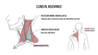
Explore the posterior triangle of the neck—its borders, subdivisions, key nerves and vessels, and critical clinical implications. In this concise anatomy video you’ll learn the precise boundaries of the posterior triangle (sternocleidomastoid, trapezius, clavicle), how the omohyoid divides it into the occipital and subclavian (omoclavicular) triangles, and which structures lie within each region. Discover the spinal accessory nerve (CN XI), supraclavicular sensory branches, superficial cervical lymph nodes, and the upper brachial plexus in the occipital triangle, and the subclavian artery and vein, brachial plexus trunks, and supraclavicular lymph nodes in the subclavian triangle. The video highlights high-yield clinical applications: recognizing accessory nerve injury signs (trapezius weakness, shoulder droop, scapular winging), performing cervical plexus or supraclavicular brachial plexus blocks, central venous access via the subclavian vein, and identifying lymphadenopathy including Virchow’s node that may signal systemic malignancy. It also explains thoracic outlet syndrome mechanics—compression between the clavicle and first rib—and why anatomic landmarks matter for safe surgery and examination. Clear visuals and step-by-step explanation make this essential for medical students, surgical trainees, and clinicians refreshing neck anatomy. Watch to confidently locate vital structures, anticipate clinical problems, and apply regional anesthesia and vascular access techniques with anatomical precision.