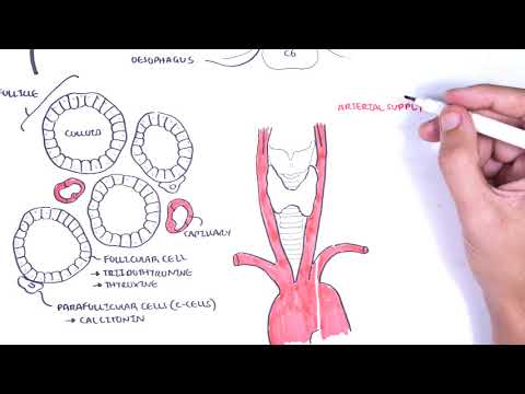
0:00 Hello, in this video we're going to talk about the clinical anatomy of the es 0:12 ophagus. 0:13 The adult human esophagus is an 18-25cm long muscular tube, extending from the 0:20 pharynx 0:21 to the stomach. 0:23 Here are your incisors, your teeth, and here is the pharynx, the throat. 0:28 The pharynx continues to the esophagus. 0:31 The esophagus passes through the diaphragm and joins with the stomach. 0:35 The esophagus has cervical, thoracic, and abdominal parts. 0:39 The cervical esophagus extends from the pharyngeal esophageal junction to the 0:44 sub-sternal 0:45 notch. 0:46 The thoracic esophagus extends from the suprasternal notch to the diaphragmatic 0:52 hiatus. 0:53 The abdominal esophagus extends from the diaphragmatic hiatus to the orifice of 0:58 the 0:58 cardio of the stomach. 1:01 Three narrow points are anatomical constriction sites of the esophagus, which 1:05 is important 1:06 to know. 1:07 I am drawing the larynx and trachea here, running in front of the esophagus. 1:14 The upper esophageal sphincter, which is the cricopharyngeus muscle, is here. 1:20 This is the first narrow point. 1:22 Second narrow point is the bifurcation of the trachea to the main bronchi and 1:27 also where 1:28 the aortic arches. 1:30 Again here is the trachea and its bifurcation in the main bronchi and behind it 1:36 , continuing 1:37 on is the esophagus. 1:39 The third anatomical constriction site is the lower esophageal sphincter, where 1:43 the esophagus 1:45 passes through the diaphragm, essentially. 1:49 If we look at the sites from a lateral view, the cricopharyngeus muscle is the 1:54 muscle attached 1:55 to the trachea and which wraps around the esophagus. 2:00 Located around the C5, C6 vertebral level, sorry you cannot see the numbers 2:06 clearly here. 2:08 The esophagus runs behind the respiratory tract. 2:12 The bifurcation of the trachea and where the aortic arches are located is the 2:17 level of 2:17 the sternal angle roughly which is between T4 and T5 vertebral level. 2:24 The last anatomical constriction site is where the esophagus passes through the 2:29 diaphragm 2:29 at level T10. 2:39 Two high pressure zones prevent the backflow of food. 2:44 These are the upper and lower esophageal sphincters. 2:48 These functional zones are located at the upper and lower ends of the esophagus 2:54 . 2:54 These are physiological constriction sites as well as anatomical as we have 3:02 learned. 3:02 Some clinical anatomy, gastroesophageal reflux disease, gourd, also known as g 3:08 ourd, is a 3:09 condition in which reflux of the stomach content into the esophagus results in 3:13 symptoms such 3:14 as heartburn, usually due to an incompetent or dysfunctional lower esophageal 3:21 sphincter. 3:22 An example of what could cause reflux is alcohol, because alcohol is known to 3:28 relax the lower 3:29 esophageal sphincter, allowing food content and then the acid to flow backwards 3:34 , causing 3:35 irritation of the esophagus. 3:38 Again, clinical anatomy, the esophageal constriction sites as we talked about, 3:43 the sites at which 3:45 you might expect to swallow foreign bodies and these foreign bodies can get 3:49 impacted and 3:50 strictures can occur after swallowing corrosive fluids, for example. 3:55 Not only that, these sites can be potentially difficult sites for gastroscopy 4:00 to pass through. 4:02 Strictures as mentioned may develop after ingestion of corrosive agents and 4:06 lastly as 4:06 mentioned foreign bodies may get stuck in these locations. 4:13 Clinical anatomy, zenke's diverticulum, also known as the pharyngeal pouch, is 4:18 the 4:18 out-pouching of the hypopharynx. 4:21 So drawing it out, the larynx and trachea here on the left, you have the esoph 4:26 agus running 4:27 behind it. 4:29 There are important muscles, the cricopharyngeus and the inferior pharyngeal 4:34 constrictor, which 4:35 wraps around the esophagus, the cricopharyngeus, as we know, is the upper esoph 4:43 ageal sphincter. 4:45 If there is a weakened layer around the pharyngeal esophageal junction, the muc 4:51 osa and sub mucosa 4:52 layers originating here can pop out, resulting in an out-pouching sac. 5:05 Here is an image, a barium swallowing study showing a pharyngeal pouch. 5:15 The clinical presentation of zenke's diverticulum includes a globus sensation, 5:22 dysphagia, halitosis 5:25 and regurgitation. 5:30 The anatomical relationship of the esophagus, we already learned a bit about it 5:35 . 5:35 The trachea is anterior to the esophagus, bifurcation of the trachea, aortic 5:42 arch anterior 5:43 to the chukia level at the sternal angle, about T4 and T5 vertebral level. 5:49 The arch of the aorta wraps over the left main bronchus and it travels 5:55 posterior to the esophagus. 5:58 You have the right vagus nerve and the left vagus nerve on both sides of the es 6:03 ophagus, 6:04 which supplies the esophagus, forming the esophageal plexus. 6:09 The thoracic ducts travel left of the esophagus. 6:16 Imagine cutting a cross section now of the thorax. 6:20 Imagine this is a CT scan. 6:23 Orientate ourselves, here we have the right lung, the sternum, here is the es 6:29 ophagus at 6:30 the back. 6:31 Now this is where we introduce the mediastinum. 6:34 The mediastinum is the area of the thorax, which can be divided into the 6:38 superior and 6:39 inferior compartments. 6:42 The inferior compartments can be further divided into the posterior compartment 6:47 , where we find 6:48 the esophagus, the middle mediastinum, where we can find the heart, and then 6:53 the anterior 6:54 part of the mediastinum, where we find the sternum. 6:59 Let's zoom into this area here. 7:03 Again, here is the right lung, and then you have your left lung. 7:09 This is about the T8, a tuba level. 7:13 You have the esophagus, and then you have the thoracic aorta to the left of the 7:18 esophagus. 7:20 The thoracic aorta will have branches called the posterior intercostal arteries 7:25 coming off 7:27 that will supply the ribs and the spinal cord. 7:32 The azigos vein is posterior and to the right of the esophagus, and will drain 7:37 blood into 7:38 the superior vena cava, which leads us to the venous drainage of the esophagus. 7:44 You have the esophagus vein, which drains the majority of the esophagus. 7:49 The hemi azigos vein runs left of the esophagus and joins with the azigos vein 7:53 on the right. 7:55 The azigos vein, as mentioned, drains into the superior vena cava. 7:59 I have not drawn the heart, but imagine it is sitting on the diaphragm in the 8:03 middle. 8:04 Now the esophageal vein also anastomoses with the left gastric vein originating 8:09 below 8:10 the diaphragm. 8:11 The left gastric vein drains into the portal vein. 8:16 The portal vein, as we know, drains into the liver. 8:20 The portal vein is super important because it drains most of the 8:28 gastrointestinal organs. 8:29 It drains the spleenic vein, inferior mesenteric vein, and the superior mes 8:35 enteric vein. 8:36 Important to realize here, the left gastric vein and esophageal vein anastom 8:42 oses and 8:43 is a connection between the portal and systemic venous system and is a 8:46 potential site for 8:47 the development of esophageal varices in portal hypertension. 8:52 So clinical anatomy, esophageal varices, anastomoses between the azigos and 8:57 left gastric 8:58 vein may develop varices from portal hypertension. 9:04 So drawing this out, here we have the portal vein. 9:07 You can imagine a liver cirrhosis where the liver becomes fibros. 9:11 This increases resistance in the portal vein causing portal hypertension. 9:16 This means that blood can get pushed back, back to the spleenic vein, superior 9:21 mesenteric 9:22 vein, and also the left gastric vein causing esophageal varices. 9:30 Here is an endoscopic picture of the esophagus where you can see esophageal var 9:37 ices. 9:37 Varices is an abnormal dilated vessel with a torturous cause. 9:43 And these varices can rupture, hemorrhage, you get bleeding. 9:47 This patient can eventually present with malena which is a black stool as well 9:55 as hematemesis 9:56 vomiting blood. 9:59 And this brings us to the blood supply to the esophagus, there are three main 10:03 ones. 10:03 And this also is good because you can divide the esophagus into three parts. 10:09 The first are the branches of the inferior thyroid artery which provides arter 10:13 ial supplier 10:14 to the cervical esophagus. 10:16 The paired aortic esophageal arteries supply the thoracic esophagus and then 10:20 the left gastric 10:22 artery supplies the abdominal esophagus. 10:25 When talking about the histology or the pathology of the esophagus, it is good 10:30 to divide the 10:31 esophagus into the upper two thirds and lower thirds. 10:35 By dividing it like this we can easily differentiate the cell types found in 10:39 the esophagus. 10:40 Let's focus on the upper two thirds and zoom into this area and look at the 10:44 layers 10:45 of the esophagus from the most inner layer first which is the mucosa. 10:50 The upper two thirds contain stratified squamous epithelial cells. 10:56 And these cells can develop into squamous carcinoma. 11:00 Below it you have the submucosa which contains mucous glands, goblet cells. 11:05 The muscle layer is next and you have an inner striated circular voluntary 11:09 muscle and then 11:09 the external longitudinal muscle. 11:13 The outermost layer of the esophagus is the adventitia. 11:18 Limpatic drainage is to the neck and the mediastinal nodes. 11:24 Unlike other areas of the GI tract, the esophagus does not have a distinct ser 11:29 osal covering. 11:30 This allows esophageal tumors to spread more easily and makes them harder to 11:38 treat surgically. 11:40 The lower third of the esophagus, the mucosa layer, you have transition from 11:45 stratified 11:46 squamous epithelium to columnar epithelium, simple. 11:51 And these guys can actually develop into adenocarcinomas. 11:56 The submucosa contain the mucous glands, the goblet cells. 12:00 The muscle layer here we can find transition of the inner striated muscles to 12:07 involuntary 12:08 smooth muscles. 12:09 The external or the outer muscle layer is still the longitudinal muscle. 12:15 The most outer layer is the adventitia, remember there is no serosal layer in 12:20 the esophagus. 12:22 Limpatic drainage at the lower third is to the gastric and paraeortic nodes. 12:31 Let's talk about some embryology of the esophagus. 12:36 Week 4 of gestation, the respiratory tube and the esophageal tube are forming 12:40 from the 12:41 same bud. 12:43 They separate respiratory tube moving anteriorly, forming slowly the bronchi 12:50 and its branches. 12:52 And then the esophagus is initially very short, but elongates rapidly, reaching 12:57 its final 12:58 relative length by about week 7. 13:01 By week 5 and 6, the respiratory tube and esophageal tube have separated. 13:07 Now you can imagine that if the respiratory tube and the esophageal tube does 13:11 not separate 13:12 properly, problems can arise. 13:15 Some clinical anatomy, tracheo esophageal fistula. 13:20 It is a pathological connection between the esophagus and the trachea. 13:23 It is a development abnormality and there are many types. 13:34 Signs and symptoms of tracheo esophageal fistula include sinosis, difficulty 13:39 breathing and 13:40 also coughing and vomiting episodes for the infant. 13:44 Because we talked about gourd, gourd, which is where you have an incompetent 13:49 lower esophageal 13:50 sphincter, we need to talk about esophageal aclesia, where the lower esophageal 13:55 sphincter 13:56 is too competent or stiff. 13:59 It doesn't allow things to pass through smoothly, causing symptoms of regurg 14:03 itation and heartburn. 14:05 Asophageal aclesia is where food accumulates in the esophagus, and the esoph 14:09 agus becomes 14:10 dilated. 14:11 It is due to increased tone to the lower esophageal sphincter, failure to relax 14:17 . 14:17 And whereas in gourd, remember it is too relaxed, you can say it. 14:22 Here is a barium swallowing study of a patient with esophageal aclesia. 14:27 As you can see, the barium stops at a tip of constriction. 14:32 This is where the lower esophageal sphincter is. 14:35 And you can see proximal to that tip, the esophagus is dilated, forming what is 14:40 known 14:41 as a bird beak appearance. 14:44 Thank you for watching this video. 14:45 I hope you enjoyed it on the esophagus clinical anatomy.