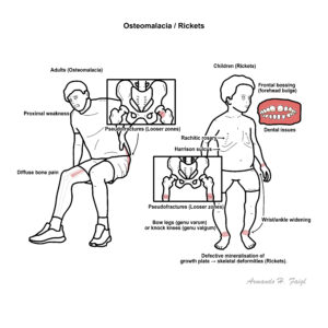
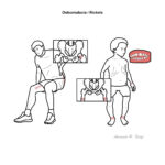
Osteomalacia (adults) and rickets (children) are disorders of impaired bone mineralisation leading to soft, poorly mineralised bone. It arises when there is insufficient calcium–phosphate supply to the mineralising matrix due to vitamin D deficiency, calcium deficiency, phosphate deficiency (often FGF23-mediated). Nutritional rickets still occurs worldwide, while hypophosphataemic forms (e.g., XLH, tumour-induced osteomalacia) are rarer but important. Major complications include growth failure and limb deformity in children, pseudofractures and fragility fractures in adults, bone pain, proximal myopathy, and impaired quality of life.1,3,8,11
Definition
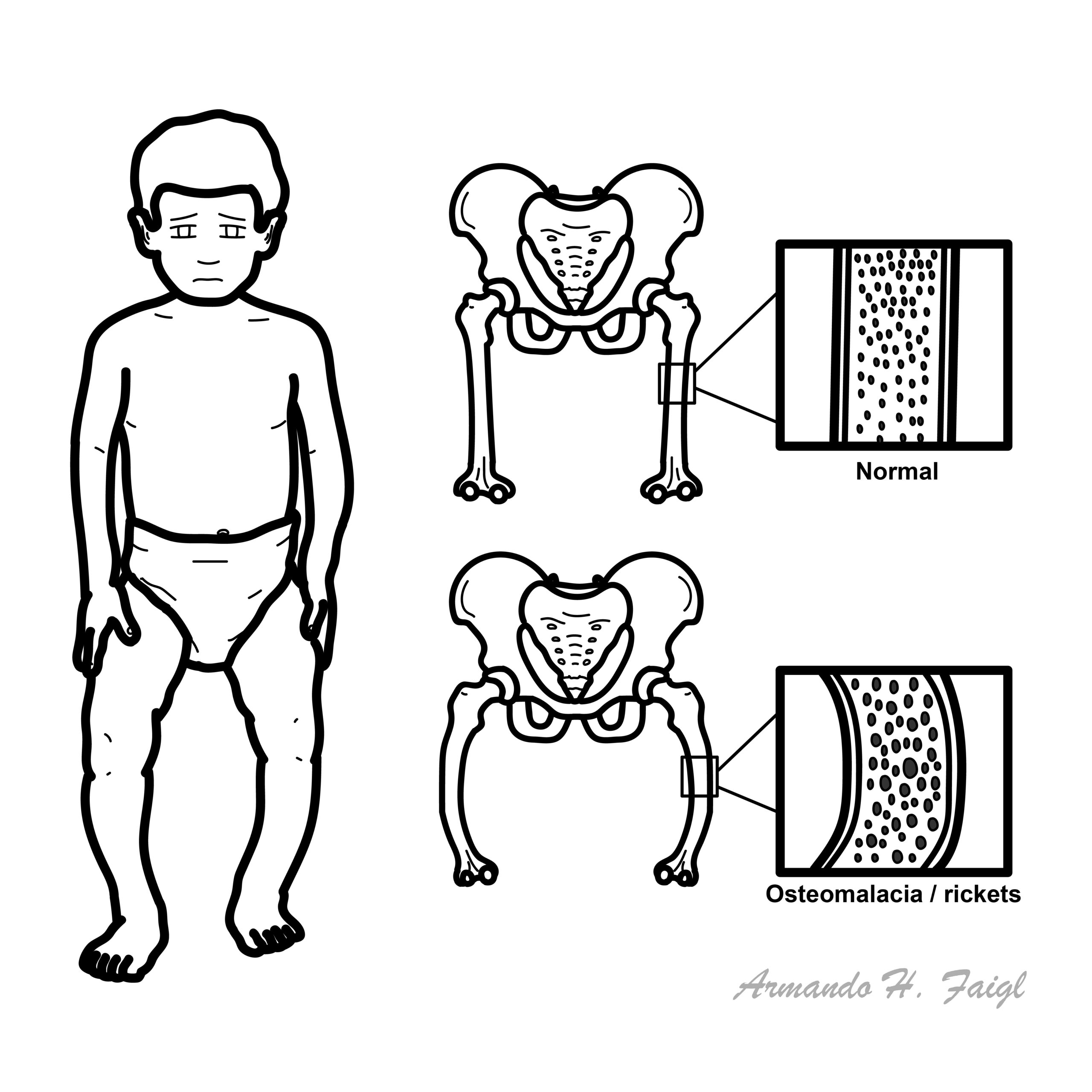
Osteoid: unmineralised organic bone matrix; excess osteoid with delayed mineralisation is the hallmark of osteomalacia (histomorphometry). Looser zones (pseudofractures): transverse lucencies with sclerotic margins at sites of tensile stress (ribs, pubic rami, femoral neck) pathognomonic of osteomalacia. FGF23: phosphaturic hormone that reduces renal phosphate reabsorption and 1α-hydroxylation of vitamin D; excess causes hypophosphataemic rickets/osteomalacia.3,8 Rickets vs osteomalacia: rickets = defective mineralisation of growth plate cartilage in children; osteomalacia = defective mineralisation of bone matrix in all ages; in children rickets and osteomalacia usually coexist.
Anatomy & Physiology
Bone remodelling units:
osteoclasts resorb
osteoblasts lay osteoid
mineralisation requires adequate Ca2+×PO4 product and alkaline phosphatase activity.
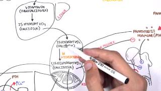
Vitamin D pathway: skin 7-dehydrocholesterol → cholecalciferol (UVB) → 25-OH-D (liver) → 1,25-(OH)2D (kidney via CYP27B1); 1,25-(OH)2D increases intestinal calcium and phosphate absorption and, with PTH, regulates bone turnover.
Phosphate handling: ~80% reabsorbed in proximal tubule via NaPi2a/2c co-transporters; FGF23 (via Klotho) down-regulates these transporters and suppresses renal 1α-hydroxylase, lowering calcitriol.3,8
Calcium–phosphate homeostasis: PTH raises Ca2+ (bone resorption, renal Ca reabsorption) and lowers phosphate (phosphaturia); calcitriol raises both; FGF23 lowers phosphate and calcitriol.
A sustained fall in either intestinal supply (vitamin D/calcium) or renal phosphate reabsorption (FGF23 excess) prevents attainment of the Ca×PO4 ion product necessary for crystal deposition.
Dark skin, veiling/covering, high latitude, winter season, indoor living; pregnancy/lactation; exclusive breastfeeding without maternal/infant supplementation
In adults with diffuse bone pain + proximal weakness + low phosphate, ask about dental abscesses (In X-linked phosphataemia) and search for tumour induced osteomalacia (TIO).
Bone densitometry: BMD may be low but nonspecific; do not confuse with osteoporosis
Bone biopsy (rare/definitive): trans-iliac biopsy with double tetracycline labels shows ↑osteoid volume and prolonged mineralisation lag time; useful when diagnosis unclear or before treating suspected adynamic bone disease
Other
In FGF23-mediated forms—low phosphate, high/normal FGF23, low/normal 1,25-(OH)2D, ↓TmP/GFR
Hyperparathyroidism: high Ca, low phosphate, subperiosteal resorption not Looser zones (DIF).
Osteitis fibrosa/renal osteodystrophy: CKD context; PTH very high; bone biopsy pattern (DIF).
Treatment
General principles: correct biochemical abnormalities, relieve pain and myopathy, heal fractures, and treat the cause
Nutritional vitamin D deficiency (adults): cholecalciferol 3000–5000 IU daily or 50,000 IU weekly for 6–8 weeks, then maintenance 800–2000 IU/day; add elemental calcium 1–1.5 g/day if dietary intake low; monitor ALP, Ca, PO4, PTH at 8–12 week.11
Children (nutritional rickets): treat per Global Consensus—age-specific vitamin D and calcium dosing; ensure ongoing maintenance and prevention strategies
Malabsorption/bariatric: higher/parental vitamin D dosing; treat underlying disease; ensure calcium and magnesium repletion.11
Hypophosphataemic forms (FGF23-mediated):
XLH: first-line burosumab (anti-FGF23 mAb) improves phosphate, rickets severity, pain and physical function; alternative/legacy therapy = divided oral phosphate + active vitamin D (calcitriol/alfacalcidol) with monitoring for nephrocalcinosis
Tumour-induced osteomalacia: localise and resect tumour (curative); if unresectable or occult, burosumab or phosphate + calcitriol therapy.2
Renal disease: treat CKD-MBD per nephrology guidance, active vitamin D analogues as indicated; correct acidosis.
Pain/fractures: protected weight-bearing, physiotherapy for proximal weakness; manage fractures (often heal rapidly after repletion).
Avoid high-dose intermittent (“stoss”) vitamin D in infants/children unless per protocol; ongoing prevention via supplementation/fortification for high-risk groups is guideline-endorsed.1,12
Persistently high ALP after normalisation of 25-OH-D suggests unrecognised phosphate wasting—check phosphate and TmP/GFR.
Complications and Prognosis
Complications
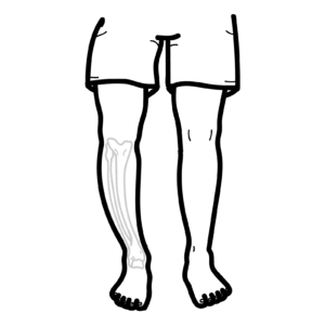
Children: permanent skeletal deformities (genu varum/valgum), short stature, dental enamel defects/abscesses, hypocalcaemic seizures, cardiomyopathy in severe cases
Adults: recurrent insufficiency fractures/pseudofractures, chronic pain, proximal myopathy, falls, impaired function; nephrocalcinosis with phosphate-calcitriol therapy; kidney stones if overtreatment
Nutritional deficiency: excellent with timely repletion; radiographic healing in months, strength improves over weeks; relapse without maintenance or if risk factors persist
FGF23-mediated disease: better growth/function and pain with burosumab; poorer outcomes with delayed diagnosis, longstanding deformity, CKD, or unresectable TIO
Poor prognostic factors: late presentation, persistent hypophosphataemia, severe deformity, coexistent CKD/malabsorption, inadequate adherence to supplementation
Table 1. Biochemical patterns (simplified)
Pattern
Ca
PO4
ALP
PTH
25-OH-D
1,25-(OH)2D
FGF23
TmP/GFR
Likely mechanism
Nutritional Vit D deficiency
low/normal
low/normal
↑
↑
↓
low/normal
normal
normal
reduced calcitriol substrate
Calcium deficiency
low
low/normal
↑
↑
normal
↑
normal
normal
low calcium intake
XLH/TIO (FGF23-mediated)
normal
low
↑
normal/↑
normal
low/normal
↑
low
renal phosphate wasting 3,8
HHRH (FGF23-independent)
normal/high Ca
low
↑
low/normal
normal
↑
low
low
SLC34A3; hypercalciuria
References
Munns CF, Shaw N, Kiely M, et al. Global consensus recommendations on prevention and management of nutritional rickets. J Clin Endocrinol Metab. 2016;101(2):394-415. (Oxford Academic)
Florenzano P, Hartley IR, Jimenez M, et al. Diagnosis and management of tumor-induced osteomalacia: Perspectives from clinical practice. J Endocr Soc. 2021;5(9):bvab099. (Oxford Academic)
Carpenter TO, Whyte MP, Imel EA, et al. FGF23, hypophosphatemia, and emerging treatments. JBMR Plus. 2019;3(8):e10190. (Oxford Academic)
Holick MF, Binkley NC, Bischoff-Ferrari HA, et al. Evaluation, treatment, and prevention of vitamin D deficiency: an Endocrine Society clinical practice guideline. J Clin Endocrinol Metab. 2011;96(7):1911-30. (historical background)
Shoback DM, Rosen CJ. Vitamin D for the prevention of disease: an Endocrine Society Clinical Practice Guideline. J Clin Endocrinol Metab. 2024;109(8):1907-1932. (Oxford Academic)
Bhan A, Rao SD. Osteomalacia and vitamin D status: a clinical update. JBMR Plus. 2020;4(8):e10447. (Wiley Online Library)
Takeuchi Y, Fukumoto S. FGF23-related hypophosphataemic rickets/osteomalacia: diagnosis and new treatment. J Mol Endocrinol. 2021;66(2):R57-R65. (jme.bioscientifica.com)
Endocrine Society. Vitamin D for the Prevention of Disease Guideline page (summary). 2024. (Endocrine)
ANZSPED copy of the Global Consensus PDF (for algorithms). 2016. (media.anzsped.org)


Discussion