

Rheumatoid Arthritis-Associated Interstitial Lung Disease (RA-ILD)

Overview
Rheumatoid arthritis-associated interstitial lung disease (RA-ILD) is a serious extra-articular manifestation of RA, affecting up to 10% of RA patients clinically, though subclinical involvement may be present in up to 60% on HRCT. It typically presents in the 5th–6th decade, with a male predominance and is a leading cause of RA-related mortality. The most common histopathological subtype is usual interstitial pneumonia (UIP), which carries a worse prognosis than nonspecific interstitial pneumonia (NSIP). Risk factors include smoking, high RA disease activity, anti-CCP positivity, and MUC5B promoter variant. Complications include progressive pulmonary fibrosis and respiratory failure.
Definition
Interstitial Lung Disease (ILD): A group of disorders characterized by inflammation and fibrosis of the lung interstitium.
Usual Interstitial Pneumonia (UIP): A histologic pattern of ILD marked by patchy fibrosis and honeycombing, often seen in RA-ILD.
Anti-Cyclic Citrullinated Peptide (anti-CCP): Autoantibody highly specific for RA, associated with extra-articular manifestations.
High-Resolution Computed Tomography (HRCT): Imaging modality of choice for detecting ILD patterns.
Anatomy & Physiology
- Respiratory System: The lungs are composed of airways (bronchi, bronchioles) and alveoli, which are tiny air sacs responsible for gas exchange. The interstitium is the space between the alveolar epithelial cells and the endothelial cells of the capillaries.
- Gas Exchange: Oxygen diffuses from the alveoli into the capillaries, and carbon dioxide diffuses from the capillaries into the alveoli. The thinness of the alveolar-capillary membrane is crucial for efficient gas exchange.
- Immune System in the Lung: The lung possesses its own immune cells (e.g., alveolar macrophages, lymphocytes) that provide defense against inhaled pathogens and irritants.
- Fibroblasts: These are connective tissue cells that produce collagen and other fibers. In fibrotic lung diseases, fibroblasts become overactive and produce excessive amounts of extracellular matrix, leading to scarring.
Aetiology & Risk Factors
Aetiology
- Autoimmune inflammation from RA targeting lung parenchyma
- Genetic predisposition: MUC5B promoter variant
- Environmental triggers: smoking, pollutants
- Drug-induced (e.g., methotrexate, leflunomide — controversial)
Risk Factors
- Male sex
- Age >60
- Smoking history
- High RA disease activity (DAS28)
- Anti-CCP and RF positivity
- HLA-DRB1 shared epitope
Anti-CCP positivity is a stronger predictor of RA-ILD than RF alone.
Pathophysiology
- RA triggers systemic immune activation → cytokine release (TNF-α, IL-6)
- Lung epithelial injury → activation of alveolar macrophages and dendritic cells
- Recruitment of T cells and B cells → autoantibody production
- Fibroblast activation → extracellular matrix deposition
- Progressive fibrosis → impaired gas exchange and restrictive lung physiology
Fibrosis in RA-ILD is not merely a consequence of inflammation but involves aberrant wound healing and epithelial–mesenchymal transition.
Clinical Manifestations
- Dyspnea on exertion
- Nonproductive cough
- Bibasilar crackles on auscultation
- Clubbing (less common than in IPF)
- Hypoxia (especially during exertion)
- Reduced exercise tolerance
- Restrictive pattern on spirometry
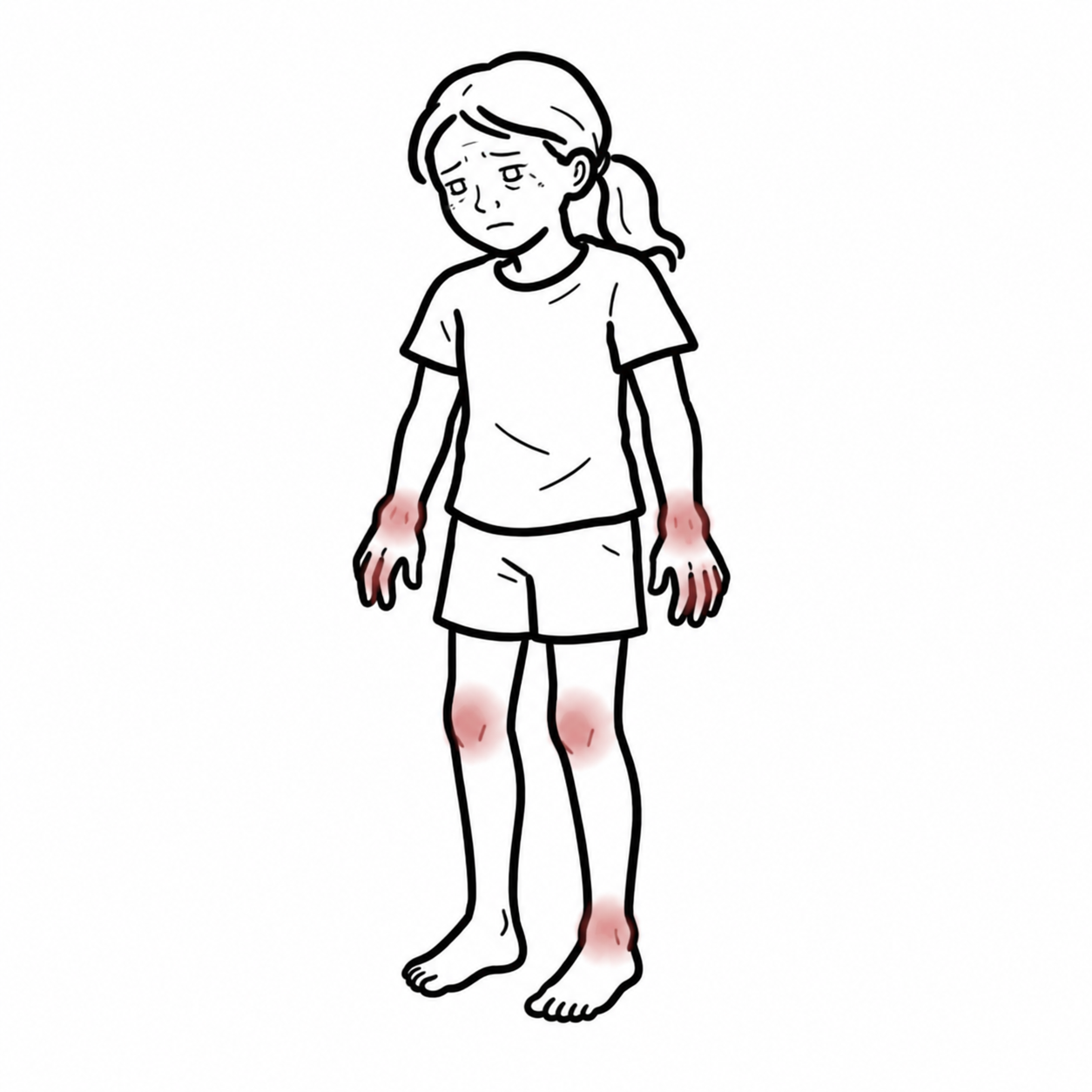
- Features of Rheumatoid arthritis
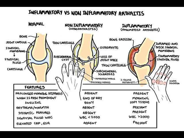
- Polysynovitis of small and/or large joints
- Positive RF/CCP ab
Diagnosis
- RA diagnosis
- HRCT evidence of ILD
- Exclusion of other ILD causes
Investigations
- HRCT: gold standard
- Identifies UIP (honeycombing, subpleural reticulation) vs NSIP (ground-glass opacities)
- Pulmonary Function Tests: restrictive pattern, ↓ DLCO
- Serology: RF, anti-CCP
- BAL (bronchoalveolar lavage): lymphocytosis in NSIP
- Lung biopsy: rarely needed, reserved for atypical cases
| Differential Diagnosis | Differentiating Feature |
| IPF | Older age, absence of RA, rapid progression |
| Hypersensitivity pneumonitis | Exposure history, centrilobular nodules |
| Drug-induced ILD | Temporal relation to medication |
| Sarcoidosis | Noncaseating granulomas, hilar lymphadenopathy |
Classification
Based on HRCT/histopathology
- Usual Interstitial Pneumonia (UIP) – most common
- Nonspecific Interstitial Pneumonia (NSIP)
- Organizing Pneumonia (OP)
- Lymphoid Interstitial Pneumonia (LIP)
UIP subtype in RA-ILD mirrors IPF and carries worse prognosis.
UIP is common in RA-ILD, the other rheumatological conditions typically develop NSIP pattern.
Treatment
Immunomodulators
- Methotrexate
- Mycophenolate mofetil
- Azathioprine
- Rituximab (especially in UIP)
Anti-fibrotics
- Nintedanib (approved for progressive fibrosing ILD)
Corticosteroids
- limited role, more effective in NSIP
Supportive
- Oxygen therapy
- Pulmonary rehab
- Vaccinations (influenza, pneumococcus)
Nintedanib slows FVC decline in progressive RA-ILD.
Complications & Prognosis
Complications
- Progressive pulmonary fibrosis
- Respiratory failure
- Pulmonary hypertension
- Superimposed infections
- Drug-induced lung injury
Prognosis
- UIP subtype: median survival ~3–5 years
- NSIP subtype: better prognosis
- Poor prognostic factors:
- Older age
- Male sex
- UIP pattern
- Low DLCO
- High RA activity
Early detection and subtype identification are key to prognosis and treatment planning.
References
- Kadura S, Raghu G. Rheumatoid arthritis-interstitial lung disease: manifestations and current concepts in pathogenesis and management. Eur Respir Rev. 2021;30(160):210011.
- Dai Y, Wang W, Yu Y, Hu S. Rheumatoid arthritis–associated interstitial lung disease: an overview of epidemiology, pathogenesis and management. Clin Rheumatol. 2021;40:1211–1220.
- Kim Y, Yang H-I, Kim K-S. Etiology and Pathogenesis of Rheumatoid Arthritis-Interstitial Lung Disease. Int J Mol Sci. 2023;24(19):14509.
- Radiopaedia.org. Rheumatoid arthritis associated interstitial lung disease. Accessed 2025.
- Sparks JA et al. Rheumatoid arthritis disease activity and risk of ILD. Springer. 2020.
- European Respiratory Society. RA-ILD review. Eur Respir Rev. 2021.
- Flaherty KR et al. Nintedanib in progressive fibrosing ILDs. N Engl J Med. 2019;381:1718–1727.
- Kim EJ et al. Epithelial–mesenchymal transition in RA-ILD. J Immunol Res. 2022.












Members only discussions coming soon…