

Tarsal Tunnel Syndrome
Overview
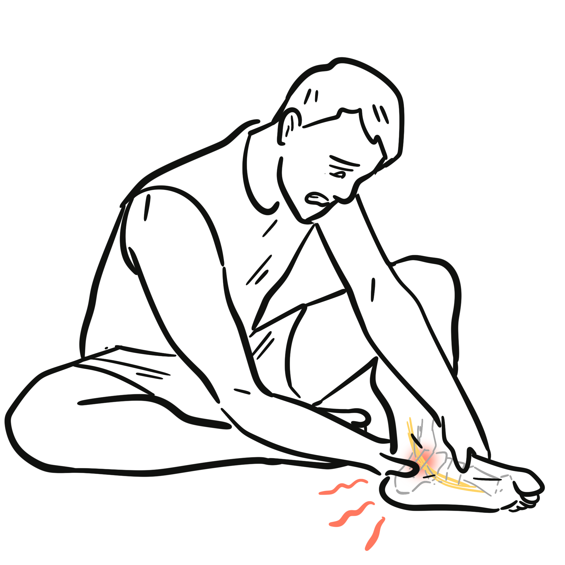
Tarsal Tunnel Syndrome (TTS), also known as posterior tibial nerve neuralgia, is an entrapment neuropathy caused by compression of the tibial nerve or its terminal branches within the fibro-osseous tarsal tunnel beneath the flexor retinaculum on the medial side of the ankle. It represents the lower extremity analogue of Carpal Tunnel Syndrome.
Because the tarsal tunnel is a rigid, non-yielding anatomical space, any intra-tunnel space-occupying lesion, localized tissue edema, or biomechanical alignment abnormality can elevate intraneural pressure. This leads to microvascular ischemia, paresthesias, burning pain, and numbness across the plantar aspect of the foot. Diagnosing TTS requires careful clinical evaluation and neurophysiological testing to differentiate it from more common causes of foot pain, such as plantar fasciitis and lumbar radiculopathy.
Tarsal tunnel syndrome is the lower extremity equivalent of Carpal Tunnel Syndrome, caused by compression of the tibial nerve beneath the flexor retinaculum behind the medial malleolus.
Definition
- Flexor Retinaculum (Laciniate Ligament)
- A strong fibrous band extending from the medial malleolus above to the medial tubercle of the calcaneus below, forming the rigid roof of the tarsal tunnel.
- Tinel Sign at the Ankle
- Percussion directly over the posterior tibial nerve inferior and posterior to the medial malleolus reproduces tingling, electric shocks, or paresthesias radiating into the sole of the foot.
- Trepman Test (Dorsiflexion-Eversion Test)
- Passive maximal ankle dorsiflexion combined with foot eversion and toe extension, which increases intraneural pressure within the tarsal tunnel and reproduces symptoms.
- Valleix Sign
- Retrograde (proximal) radiation of pain or paresthesias up the medial calf along the path of the sciatic/tibial nerve, triggered by distal nerve compression.
Why does TTS pain frequently worsen at night?
During sleep, venous stasis and tissue edema increase within the lower extremity. In the enclosed, rigid tarsal tunnel, this venous engorgement raises tissue pressure, causing microvascular ischemia of the nerve and triggering nocturnal dysesthesias.
Anatomy
The tarsal tunnel is a narrow fibro-osseous canal located posterior and inferior to the medial malleolus.
Anatomical Boundaries
- Roof: Flexor retinaculum (laciniate ligament).
- Floor: Medial surfaces of the talus, calcaneus, and distal tibia.
- Distal Division: The tunnel terminates distally where the tibial nerve bifurcates into the Medial Plantar Nerve, Lateral Plantar Nerve, and Medial Calcaneal Nerve.
Contents of the Tarsal Tunnel
From anterior to posterior (medial to lateral), the structures pass in a specific order:
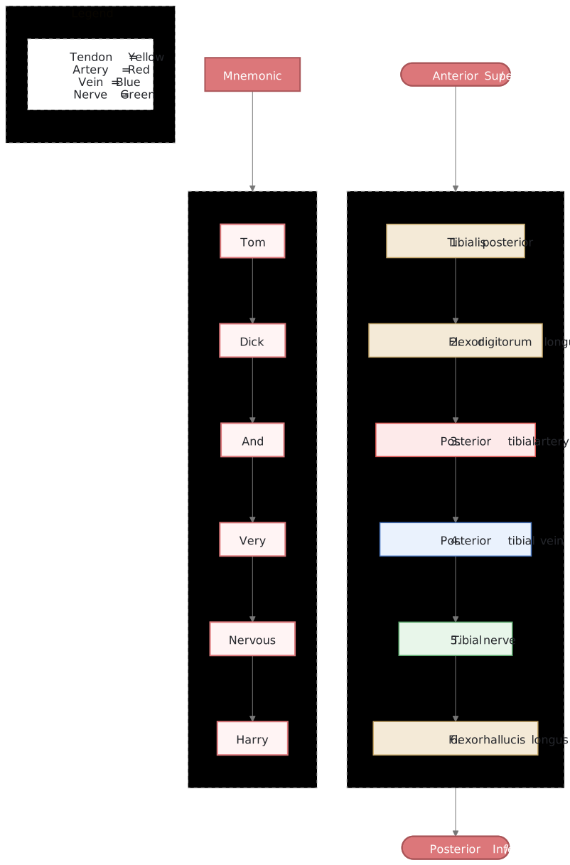
Tarsal tunnel contents arranged from anterior-superior to posterior-inferior, paired with the mnemonic “Tom, Dick, And Very Nervous Harry”.
What to notice
Detailed image description
The ordered contents of the tarsal tunnel are shown from anterior-superior to posterior-inferior: tibialis posterior, flexor digitorum longus, posterior tibial artery, posterior tibial vein, tibial nerve and flexor hallucis longus. A parallel mnemonic column reinforces the sequence: Tom, Dick, And Very Nervous Harry.
The classic anatomical mnemonic to remember the contents of the tarsal tunnel from anterior to posterior is “Tom, Dick, And Very Nervous Harry” (Tibialis posterior, Flexor Digitorum longus, Artery, Vein, Nerve, Flexor Hallucis longus).
Aetiology & Risk Factors
Extrinsic Mechanical Factors (~40% of cases)
- Biomechanical Foot Deformities: Excessive pes planus (flat feet) and hyperpronation strain and stretch the nerve over the sustentaculum tali while tightening the flexor retinaculum.
- Trauma: Post-traumatic scarring following ankle sprains, calcaneal or medial malleolar fractures, or talar dislocations.
- Footwear: Tight-fitting boots, high heels, or restrictive athletic straps compressing the medial ankle.
Intrinsic Space-Occupying Lesions (~40% of cases)
- Benign Lesions: Ganglion cysts, lipomas, neurilemomas (schwannomas), or neurofibromas within the canal.
- Vascular Anomalies: Varicose veins, venous plexus engorgement, or posterior tibial artery aneurysms.
- Tenovaginitis / Tendinopathy: Hypertrophy or inflammation of surrounding flexor tendons.
- Anatomical Variants: Accessory muscles (e.g., accessory flexor digitorum longus or abductor hallucis hypertrophy).
Systemic & Inflammatory Factors (~20% of cases)
- Systemic disorders causing peripheral nerve vulnerability or localized fluid retention: Diabetes mellitus, Rheumatoid arthritis, Hypothyroidism, Gout, and Pregnancy.
Pathophysiology
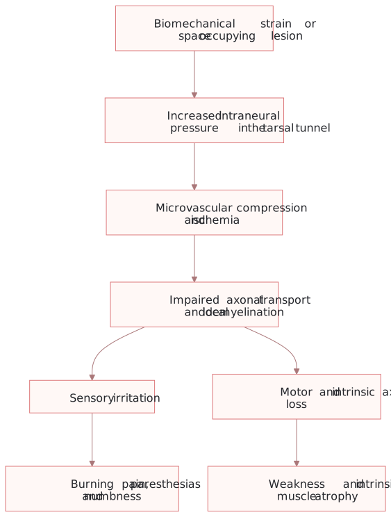
Tarsal tunnel pathophysiology: increased intraneural pressure produces microvascular ischaemia, impaired axonal transport and demyelination, leading to sensory and motor dysfunction.
What to notice
Detailed image description
Biomechanical strain or a space-occupying lesion increases pressure on the tibial nerve within the tarsal tunnel. Microvascular compression and ischaemia impair axonal transport and cause local demyelination. Sensory irritation leads to burning pain, paresthesias and numbness, while motor and intrinsic axonal loss can lead to weakness and intrinsic foot muscle atrophy.
- Mechanical Compression: Direct pressure or traction stretches the nerve against the unyielding flexor retinaculum or bone.
- Ischemic Cascade: Intraneural venous pressure exceeds normal limits (> 20–30 mmHg), reducing capillary blood supply. Persistent ischemia leads to breakdown of the blood-nerve barrier, local edema, and intra-axonal swelling.
- Demyelination & Axonal Degeneration: Chronic compression damages the myelin sheath, causing localized conduction block, persistent ectopic nerve firing (burning pain), and eventual distal axonal degeneration.
Clinical Manifestations
- Pain & Dysesthesias: Burning, sharp, stabbing, or aching pain localized to the plantar surface of the foot, heel, or toes.
- Paresthesias & Numbness: Tingling or altered sensation along the distribution of the medial or lateral plantar nerves.
- Diurnal Pattern: Aggravated by prolonged weight-bearing, walking, or standing; classic nocturnal exacerbation that awakens the patient from sleep.
- Proximal Radiation: Pain may radiate proximally up the medial calf (Valleix phenomenon).
Clinical Examination
- Palpation & Tenderness: Focal tenderness directly posterior to the medial malleolus.
- Tinel Sign: Positive in up to 50–80% of true cases (tingling radiated to sole upon tapping over the tarsal tunnel).
- Provocative Tests:
- Dorsiflexion-Eversion Test: Passive dorsiflexion and eversion held for 30 seconds reproduces symptoms.
- Direct Manual Compression: Sustained pressure over the nerve for 60 seconds reproduces distal burning.
- Motor Deficits (Late Stage): Weakness of intrinsic foot muscles (abductor hallucis, flexor digitorum brevis), clawing of the toes, and atrophy of the abductor hallucis muscle belly creating a sunken arch contour.
Diagnosis
Tarsal Tunnel Syndrome is a clinical diagnosis supported by electrodiagnostic testing and neuroimaging.
Diagnostic Comparison: TTS vs. Plantar Fasciitis vs. L5/S1 Radiculopathy
| Feature | Tarsal Tunnel Syndrome | Plantar Fasciitis | L5 / S1 Radiculopathy |
| Primary Site of Pain | Entire sole / arch / toes | Medial calcaneal tubercle | Lumbar spine, buttock, lateral/posterior leg |
| First Steps in Morning | Variable | Extremely severe (“first-step pain”) | Unaffected |
| Sensory Deficits | Present (Numbness / Burning) | Absent | Present (Dermatomal distribution) |
| Tinel Sign at Ankle | Positive | Negative | Negative |
| Back Pain / SLR | Negative | Negative | Positive Straight Leg Raise (SLR) |
The absence of sensory loss or paresthesias strongly points away from Tarsal Tunnel Syndrome and toward Plantar Fasciitis.
Investigations
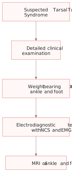
Investigation pathway for suspected tarsal tunnel syndrome: clinical examination followed by targeted imaging and electrodiagnostic assessment.
What to notice
Detailed image description
Suspected tarsal tunnel syndrome is assessed with detailed clinical examination, followed by weight-bearing X-rays of the ankle and foot, electrodiagnostic testing with nerve conduction studies and electromyography, and MRI of the ankle or foot to identify structural or soft-tissue causes.
- Electrodiagnostic Studies (NCS / EMG):
- Nerve Conduction Studies: Demonstrates prolonged distal sensory and motor latencies, along with decreased Nerve Action Potential (NAP) amplitudes across the medial and lateral plantar nerves.
- Electromyography: Evaluates for denervation changes (fibrillations, positive sharp waves) in the abductor hallucis or abductor digiti minimi muscles.
- Magnetic Resonance Imaging (MRI): Gold standard neuroimaging. Essential for identifying space-occupying intrinsic lesions (ganglions, lipomas, varicosities) and mapping surgical anatomy.
- Diagnostic Ultrasound: Highly sensitive dynamic modality to evaluate tendon sheath effusion, ganglion cysts, and nerve thickening (increased cross-sectional area).
When is an MRI mandatory in suspected TTS?
An MRI should be performed whenever an intrinsic space-occupying lesion is clinically suspected, in patients who fail initial conservative therapy, or prior to operative intervention to define the structural cause.
Treatment
Conservative Management (First-Line)
Indicated for patients without acute progressive motor deficits or structural space-occupying lesions:
- Biomechanical Correction: Custom-molded orthotics with medial arch support and heel varus posting to minimize pronation and reduce nerve traction.
- Immobilization & Activity Modification: Short-term immobilization in a removable walking boot or night splint to maintain neutral ankle alignment.
- Pharmacotherapy:
- Anti-inflammatory: Oral NSAIDs to reduce local tenosynovial edema.
- Neuropathic Agents: Gabapentin, Pregabalin, or Duloxetine for persistent burning dysesthesias.
- Targeted Corticosteroid Injection: Injection of local anesthetic and corticosteroid into the tarsal tunnel under ultrasound guidance to reduce soft-tissue inflammation.
Surgical Management (Tarsal Tunnel Release)
- Indications: Failure of conservative management (after 3–6 months), presence of a structural space-occupying lesion (ganglion, tumor), or progressive motor weakness/atrophy.
- Procedure: Open incision made 1 cm posterior to the medial malleolus, releasing the entire flexor retinaculum from proximal to distal and releasing individual fibrous septa around the medial plantar, lateral plantar, and calcaneal branches.
Surgical decompression outcomes are highest (up to 85–90% success) when a clear space-occupying structural lesion is identified on pre-operative MRI. Idiopathic cases have more variable success rates.
Complications & Prognosis
Complications
- Irreversible Neuropathy: Permanent plantar sensory loss, chronic pain, and severe intrinsic muscle atrophy from delayed diagnosis.
- Complex Regional Pain Syndrome (CRPS Type II): Causalgia developing post-trauma or post-operatively.
- Surgical Complications: Incomplete retinacular release, painful cutaneous scar neuroma (medial calcaneal branches), wound dehiscence, or recurrent fibrosis.
Prognosis
- Prognosis is generally favorable when diagnosed early and treated appropriately.
- Patients with flexible flat feet responding to orthotics or those with discrete, resectable space-occupying lesions achieve the best long-term outcomes.
Entrapment of the first branch of the lateral plantar nerve (known as Baxter’s Nerve) causes severe heel pain that is frequently misdiagnosed as chronic plantar fasciitis. Baxter’s neuropathy accounts for up to 20% of chronic recalcitrant heel pain cases.
References
- McSweeney SC, Cichowitz A. Tarsal tunnel syndrome: A narrative review of anatomy, etiology, diagnosis, and management. Foot Ankle Surg. 2016;22(3):170-176. doi:10.1016/j.fas.2015.08.008
- Kiel J, Kaiser K. Tarsal Tunnel Syndrome. In: StatPearls. Treasure Island (FL): StatPearls Publishing; 2024. Available from: https://www.ncbi.nlm.nih.gov/books/NBK513273/
- Ahmad M, Tsang K, Mackenney PJ, Adedapo AO. Tarsal tunnel syndrome: A literature review. Foot Ankle Surg. 2012;18(3):149-152. doi:10.1016/j.fas.2011.09.006












Members only discussions coming soon…