

Adhesive Capsulitis (Frozen Shoulder)
Overview
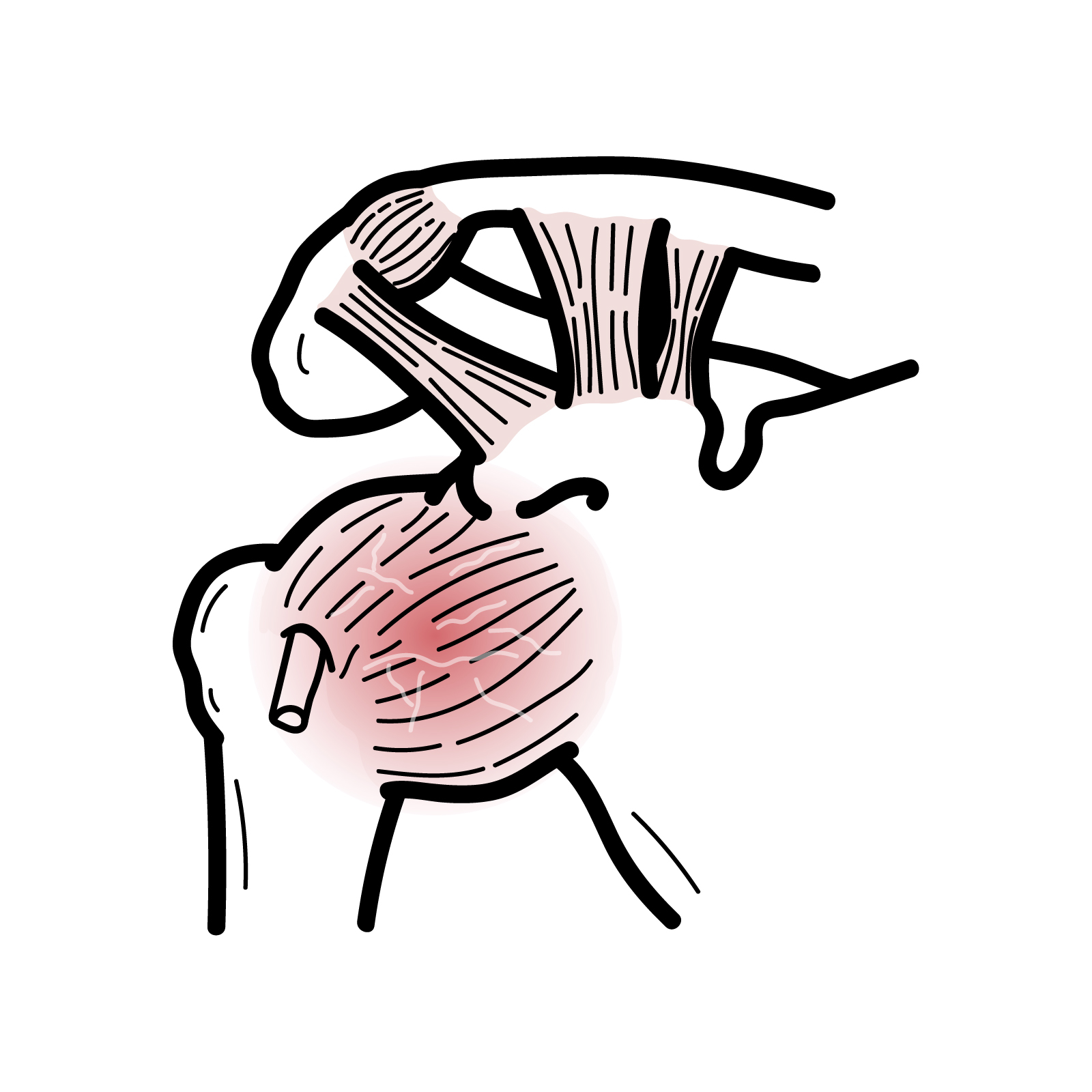
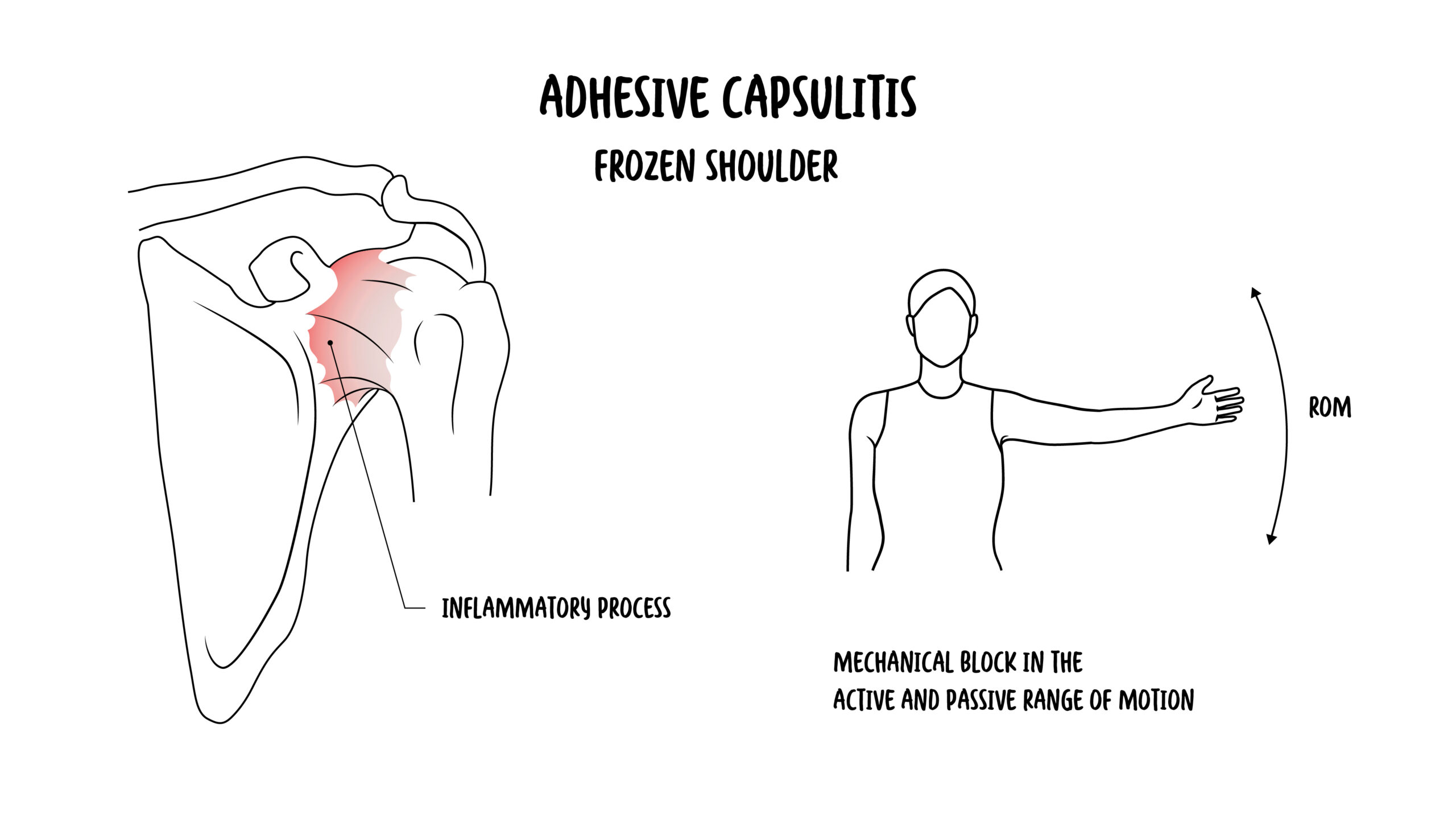
Adhesive capsulitis, commonly called frozen shoulder, is a painful disorder characterised by progressive restriction of both active and passive glenohumeral movement. External rotation is typically affected earliest and most prominently.
The disorder develops through inflammation, fibrosis and contracture of the glenohumeral joint capsule and surrounding ligaments. Symptoms can substantially impair sleep, dressing, grooming, reaching overhead and other everyday activities. Although adhesive capsulitis often improves over time, recovery may take months to years and may remain incomplete in some patients.
Definition
- Adhesive capsulitis
- A painful condition in which fibrosis and contracture of the glenohumeral joint capsule progressively restrict active and passive shoulder movement.
- Frozen shoulder
- The commonly used clinical term for adhesive capsulitis.
- Capsular restriction
- Loss of movement caused by reduced extensibility of the joint capsule rather than weakness or pain alone.
- Glenohumeral joint
- The ball-and-socket articulation formed by the humeral head and glenoid cavity of the scapula.
- Capsular release
- A surgical procedure that divides contracted portions of the glenohumeral capsule to restore movement.
Anatomy & Physiology
The glenohumeral joint is formed by the humeral head and the shallow glenoid cavity. Its large range of movement depends on a relatively loose fibrous capsule supported by the glenohumeral ligaments, coracohumeral ligament, rotator cuff muscles and surrounding soft tissues.
The joint capsule attaches around the glenoid and anatomical neck of the humerus. Inferiorly, the capsule forms the axillary recess, which unfolds during shoulder elevation.
The rotator interval is the triangular area between the supraspinatus and subscapularis tendons. It contains the coracohumeral ligament, superior glenohumeral ligament and long head of the biceps tendon. Normal flexibility within this region is particularly important for external rotation.
Coordinated glenohumeral and scapulothoracic movement permits shoulder elevation. When glenohumeral movement becomes restricted, increased scapular movement may partially compensate but cannot restore normal shoulder function.
Aetiology & Risk Factors
Aetiology
Adhesive capsulitis may be classified according to whether an identifiable precipitating factor is present.
Primary adhesive capsulitis develops without a clear local cause and is considered idiopathic.
Secondary adhesive capsulitis develops in association with:
- Shoulder trauma
- Shoulder or chest surgery
- Prolonged upper-limb immobilisation
- Rotator cuff disease
- Other painful shoulder conditions that reduce movement
- Systemic disorders associated with capsular fibrosis
Reduced shoulder movement following injury or surgery may contribute to secondary disease, but not every stiff or painful shoulder represents adhesive capsulitis.
Risk Factors
Recognised associations include:
- Diabetes mellitus
- Thyroid disease
- Female sex
- Age between approximately 40 and 60 years
- Previous adhesive capsulitis in the opposite shoulder
- Prolonged shoulder immobilisation
- Previous shoulder trauma or surgery
- Neurological disease associated with impaired upper-limb movement
Diabetes is one of the strongest recognised systemic associations. People with diabetes have an increased prevalence of adhesive capsulitis and may experience more prolonged symptoms or incomplete recovery.
Adhesive capsulitis is more common in people with diabetes and thyroid disease.
Pathophysiology
The pathological process usually begins with inflammation of the glenohumeral synovium and joint capsule. This is followed by fibroblast proliferation, collagen deposition, capsular thickening and progressive contracture.
Important pathological changes include:
- Thickening and contracture of the coracohumeral ligament
- Fibrosis of the rotator interval
- Thickening of the anterior and inferior joint capsule
- Reduced capacity of the axillary recess
- Adhesion of capsular folds
- Reduced capsular compliance and joint volume
Early inflammation contributes to pain, particularly at night and at the limits of movement. Progressive fibrosis then produces mechanical restriction of the humeral head within the contracted capsule.
Contracture of the rotator interval and coracohumeral ligament particularly restricts external rotation. Inferior capsular involvement limits elevation and abduction.
Why is passive movement restricted in adhesive capsulitis?
The primary abnormality is fibrosis and contracture of the joint capsule. The examiner therefore cannot restore a normal range by moving the relaxed patient’s arm. This differs from isolated weakness, in which active movement may be limited while passive movement remains relatively preserved.
Clinical Phases
The clinical course is traditionally described in three overlapping phases. The duration and severity of each phase vary considerably.
Freezing phase
Pain is prominent and progressively worsens, often disturbing sleep. Movement gradually becomes more restricted.
Frozen phase
Stiffness and functional restriction predominate. Pain may lessen at rest but persists at the limits of movement.
Thawing phase
Movement gradually improves as capsular restriction resolves. Recovery may be slow and is not always complete.
The phases are descriptive rather than rigid. Patients may have substantial pain and stiffness simultaneously and should not be denied appropriate treatment because they do not fit neatly into one phase.
Clinical Manifestations
Adhesive capsulitis causes progressive shoulder pain and stiffness with restriction of both active and passive movement.
The onset is usually gradual. Patients may report:
- Diffuse shoulder pain, often poorly localised
- Progressive stiffness over weeks or months
- Night pain and difficulty sleeping on the affected side
- Pain at the limits of shoulder movement
- Difficulty reaching overhead
- Difficulty reaching behind the back
- Difficulty dressing, fastening a bra or placing an arm into a sleeve
- Difficulty washing or combing the hair
- Difficulty reaching a back pocket
- Reduced ability to perform work, sport or household activities
Pain may radiate into the upper arm but usually does not extend below the elbow unless another disorder is present.
Clinical Examination
- Restriction of both active and passive glenohumeral movement
- Marked restriction of external rotation with the arm by the side
- Restriction of abduction and internal rotation
- Pain at the end of the available range
- Compensatory scapular movement during attempted elevation
- Relatively preserved strength when tested within the available painless range
- No focal neurological deficit attributable to adhesive capsulitis
The shoulder may be difficult to examine during the pain-dominant phase because pain and guarding can exaggerate the apparent loss of movement.
Restriction of passive external rotation is the key clinical feature of adhesive capsulitis.
If passive movement is relatively preserved despite limited active movement, consider rotator cuff weakness, pain inhibition or neurological disease rather than true capsular restriction.
The condition typically progresses through painful, stiffening, and recovery phases.
Diagnosis
Adhesive capsulitis is primarily a clinical diagnosis. It is supported by gradual shoulder pain and stiffness together with restriction of both active and passive movement, particularly external rotation.
No single blood test or imaging finding confirms the diagnosis. Investigations are mainly used to exclude alternative causes of shoulder stiffness.
Plain Radiography
Plain shoulder radiographs are commonly obtained when the diagnosis is uncertain or symptoms are atypical. Radiographs are usually normal in adhesive capsulitis
Ultrasound
Ultrasound is not required to diagnose adhesive capsulitis. It may be useful when rotator cuff disease, subacromial bursitis or another soft-tissue disorder is suspected. It can also guide intra-articular corticosteroid injection.
Magnetic Resonance Imaging
MRI is not routinely required. Findings may include capsular thickening, coracohumeral ligament thickening, rotator interval fibrosis and oedema within the axillary recess. These findings are supportive rather than independently diagnostic.
MRI may be appropriate when the presentation is atypical or when significant rotator cuff disease, tumour, osteonecrosis or another structural disorder is suspected.
Laboratory Investigations
Routine laboratory testing is unnecessary in an otherwise typical presentation. Testing for diabetes or thyroid dysfunction may be considered when clinically indicated by the history, examination or absence of previous screening.
Differential Diagnosis
Important differential diagnoses include:
- Glenohumeral osteoarthritis
- Rotator cuff tear or tendinopathy
- Subacromial pain syndrome
- Calcific tendinopathy
- Cervical radiculopathy
- Parsonage–Turner syndrome
- Previous fracture or dislocation
- Septic arthritis
- Inflammatory arthritis
- Osteonecrosis of the humeral head
- PMR
Fever, systemic illness, significant trauma, deformity, rapidly progressive symptoms, neurological deficit or a history of malignancy should prompt investigation for an alternative diagnosis.
The term “frozen shoulder” should not be applied to every painful shoulder with reduced active movement.
Adhesive capsulitis requires genuine passive restriction. Severe pain, rotator cuff disease or neurological weakness may reduce active movement without producing the characteristic capsular limitation.
Treatment
Treatment aims to control pain, preserve function and gradually restore shoulder movement. Management should consider symptom severity, disease phase, functional requirements and associated conditions.
Education and Activity Modification
Patients should be advised that:
- Recovery is often gradual
- Normal daily use should continue within tolerable limits
- Complete immobilisation can worsen stiffness
- Exercises should be regular but should not repeatedly provoke severe or prolonged pain
- Diabetes and other associated disorders should be appropriately managed
Most cases adhesive capsulitis improve gradually over months to years.
Analgesia
Paracetamol or a non-steroidal anti-inflammatory drug may be considered for short-term pain relief when clinically appropriate and not contraindicated. Analgesia may improve sleep and allow participation in rehabilitation but does not directly reverse capsular fibrosis.
Physiotherapy and Exercise
Physiotherapy may include:
- Education about the expected course
- Gentle active and passive range-of-motion exercises
- Stretching within tolerable limits
- Scapular control exercises
- Progressive strengthening as pain and movement improve
- A structured home-exercise program
Aggressive stretching during a highly painful phase may worsen symptoms. The intensity of mobilisation should therefore be adjusted to pain irritability and the stage of the disorder.
Intra-articular Corticosteroid Injection
Glenohumeral corticosteroid injection can provide short-term improvement in pain and function, particularly during the early pain-dominant phase. Evidence supports intra-articular corticosteroid injection as an effective non-operative treatment, with additional benefit when combined with an exercise or physiotherapy program.4
Treatment should reflect the dominant clinical problem.
During the pain-dominant phase, prioritise pain control and gentle movement. As pain settles and stiffness predominates, progressively increase stretching, mobilisation and functional rehabilitation.
Hydrodilatation
Hydrodilatation involves image-guided injection of fluid into the glenohumeral joint to distend the contracted capsule, usually with local anaesthetic and corticosteroid.
It may provide short-term improvement in disability and passive external rotation. However, studies vary in technique, and its additional benefit over intra-articular corticosteroid injection alone remains uncertain.5
Manipulation Under Anaesthesia
Manipulation under anaesthesia uses controlled force to disrupt capsular adhesions and improve movement. It is generally reserved for persistent, functionally limiting disease.
Arthroscopic Capsular Release
Arthroscopic capsular release permits controlled division of contracted capsular structures under direct visualisation. It may be considered when substantial pain and restriction persist despite appropriate conservative treatment.
Most patients are managed non-operatively.
Manipulation under anaesthesia and arthroscopic capsular release are escalation options for persistent, functionally limiting symptoms—not routine first-line treatment.
Complications & Prognosis
Complications
Adhesive capsulitis may result in:
- Persistent pain
- Chronic restriction of shoulder movement
- Sleep disturbance
- Reduced ability to work, exercise or perform self-care
- Compensatory scapular or cervical discomfort
- Loss of upper-limb strength through disuse
- Psychological distress related to prolonged pain and disability
Treatment-related complications may occur following corticosteroid injection, hydrodilatation, manipulation under anaesthesia or surgery.
Prognosis
Most patients improve with time and non-operative management, but the course is variable. Recovery may require many months or several years, and some patients retain residual pain or movement restriction.1,3
A previous assumption that every untreated frozen shoulder resolves completely within a predictable period is not supported by the available evidence. Earlier recognition and appropriate symptom-directed treatment may reduce pain and disability while recovery occurs.
Diabetes is associated with a greater likelihood of persistent symptoms, reduced final range of movement and a less favourable treatment response.2 Adhesive capsulitis may subsequently affect the opposite shoulder, although recurrence in the same shoulder is uncommon.
References
- Le HV, Lee SJ, Nazarian A, Rodriguez EK. Adhesive capsulitis of the shoulder: review of pathophysiology and current clinical treatments. Shoulder Elbow. 2017;9(2):75–84. doi:10.1177/1758573216676786
- Zreik NH, Malik RA, Charalambous CP. Adhesive capsulitis of the shoulder and diabetes: a meta-analysis of prevalence. Muscles Ligaments Tendons J. 2016;6(1):26–34. doi:10.11138/mltj/2016.6.1.026
- Rangan A, Goodchild L, Gibson J, Brownson P, Thomas M, Rees J, et al. Frozen shoulder. Shoulder Elbow. 2015;7(4):299–307. doi:10.1177/1758573215601779
- Challoumas D, Biddle M, McLean M, Millar NL. Comparison of treatments for frozen shoulder: a systematic review and meta-analysis. JAMA Netw Open. 2020;3(12):e2029581. doi:10.1001/jamanetworkopen.2020.29581
- Poku D, Hassan R, Migliorini F, Maffulli N. Efficacy of hydrodilatation in frozen shoulder: a systematic review and meta-analysis. Br Med Bull. 2023;147(1):121–147. doi:10.1093/bmb/ldad018








Members only discussions coming soon…