

Neck of Femur Fracture
Overview
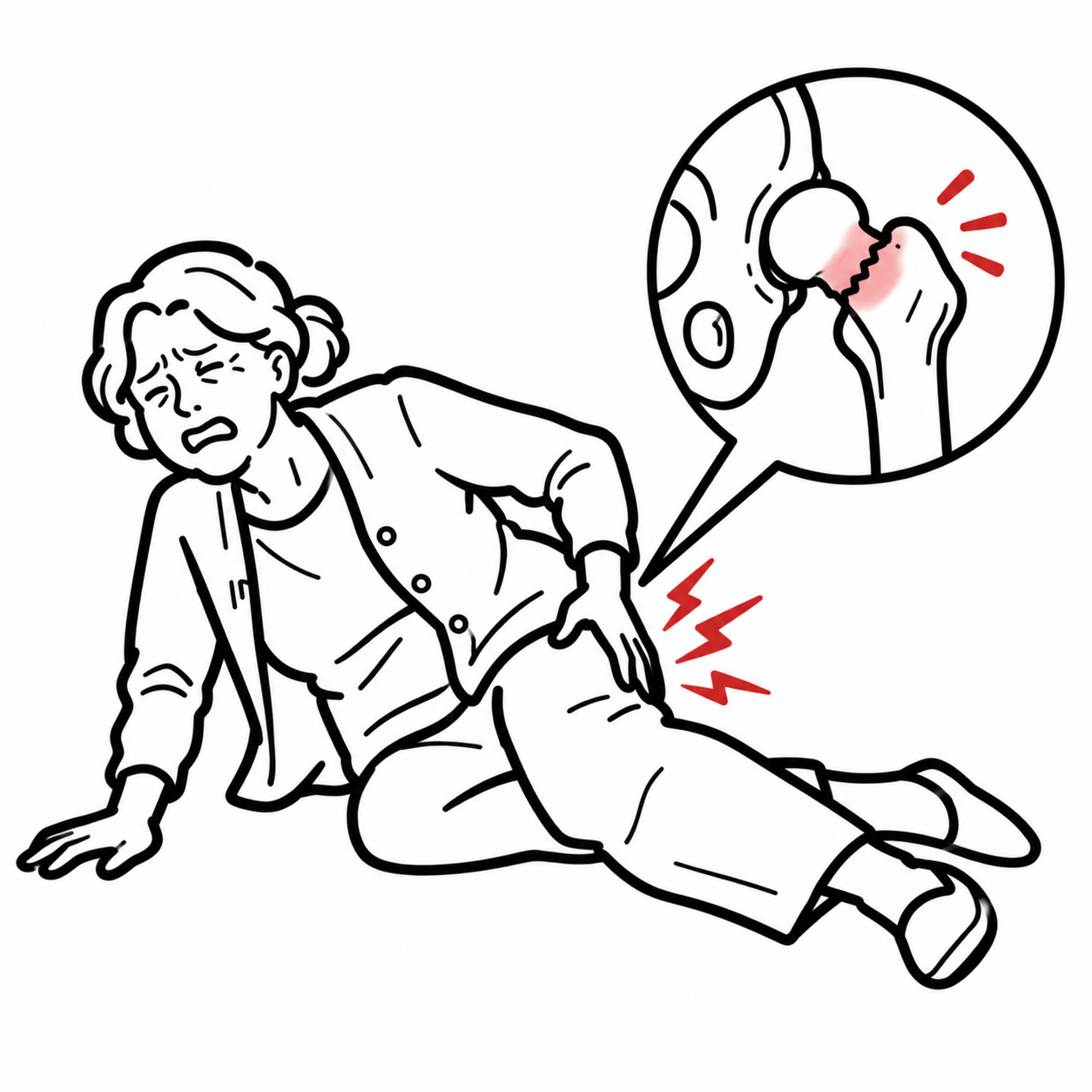
A neck of femur fracture is a fracture through the region connecting the femoral head to the femoral shaft. It is an intracapsular fracture because most of the femoral neck lies within the hip joint capsule.
Neck of femur fractures most commonly occur in older adults following a low-energy fall, often in the presence of osteoporosis, frailty and impaired balance. In younger people, they usually result from high-energy trauma or, less commonly, repetitive loading causing a stress fracture.
These fractures are clinically important because displacement can disrupt the blood supply to the femoral head, resulting in avascular necrosis or nonunion. In older adults, the injury is also associated with pain, immobility, delirium, venous thromboembolism, loss of independence and increased mortality.
Most patients require operative treatment. Management depends on the patient’s age, pre-fracture function, comorbidities, bone quality and the location and displacement of the fracture.
Definition
- Neck of femur fracture
- A fracture involving the femoral neck between the femoral head and the trochanteric region.
- Intracapsular fracture
- A fracture occurring within the hip joint capsule.
- Impacted fracture
- A fracture in which one fragment is driven into another, potentially producing little deformity.
- Internal fixation
- Stabilisation of the fracture using implants such as screws, a sliding hip screw or a femoral neck system.
- Hemiarthroplasty
- Replacement of the femoral head and neck while retaining the native acetabulum.
- Total hip arthroplasty
- Replacement of both the femoral head and the acetabular surface.
- Avascular necrosis
- Death of bone caused by interruption of its blood supply.
- Nonunion
- Failure of a fracture to unite within the expected period.
Terminology
“Hip fracture” is a broader term that includes femoral neck, intertrochanteric and subtrochanteric fractures. A true neck of femur fracture is usually intracapsular, whereas intertrochanteric and subtrochanteric fractures are extracapsular.
Anatomy & Physiology
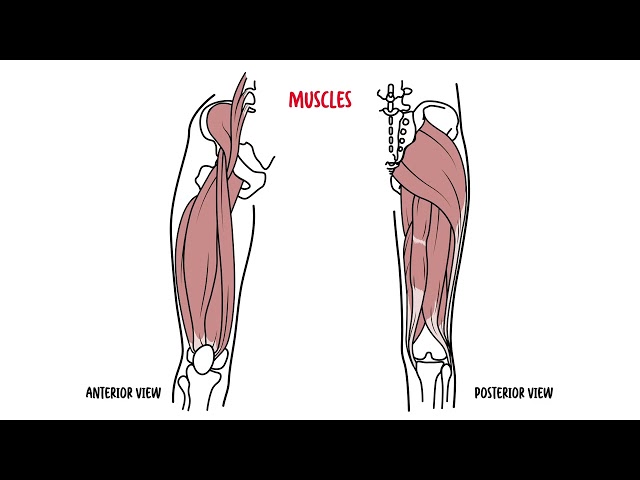
The hip is a ball-and-socket synovial joint formed by the femoral head and the acetabulum. The femoral neck joins the femoral head to the proximal femoral shaft and transmits forces between the pelvis and lower limb.
The femoral neck is largely intracapsular and is covered by synovium. It has relatively little periosteum, which limits external callus formation following fracture.
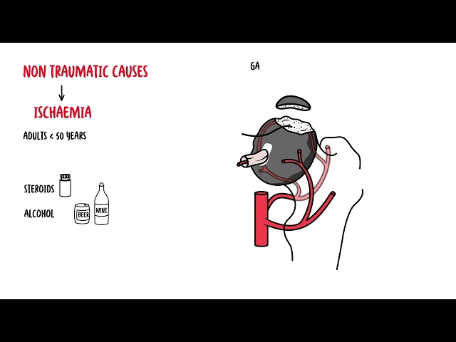
The principal blood supply to the adult femoral head arises from retinacular branches of the medial femoral circumflex artery. These vessels travel along the femoral neck beneath the joint capsule before entering the femoral head.
The lateral femoral circumflex artery and the artery within the ligamentum teres may provide additional blood flow, but the ligamentum teres artery usually makes only a minor contribution in adults.
A displaced femoral neck fracture can tear or compress the retinacular vessels. This places the femoral head at risk of ischaemia, avascular necrosis and subsequent collapse.
Why are displaced neck of femur fractures particularly prone to avascular necrosis?
The principal vessels supplying the femoral head travel along the femoral neck. Fracture displacement can disrupt these retinacular vessels, leaving the femoral head with an inadequate blood supply. The risk increases with greater displacement and vascular injury.
Aetiology & Risk Factors
Low-Energy Fragility Fractures
Most neck of femur fractures occur in older adults after a fall from standing height or less. The injury usually reflects a combination of reduced bone strength and an increased tendency to fall.
Important risk factors include:
- Osteoporosis or osteopenia
- Increasing age
- Female sex
- Previous fragility fracture
- History of falls
- Frailty
- Sarcopenia and impaired balance
- Visual impairment
- Cognitive impairment or dementia
- Neurological disease
- Vitamin D deficiency
- Low body weight or malnutrition
- Smoking
- Excessive alcohol consumption
- Long-term corticosteroid treatment
- Sedative, antihypertensive or other medicines that increase falls risk
- Environmental hazards
- Residence in an aged-care facility
High-Energy Trauma
In younger adults, femoral neck fractures typically follow substantial trauma, such as:
- Motor vehicle or motorcycle collision
- Fall from height
- Sporting injury
- Direct high-energy impact
These patients should also be assessed for associated pelvic, acetabular, femoral shaft and other traumatic injuries.
Stress and Insufficiency Fractures
Femoral neck stress fractures may occur following repetitive loading, particularly in:
- Distance runners
- Military recruits
- Athletes with a sudden increase in training load
- People with low energy availability
- People with menstrual dysfunction
- People with reduced bone mineral density
An insufficiency fracture occurs when normal physiological loading is applied to structurally weakened bone.
Pathological Fractures
Less commonly, a fracture may occur through bone weakened by:
- Primary bone tumour
- Metastatic malignancy
- Multiple myeloma
- Metabolic bone disease
- Previous radiotherapy
- Infection
Pathophysiology
In older adults, age-related loss of bone mass and deterioration of bone microarchitecture reduce the ability of the proximal femur to withstand loading. A sideways fall commonly produces direct impact over the greater trochanter, transmitting force through the femoral neck.
Following an intracapsular fracture:
- The femoral neck loses structural continuity.
- Displacement may disrupt the retinacular blood vessels.
- Intracapsular bleeding may increase pressure within the joint.
- Fracture healing is impaired by limited periosteum and synovial fluid at the fracture site.
- Shearing forces may produce displacement, varus collapse or fixation failure.
- Compromised perfusion may result in avascular necrosis of the femoral head.
Vertical fractures are subjected to greater shearing forces than more horizontal fractures. They are therefore more mechanically unstable and have a higher risk of displacement and nonunion.
Classification
Anatomical Classification
Femoral neck fractures may be described according to their location:
- Subcapital: Immediately below the femoral head
- Transcervical: Through the middle of the femoral neck
- Basicervical: At the base of the femoral neck near the intertrochanteric region
Garden Classification
The Garden classification describes fracture displacement on anteroposterior radiographs:
- Garden I: Incomplete, valgus-impacted fracture
- Garden II: Complete but nondisplaced fracture
- Garden III: Complete fracture with partial displacement
- Garden IV: Complete fracture with full displacement
Garden I and II fractures are generally considered nondisplaced. Garden III and IV fractures are considered displaced.
Pauwels Classification
The Pauwels classification is based on the angle of the fracture line relative to the horizontal:
- Type I: Less than 30°
- Type II: 30–50°
- Type III: Greater than 50°
A more vertical fracture line produces greater shear stress and mechanical instability.
Garden classification describes displacement, whereas Pauwels classification describes the inclination and mechanical stability of the fracture line.
Clinical Manifestations
Patients commonly report:
- Sudden groin or hip pain following a fall
- Inability to stand or bear weight
- Pain with attempted movement of the hip
- Pain radiating into the thigh or knee
- Reduced mobility
- A history of a fall that may be unclear or unwitnessed
- Increasing pain during weight-bearing in a stress fracture
In older adults with cognitive impairment, the presentation may be nonspecific. New immobility, agitation, delirium or apparent knee pain may be the main features.
An impacted or nondisplaced fracture may cause less severe pain. Some patients remain able to stand or walk, although this does not exclude a fracture.
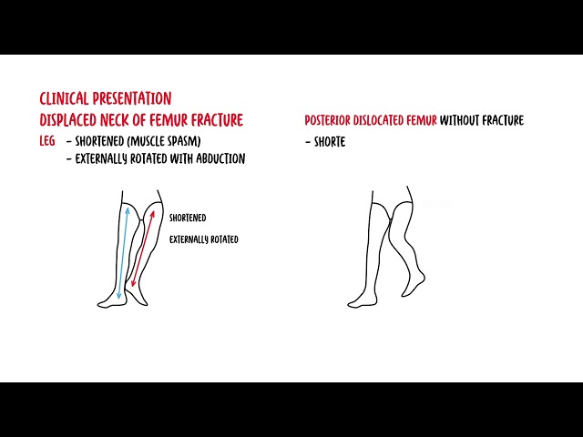
Patients typically present with hip pain, inability to weight-bear, and a shortened externally rotated leg.
Clinical Examination
Typical examination findings include:
- The affected leg held shortened and externally rotated
- Inability to straight-leg raise
- Pain on axial loading of the limb
- Pain with gentle hip rotation
- Groin tenderness
- Reduced hip movement
- Inability to bear weight
- Bruising or swelling, although these may be minimal in an intracapsular fracture
The limb may appear normal in an impacted or nondisplaced fracture.
The classic shortened, externally rotated leg strongly suggests a displaced hip fracture. However, its absence does not exclude an impacted, nondisplaced or occult neck of femur fracture.
Diagnosis
Plain Radiography
Initial imaging generally includes:
- Anteroposterior pelvis radiograph
- Lateral view of the affected hip, usually a cross-table lateral view
The entire pelvis should be reviewed for associated pelvic fractures, contralateral injury and other abnormalities.
Occult Fracture
A normal initial radiograph does not exclude a femoral neck fracture when clinical suspicion remains high.
MRI is the preferred investigation for a suspected occult fracture because it is highly sensitive for trabecular injury and bone marrow oedema. CT may be used when MRI is unavailable or contraindicated, although a negative CT does not always exclude an occult fracture.
Until the diagnosis is clarified, the patient should generally be treated as having a fracture and should avoid unsupported weight-bearing.
Why can a patient with a femoral neck fracture sometimes still walk?
An impacted or nondisplaced fracture may retain temporary mechanical stability. Pain may be mild and there may be no obvious shortening or external rotation. Continued weight-bearing can displace the fracture, so persistent post-fall hip or groin pain requires further investigation even when the patient can walk.
MRI is the most sensitive test if a fracture is suspected despite normal X-rays.
Laboratory Investigations
Preoperative investigations commonly include:
- Full blood count
- Electrolytes, urea and creatinine
- Liver function tests where indicated
- Coagulation studies
- Group and screen or crossmatch
- Blood glucose
- Electrocardiogram
- Additional investigations guided by the clinical presentation
Investigations should identify correctable medical problems without causing unnecessary delay to surgery.
A fragility hip fracture should prompt assessment and treatment for osteoporosis and falls risk.
Treatment
Neck of femur fracture is generally managed through a coordinated orthopaedic, anaesthetic, geriatric, nursing and allied-health pathway.
Immediate Management
Analgesia may include paracetamol, carefully titrated opioid analgesia and a regional nerve block such as a fascia iliaca or femoral nerve block where appropriate.
Routine preoperative traction is not generally required.
Priorities at Presentation
Treat pain early, identify reversible medical problems, prevent delirium and pressure injury, and prepare the patient for timely surgery. Medical assessment should facilitate surgery rather than introduce avoidable delays.
Timing of Surgery
For most older adults, surgery should occur as soon as the patient is medically ready. Australian standards recommend surgery within 36 hours of first presentation to hospital when there is no clinical contraindication.
Stable chronic medical conditions should not usually delay surgery unnecessarily.
Hip fracture surgery is urgent. Aim for surgery within 36 hours of first hospital presentation unless a clear clinical contraindication requires stabilisation.
Operative Management
Nondisplaced or Impacted Fractures
Internal fixation preserves the native femoral head but carries risks of secondary displacement, nonunion and avascular necrosis. Internal fixation includes:
- Multiple cannulated screws
- Sliding hip screw with an antirotation screw
- Femoral neck system
Displaced Fractures in Older Adults
Options include:
- Hemiarthroplasty: Commonly used for patients with lower functional demand, cognitive impairment, frailty or limited life expectancy.
- Total hip arthroplasty: May be considered for independently mobile, cognitively intact and medically suitable patients, particularly when there is pre-existing symptomatic hip osteoarthritis or a high functional demand.
Cemented femoral components are commonly used for arthroplasty in older adults.
Fractures in Younger Adults
In younger patients, preservation of the native femoral head is usually prioritised. Management generally involves urgent anatomical reduction and stable internal fixation.
The quality of reduction is a major determinant of healing and fixation success. Vertical or highly unstable fractures may require a more mechanically stable fixation construct.
Nonoperative Treatment
Nonoperative treatment is uncommon and generally reserved for:
- Patients who cannot tolerate any surgical intervention
- Patients who are terminally ill
- Selected patients receiving palliative care
- Rare stable stress fractures managed under specialist guidance
The goals are pain control, nursing care, pressure-area protection and mobilisation within the limits of comfort and safety.
Secondary Fracture Prevention
A neck of femur fracture following minimal trauma should prompt assessment and management of osteoporosis and falls risk.
Secondary prevention may include:
- Osteoporosis evaluation
- Calcium and vitamin D assessment where appropriate
- Antiresorptive or anabolic osteoporosis treatment when indicated
- Falls assessment
- Vision and footwear review
- Medication review
- Balance and strengthening exercises
- Environmental modification
- Referral to a fracture liaison service
The Fracture Is Not the Whole Diagnosis
A low-trauma neck of femur fracture is often the first visible consequence of osteoporosis, frailty and falls risk. Definitive care should include secondary fracture prevention rather than ending when the hip has been repaired.
Complications & Prognosis
Local and Mechanical Complications
Complications specific to femoral neck fractures include:
- Avascular necrosis of the femoral head
- Nonunion
- Malunion
- Varus collapse
- Secondary fracture displacement
- Fixation failure
- Implant cut-out or loosening
- Limb-length discrepancy
- Persistent hip pain
- Post-traumatic osteoarthritis
Avascular necrosis is more likely after a displaced fracture because displacement can disrupt the retinacular vessels supplying the femoral head. It may become radiographically apparent months or years after the injury.
Intracapsular fractures risk disruption of the femoral head blood supply and avascular necrosis.
Perioperative and Medical Complications
Older adults are at risk of:
- Delirium
- Pneumonia
- Urinary tract infection
- Pressure injury
- Deep-vein thrombosis
- Pulmonary embolism
- Anaemia
- Wound infection
- Prosthetic joint infection
- Periprosthetic fracture
- Hip dislocation following arthroplasty
- Cardiovascular events
- Acute kidney injury
- Malnutrition
- Deconditioning
- Loss of independence
Prognosis
Prognosis depends on age, frailty, pre-fracture mobility, cognition, comorbidities, fracture displacement, treatment timing and postoperative complications.
Younger patients with a well-reduced and stably fixed fracture may regain good function, although long-term surveillance is required because avascular necrosis and nonunion can occur.
In older adults, a hip fracture is a major health event. Many patients do not return fully to their previous level of mobility or independence. Some require increased community support or residential care.
Early surgery, coordinated orthogeriatric care, effective analgesia, delirium prevention, nutritional support and early mobilisation improve the likelihood of recovery.
References
- Australian Commission on Safety and Quality in Health Care. Hip Fracture Clinical Care Standard. Sydney: ACSQHC; 2023. Available from: https://www.safetyandquality.gov.au/clinical-care-standards/hip-fracture
- National Institute for Health and Care Excellence. Hip fracture: management. NICE guideline CG124. London: NICE; updated 2023. Available from: https://www.nice.org.uk/guidance/cg124
- American Academy of Orthopaedic Surgeons. Management of Hip Fractures in Older Adults: Evidence-Based Clinical Practice Guideline. Rosemont: AAOS; 2021. Available from: https://www.aaos.org/hipfxcpg
- Agency for Clinical Innovation. Hip fracture suspected: Adult Emergency Care Assessment and Treatment protocol. Sydney: NSW Health; updated 2024. Available from: https://aci.health.nsw.gov.au/ecat/adult/hip-fracture
- AO Foundation. AO Surgery Reference: Proximal femur and femoral neck fractures. Davos: AO Foundation. Available from: https://surgeryreference.aofoundation.org/orthopedic-trauma/adult-trauma/proximal-femur
- Bhandari M, Swiontkowski M. Management of acute hip fracture. N Engl J Med. 2017;377(21):2053–2062. doi:10.1056/NEJMcp1611090



Members only discussions coming soon…