

Dupuytren’s Contracture
Overview
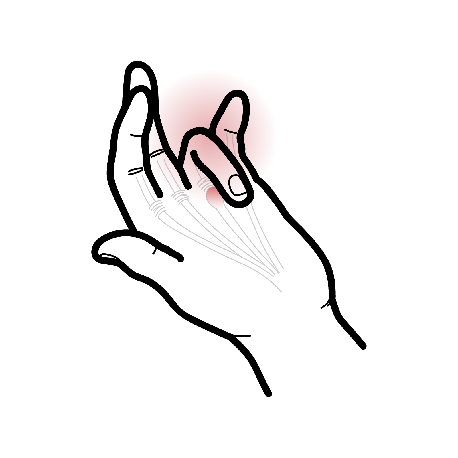
Dupuytren’s disease is a benign fibroproliferative disorder affecting the palmar fascia. Abnormal collagen deposition produces firm nodules and cords beneath the skin of the palm, which may progressively shorten and pull one or more fingers into flexion.
The ring and little fingers are most commonly affected. Contracture usually develops slowly over months or years and may eventually interfere with placing the hand flat, wearing gloves, washing the face, shaking hands or placing the hand into a pocket.
Dupuytren’s disease refers to the underlying fascial disorder, whereas Dupuytren’s contracture describes the fixed flexion deformity that develops when a pathological cord crosses a finger joint.
The condition is usually painless and does not affect the flexor tendons. Treatment is guided by functional impairment, progression and joint involvement. Procedures can improve finger extension but do not cure the underlying disease, and recurrence remains possible.
Definition
- Dupuytren’s disease
- A benign fibroproliferative disorder of the palmar fascia characterised by nodules and pathological cords.
- Dupuytren’s contracture
- A fixed flexion deformity of a finger caused by shortening of a Dupuytren cord.
- Palmar nodule
- A firm subcutaneous lump formed by proliferating fibroblasts and myofibroblasts, usually located near the distal palmar crease.
- Dupuytren cord
- A thickened longitudinal band of diseased fascia that may cross a joint and restrict finger extension.
- Tabletop test
- A clinical test in which the patient attempts to place the palm and fingers completely flat on a table.
- Fasciotomy
- Division of a pathological cord without removing it.
- Fasciectomy
- Surgical removal of part or all of the affected fascia.
Aetiology & Risk Factors
The precise cause of Dupuytren’s disease is not fully understood. Genetic susceptibility is important, and the condition is associated with altered fibroblast and myofibroblast activity within the palmar fascia.
Recognised risk factors and associations include:
- Increasing age
- Male sex
- Family history of Dupuytren’s disease
- Northern European ancestry
- Diabetes mellitus
- Cigarette smoking
- Excessive alcohol consumption
- Epilepsy, although the relationship may partly reflect associated genetic or medication factors
- Previous hand trauma or surgery
- Manual work involving repeated vibration or heavy hand use
- Bilateral disease
- Ectopic fibromatosis, including plantar fibromatosis (Ledderhose disease), penile fibromatosis (Peyronie disease) and knuckle pads
Dupuytren Diathesis
Dupuytren diathesis describes features associated with earlier onset, more extensive disease and a greater risk of progression or recurrence. These include:
- Young age at presentation
- Strong family history
- Bilateral hand involvement
- Multiple affected digits
- Ectopic disease
- Male sex
Dupuytren’s disease is strongly associated with inherited susceptibility.
Manual work may influence expression or symptoms, but the condition should not be described simply as a consequence of “wear and tear.”
Clinical Manifestations
Early Disease
- One or more firm nodules in the palm
- Pitting or tethering of the palmar skin
- Thickening of the palmar fascia
- Local tenderness during the proliferative phase
- A feeling of tightness when extending the fingers
- A palpable cord extending from the palm towards a finger
Nodules are commonly located near the distal palmar crease. They may initially be tender but often become painless as the disease matures.
Typical findings are palmar nodules and cords with progressive inability to fully extend the affected digits.
Established Contracture
Progressive shortening of a cord may produce:
- Loss of active and passive finger extension
- Fixed flexion of a metacarpophalangeal joint
- Fixed flexion of a proximal interphalangeal joint
- Difficulty placing the hand flat on a surface
The ring finger is most commonly affected, followed by the little finger. The middle finger and thumb may also be involved.
Tabletop Test
Ask the patient to place the palm and all fingers flat on a table. A positive test occurs when the palm or an affected finger cannot lie completely flat because of a flexion contracture. The test is useful for monitoring progression and identifying patients who may benefit from specialist assessment.
A positive tabletop test suggests a clinically significant extension deficit.
Treatment decisions should also consider progression, function, joint involvement and the patient’s goals.
Why is passive movement important during examination?
A patient who cannot actively straighten a finger may have tendon or nerve dysfunction. In Dupuytren’s contracture, passive extension is also restricted because a shortened fascial cord mechanically holds the joint in flexion.
Diagnosis
Diagnosis is usually clinical.
Investigations
Investigations are usually unnecessary.
- Plain radiographs: Consider when osteoarthritis, previous fracture, joint deformity or another bony abnormality is suspected.
- Ultrasound: May demonstrate nodules or cords but is not routinely required.
- MRI: Rarely indicated; may be considered when the diagnosis is uncertain or a soft-tissue mass is suspected.
- Blood tests: Do not diagnose Dupuytren’s disease but may be appropriate when investigating associated conditions such as diabetes.
Differential Diagnosis
- Trigger finger
- Flexor tendon injury or adhesion
- Camptodactyly
- Post-traumatic joint stiffness
- Osteoarthritis
- Inflammatory arthritis
- Diabetic cheiroarthropathy
- Volkmann ischaemic contracture
- Ulnar nerve palsy
- Scar contracture
- Soft-tissue tumour
- Congenital finger deformity
Dupuytren’s Disease versus Trigger Finger
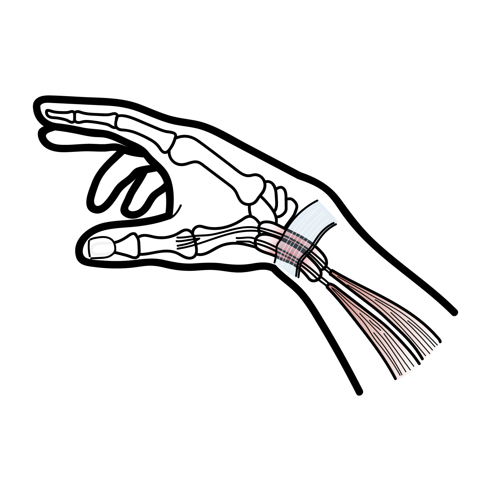
Dupuytren’s disease produces a palpable palmar cord and a progressive fixed flexion contracture. Trigger finger causes painful catching or locking as a flexor tendon passes through the A1 pulley and does not usually produce a longitudinal palmar cord.
Treatment
Treatment is guided by functional impairment, rate of progression, degree of contracture, joint involvement, skin quality, previous treatment, comorbidities, patient preference and risk of recurrence.
Observation
Observation is appropriate when:
- A nodule or cord is present without contracture
- The tabletop test remains negative
- Hand function is preserved
- Disease progression is minimal
- The patient does not wish to undergo intervention
Patients can periodically repeat the tabletop test and seek reassessment if extension deteriorates or activities become difficult.
Nonoperative Management
Nonoperative measures do not reliably remove a mature cord or permanently correct an established fixed contracture. They may include:
- Education and monitoring
- Activity modification when a tender nodule is aggravated
- Analgesia for discomfort
- Hand therapy to maintain movement and function
- Adaptive equipment for daily activities
- Corticosteroid injection for a painful early nodule in selected patients
Routine stretching or splinting alone has not been shown to prevent progression of the underlying disease. Radiotherapy has been used for early active disease in some settings, but evidence and availability vary, and it is not routinely recommended in many guidelines.
When to Consider Intervention
Referral to a hand surgeon should be considered when contracture is progressive, interferes with hand function, produces a positive tabletop test or causes a clinically important metacarpophalangeal or proximal interphalangeal extension deficit.
Proximal interphalangeal contractures should not be allowed to become very advanced before assessment because they are harder to correct and more likely to leave residual stiffness.
Percutaneous Needle Fasciotomy
A needle is passed through the skin and used to divide the pathological cord at one or more points.
Collagenase Injection
Collagenase may be injected into a suitable cord to enzymatically weaken it, followed by controlled manipulation to rupture the cord.
Limited Fasciectomy
Limited fasciectomy involves open surgical removal of the diseased fascia responsible for the contracture. It is commonly used for functionally significant contracture, progressive disease, severe or complex cords, significant proximal interphalangeal involvement, or disease unsuitable for a minimally invasive procedure.
Potential advantages include more complete correction and a lower recurrence risk than needle fasciotomy, although recovery is longer and operative complications are more frequent.
Dermofasciectomy
Dermofasciectomy removes the diseased fascia together with overlying involved skin, followed by skin grafting. It may be considered for recurrent disease, extensive skin involvement, severe Dupuytren diathesis or selected younger patients with aggressive disease.
Procedures improve extension but do not cure the disease.
Treatment releases or removes the pathological cord; it does not eliminate the biological tendency to form new Dupuytren tissue.
Treatment Comparison
| Treatment | Main role | Recovery | Important limitation |
|---|---|---|---|
| Observation | Nodules or mild disease without functional contracture | None | Disease may progress |
| Needle fasciotomy | Selected palpable cords, especially at the metacarpophalangeal joint | Usually rapid | Higher recurrence |
| Collagenase injection | Selected cords where treatment is available | Relatively rapid | Bruising, skin tears and recurrence |
| Limited fasciectomy | Functionally significant, severe or complex disease | Longer rehabilitation | Wound, nerve and stiffness risks |
| Dermofasciectomy | Recurrent or aggressive disease with skin involvement | Longest recovery | Requires skin grafting |
Complications & Prognosis
Complications
- Increasing fixed flexion deformity
- Loss of hand span
- Difficulty with gripping and personal care
- Skin maceration or hygiene problems
- Secondary joint stiffness
- Adaptive shortening of the joint capsule and surrounding tissues
- Functional disability
Prognosis
Progression is variable. Some patients have stable nodules for many years, whereas others develop progressive contractures affecting several digits.
Treatment can substantially improve finger extension and hand function, but complete correction may not be possible, particularly when a severe proximal interphalangeal contracture has been present for a long time.
Metacarpophalangeal contractures generally respond better than proximal interphalangeal contractures. Earlier assessment of progressive proximal interphalangeal involvement may reduce the risk of persistent deformity.
Recurrence can occur after any intervention. It is generally more frequent after minimally invasive cord division than after open fasciectomy, although minimally invasive procedures offer faster recovery and can be repeated.
What does recurrent deformity mean?
A recurrent flexion deformity after treatment does not necessarily mean that the original procedure failed. It may represent recurrence at the treated site, extension into a new fascial area or persistent joint stiffness after longstanding contracture.
References
- American Academy of Orthopaedic Surgeons. Dupuytren’s Disease. OrthoInfo. Available from: https://www.orthoinfo.org/diseases–conditions/dupuytrens-disease/
- British Society for Surgery of the Hand. Dupuytren’s Disease. Available from: https://www.bssh.ac.uk/patients/conditions/25/dupuytrens_disease
- Kemler MA, Houpt P, van der Horst CMAM, et al. Dutch multidisciplinary guideline on Dupuytren disease. J Hand Surg Glob Online. 2023;5(2):212–217.
- Dutta A, Jayasinghe G, Deore S, Wahed K, Bhan K, Bakti N, et al. Dupuytren’s contracture: current concepts. J Clin Orthop Trauma. 2020;11(4):590–596.
- Salari N, Heydari M, Hassanabadi M, et al. The worldwide prevalence of Dupuytren disease: a comprehensive systematic review and meta-analysis. J Orthop Surg Res. 2020;15:495.
- National Health Service. Dupuytren’s Contracture. Available from: https://www.nhs.uk/conditions/dupuytrens-contracture/







Members only discussions coming soon…