

Arrhythmogenic Right Ventricular Cardiomyopathy

Overview
Arrhythmogenic right ventricular cardiomyopathy (ARVC) is an inherited myocardial disease characterised by ventricular arrhythmias and progressive structural abnormalities, predominantly affecting the right ventricle. Left-ventricular involvement is common, and some patients have biventricular or left-dominant disease; these phenotypes are encompassed by the broader term arrhythmogenic cardiomyopathy.1,2
ARVC is an important cause of ventricular tachycardia and sudden cardiac death, particularly in young people and athletes. Clinical expression varies considerably because pathogenic variants demonstrate incomplete penetrance and variable expressivity.
Definition
- Arrhythmogenic right ventricular cardiomyopathy
- An inherited cardiomyopathy characterised by ventricular arrhythmias and structural myocardial disease predominantly affecting the right ventricle.
- Arrhythmogenic cardiomyopathy
- A broader disease spectrum that includes right-dominant, biventricular and left-dominant arrhythmogenic myocardial phenotypes.
- Penetrance
- The proportion of people carrying a disease-associated genetic variant who develop clinical features of the condition.
- Desmosome
- A specialised cell-to-cell junction that provides mechanical adhesion between cardiomyocytes.
- Epsilon wave
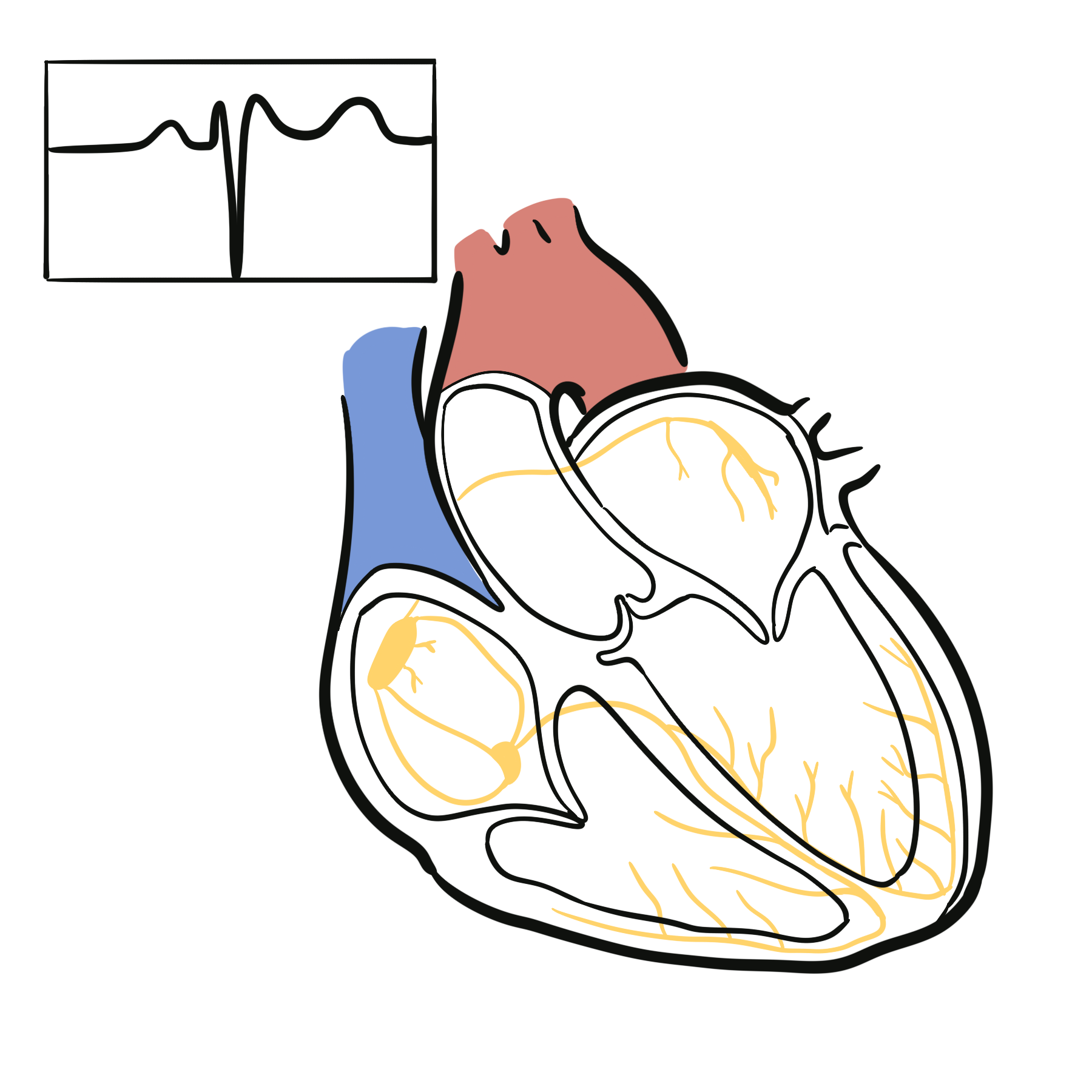
- A small terminal deflection occurring after the end of the QRS complex, typically in the right precordial leads, associated with delayed right-ventricular activation.
Anatomy & Physiology
The right ventricle is a thin-walled, crescent-shaped chamber adapted to pumping blood into the low-resistance pulmonary circulation. Its inflow tract, apex and outflow tract have complex geometry and contraction patterns.
Cardiomyocytes are mechanically connected at intercalated discs. Desmosomal proteins maintain cellular adhesion during repeated myocardial contraction. Important desmosomal proteins include:
- Plakophilin-2.
- Desmoplakin.
- Desmoglein-2.
- Desmocollin-2.
- Plakoglobin.
The cardiac conduction system normally activates the ventricles rapidly and synchronously. Loss of healthy myocardium and development of scar can produce areas of slow, heterogeneous conduction that support re-entrant ventricular arrhythmias.
Aetiology & Risk Factors
Aetiology
ARVC is most commonly genetic. Inheritance is usually autosomal dominant with age-dependent, incomplete penetrance and variable expression.
Associated genes include:
- PKP2, encoding plakophilin-2.
- DSP, encoding desmoplakin.
- DSG2, encoding desmoglein-2.
- DSC2, encoding desmocollin-2.
- JUP, encoding plakoglobin.
- TMEM43.
- PLN.
Variants involving DSP are particularly associated with left-ventricular involvement, myocardial fibrosis and inflammatory injury that may resemble myocarditis.
Rare autosomal-recessive syndromes include:
- Naxos disease: classically associated with a JUP variant, woolly hair and palmoplantar keratoderma.
- Carvajal syndrome: commonly associated with biallelic DSP variants, palmoplantar keratoderma and a left-dominant cardiomyopathy phenotype.
A clinically similar phenotype may occasionally result from inflammatory, infectious or systemic myocardial disease. These phenocopies must be distinguished from inherited ARVC.
Pathophysiology
Defective desmosomal or associated proteins weaken mechanical coupling between cardiomyocytes. Repetitive mechanical stress, particularly during intense exercise, promotes cellular injury, detachment and death.
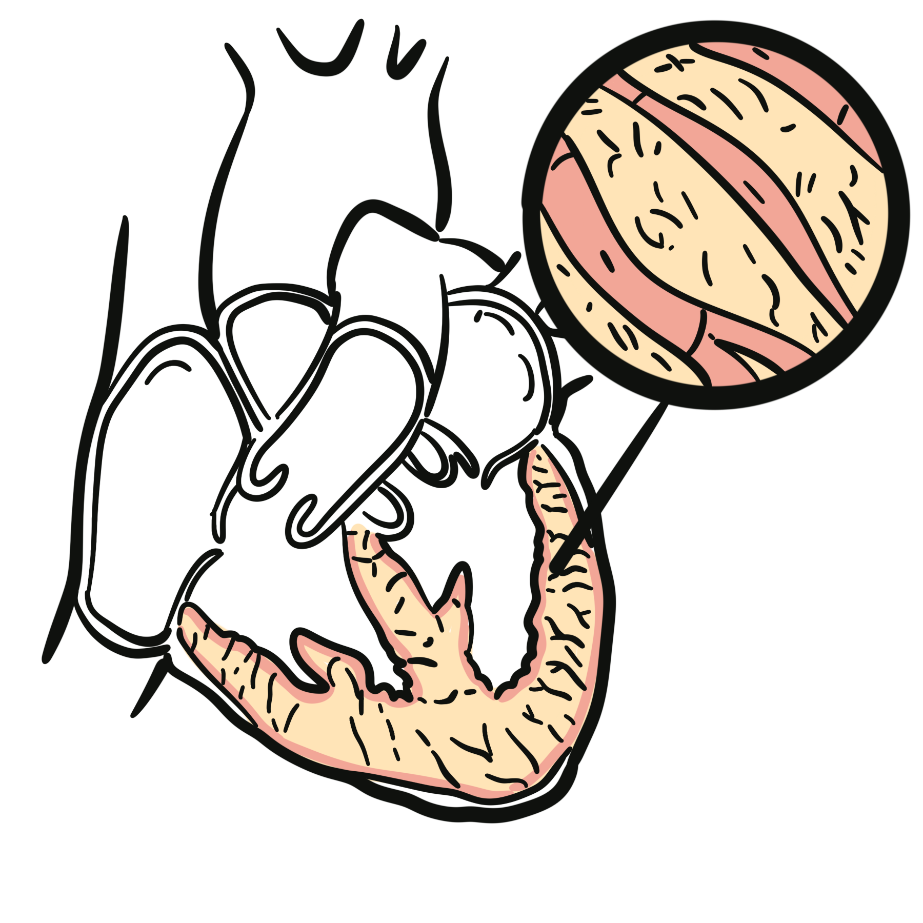
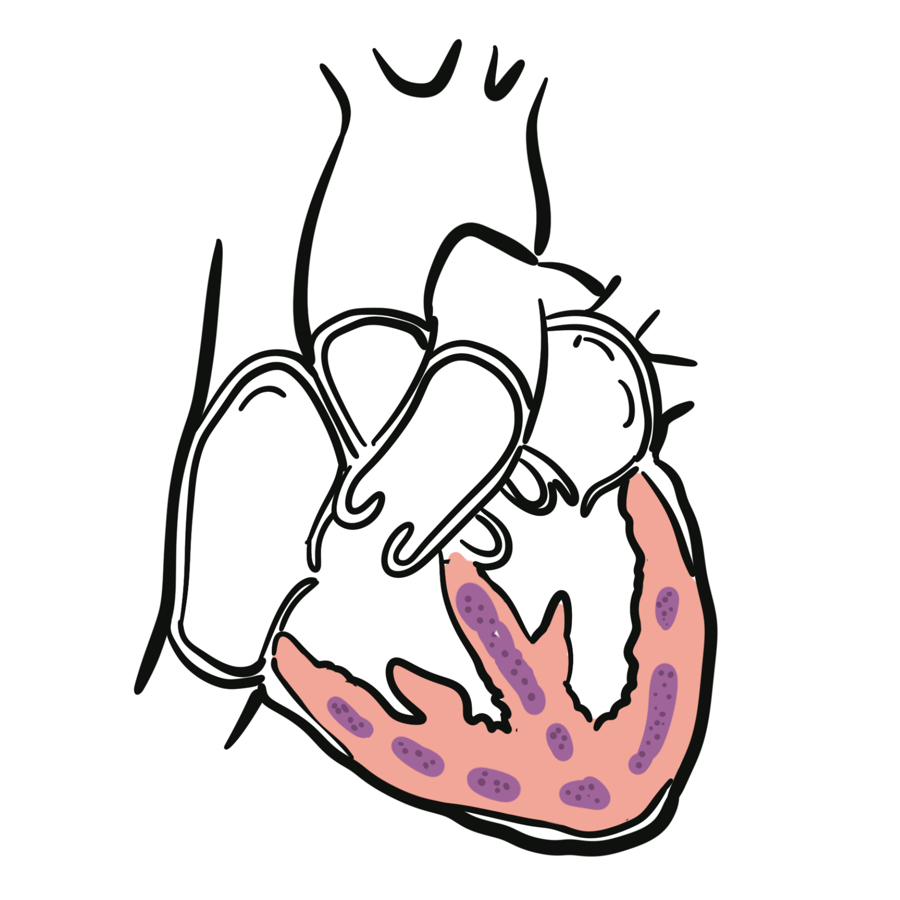
Injured myocardium is progressively replaced by fibrosis and fatty tissue. This process often begins in the subepicardium and may extend towards the endocardium. Disease distribution is patchy and may involve the right ventricle, left ventricle or both.
Structural remodelling can produce:
- Regional akinesia, dyskinesia or aneurysmal bulging.
- Right-ventricular dilatation.
- Reduced right-ventricular systolic function.
- Left-ventricular fibrosis or dysfunction.
- Progressive biventricular failure.
Fibrosis interrupts normal electrical propagation and creates slow-conduction pathways that support ventricular re-entry. Electrical abnormalities and ventricular arrhythmias may develop before obvious ventricular dilatation or systolic dysfunction.
Exercise increases ventricular wall stress and may accelerate phenotypic expression, disease progression and ventricular-arrhythmia risk in susceptible patients.2
A structurally near-normal echocardiogram does not exclude early ARVC. Electrical instability and microscopic scar may precede overt right-ventricular dilatation or systolic dysfunction, making ECG, ambulatory monitoring, cardiac MRI and family assessment important.
Clinical Manifestations
Clinical expression ranges from an asymptomatic genetic carrier state to ventricular arrhythmia, heart failure or sudden cardiac death.
Arrhythmic Manifestations
Patients may experience:
- Palpitations.
- Exertional presyncope or syncope.
- Documented premature ventricular complexes.
- Non-sustained or sustained ventricular tachycardia.
- Ventricular fibrillation.
- Resuscitated cardiac arrest.
- Sudden cardiac death.
Ventricular tachycardia arising from the right ventricle commonly has a left bundle branch block morphology. The QRS axis may help localise the ventricular origin.
Structural and Heart-Failure Manifestations
Progressive disease may cause:
- Exercise intolerance.
- Fatigue.
- Dyspnoea.
- Peripheral oedema.
- Elevated jugular venous pressure.
- Hepatic congestion.
- Ascites.
- Right-sided heart failure.
- Biventricular heart failure.
Other Presentations
ARVC may be identified following:
- Abnormal ECG screening.
- Frequent ventricular ectopy.
- Incidental cardiac imaging abnormalities.
- Investigation of an affected relative.
- Identification of a familial pathogenic variant.
- An apparent episode of myocarditis, particularly in some DSP-associated disease.
Exertional syncope, sustained ventricular tachycardia or cardiac arrest in a young person—particularly with a family history of cardiomyopathy or sudden death—requires urgent assessment for inherited arrhythmic and structural heart disease.
Diagnosis & Investigations
ARVC is a multiparametric diagnosis. No isolated ECG, imaging or genetic finding is sufficient in every patient. Investigations should be interpreted in an inherited-cardiomyopathy service when possible.1–4
Electrocardiography
Potential ECG findings include:
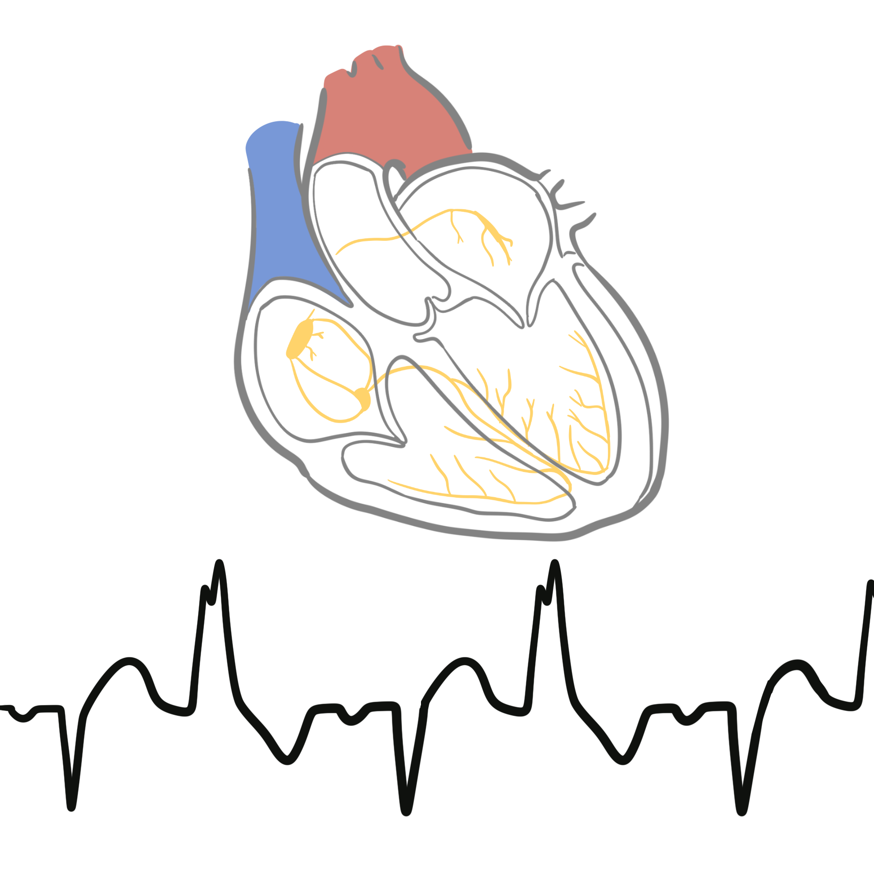
- T-wave inversion in leads V1–V3 or beyond in an appropriate age group and in the absence of complete right bundle branch block.
- Epsilon waves in the right precordial leads.
- Prolonged terminal activation duration.
- Fragmented QRS complexes.
- Premature ventricular complexes.
- Ventricular tachycardia with left bundle branch block morphology.
- Low limb-lead QRS voltages or inferolateral T-wave inversion when left-ventricular involvement is present.
An epsilon wave is relatively specific when confidently identified but has limited sensitivity and substantial interobserver variability. Its absence does not exclude ARVC.
Echocardiography
- Regional RV akinesia, dyskinesia, aneurysm/bulging
- RV dilatation and/or reduced RV systolic function
- Advanced disease may show substantial global RV dysfunction.
Cardiac Magnetic Resonance Imaging
- More sensitive assessment of RV volumes/function
- Regional RV akinesia/dyskinesia/bulging
- RV dilatation and reduced RVEF
- Contrast CMR may demonstrate myocardial fibrosis via late gadolinium enhancement (LGE) and can detect LV involvement.
Exercise Testing
Exercise testing may demonstrate:
- Exercise-induced ventricular ectopy.
- Non-sustained ventricular tachycardia.
- Abnormal blood-pressure response.
- Reduced functional capacity.
It should be performed in an appropriately monitored environment. Exercise testing is not used to prove that vigorous exercise is safe in a patient with established ARVC.
Genetic Testing
- Pathogenic/likely pathogenic variants most commonly involve desmosomal genes: PKP2, DSP, DSG2, DSC2, JUP
- Non-desmosomal genes include TMEM43, PLN, DES
- A pathogenic variant is found in up to ~60% in contemporary ARVC cohorts.
Endomyocardial Biopsy
Endomyocardial biopsy is not routinely required. Limitations include:
- Patchy disease distribution.
- Sampling error.
- Procedural risk, including perforation of the thin right-ventricular wall.
- Histological overlap with other disorders.
Biopsy may be considered when inflammatory, infiltrative or alternative myocardial disease remains a major diagnostic possibility and the result would change management.
Genetics & Family Screening
A three-generation family history should document:
- Cardiomyopathy.
- Ventricular arrhythmias.
- Implantable cardioverter-defibrillator implantation.
- Heart transplantation.
- Premature or unexplained sudden death.
- Unexplained drowning or single-vehicle accidents.
- Seizure-like episodes that may have represented arrhythmic syncope.
First-degree relatives should be offered clinical screening through an inherited-cardiac-disease service. Evaluation commonly includes:
- Medical and family history.
- Physical examination.
- Twelve-lead ECG.
- Ambulatory ECG monitoring.
- Echocardiography.
- Cardiac MRI when indicated.
- Exercise testing in selected patients.
When a pathogenic familial variant is identified, cascade genetic testing can distinguish relatives who require continued surveillance from relatives who did not inherit that variant. Clinical surveillance remains important for variant-positive relatives because penetrance is age-dependent.
The age at which screening begins and the interval between assessments should be individualised according to the familial genotype, phenotype, age of disease onset and family history.1,2
Treatment
Management aims to prevent sudden cardiac death, suppress symptomatic arrhythmia, slow disease progression and treat ventricular dysfunction.
Exercise and Lifestyle
Patients with established ARVC should avoid competitive sport and high-intensity endurance exercise because repeated intense exercise can increase arrhythmic risk and accelerate disease progression.2
Exercise advice should be individualised according to:
- Phenotype.
- Genotype.
- Previous ventricular arrhythmia.
- Ventricular function.
- Exercise intensity and duration.
- Patient values and occupation.
Regular low-intensity activity may remain appropriate in selected clinically stable patients following specialist assessment. Exercise restriction does not eliminate arrhythmic risk.
Pharmacological Treatment
Beta-blockers are commonly used to:
- Reduce adrenergic stimulation.
- Suppress ventricular ectopy.
- Reduce exercise-associated arrhythmia.
- Treat associated ventricular dysfunction.
Antiarrhythmic medicines may reduce recurrent ventricular tachycardia or implantable-cardioverter defibrillator therapies. They do not provide the same protection from sudden death as an indicated defibrillator.
Standard guideline-directed heart-failure therapy should be used when right- or left-ventricular dysfunction develops, with treatment adapted to blood pressure, renal function and ventricular phenotype.
Implantable Cardioverter-Defibrillator
An implantable cardioverter-defibrillator (ICD) is recommended for secondary prevention following:
- Resuscitated cardiac arrest.
- Ventricular fibrillation.
- Haemodynamically unstable sustained ventricular tachycardia.
Catheter Ablation
Catheter ablation may be used for:
- Recurrent monomorphic ventricular tachycardia.
- Electrical storm.
- Frequent appropriate ICD therapies.
- Ventricular tachycardia despite medication.
The arrhythmic substrate is often epicardial, and combined endocardial–epicardial ablation may be required in specialist centres. Ablation reduces arrhythmia burden but does not cure the underlying cardiomyopathy or remove the need for an otherwise indicated ICD.
Advanced Heart-Failure Therapy
Patients with progressive ventricular failure may require:
- Specialist heart-failure management.
- Diuretic therapy for congestion.
- Mechanical circulatory support in selected cases.
- Heart-transplant assessment.
Complications & Prognosis
Complications
- Sustained ventricular tachycardia.
- Ventricular fibrillation.
- Sudden cardiac death.
- Recurrent appropriate ICD therapies.
- Syncope and physical injury.
- Progressive right-ventricular failure.
- Left-ventricular or biventricular failure.
- Atrial arrhythmias.
- Intracardiac thrombus and thromboembolism.
- Medication adverse effects.
- Catheter-ablation recurrence.
- ICD infection, inappropriate shocks or lead complications.
- Need for heart transplantation.
Prognosis
The clinical course is highly variable. Some variant carriers remain phenotype-negative for many years, whereas others develop early ventricular arrhythmias, progressive myocardial dysfunction or sudden cardiac arrest.
Poor prognostic features include:
- Previous cardiac arrest or sustained ventricular tachycardia.
- Arrhythmic syncope.
- High ventricular ectopic burden.
- Non-sustained ventricular tachycardia.
- Significant right- or left-ventricular dysfunction.
- Extensive myocardial fibrosis.
- Continued high-intensity exercise.
- Early disease onset.
- High-risk genotype or multiple pathogenic variants.
Long-term follow-up is required because electrical and structural disease can progress despite initial clinical stability. Appropriate exercise modification, family screening, serial risk assessment and timely ICD implantation reduce preventable adverse outcomes.1,2
References
- Arbelo E, Protonotarios A, Gimeno JR, Arbustini E, Barriales-Villa R, Basso C, et al. 2023 ESC guidelines for the management of cardiomyopathies. Eur Heart J. 2023;44(37):3503–3626. doi:10.1093/eurheartj/ehad194.
- Towbin JA, McKenna WJ, Abrams DJ, Ackerman MJ, Calkins H, Darrieux FCC, et al. 2019 HRS expert consensus statement on evaluation, risk stratification, and management of arrhythmogenic cardiomyopathy. Heart Rhythm. 2019;16(11):e301–e372. doi:10.1016/j.hrthm.2019.05.007.
- Marcus FI, McKenna WJ, Sherrill D, Basso C, Bauce B, Bluemke DA, et al. Diagnosis of arrhythmogenic right ventricular cardiomyopathy/dysplasia: proposed modification of the Task Force Criteria. Circulation. 2010;121(13):1533–1541. doi:10.1161/CIRCULATIONAHA.108.840827.
- Corrado D, Perazzolo Marra M, Zorzi A, Beffagna G, Cipriani A, De Lazzari M, et al. Diagnosis of arrhythmogenic cardiomyopathy: the Padua criteria. Int J Cardiol. 2020;319:106–114. doi:10.1016/j.ijcard.2020.06.005.








Members only discussions coming soon…