

Patellar Tendinopathy (Jumper’s Knee)
Overview
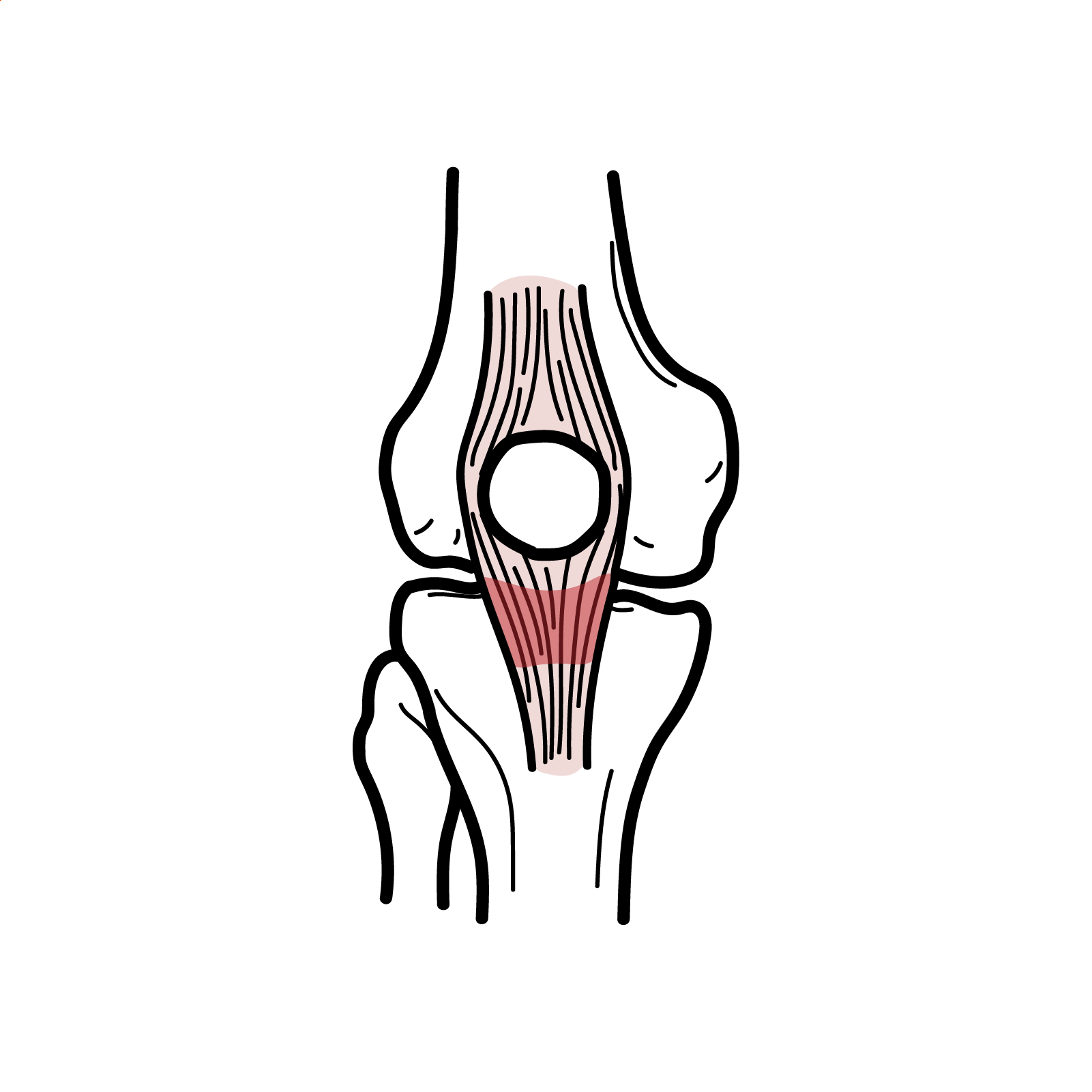
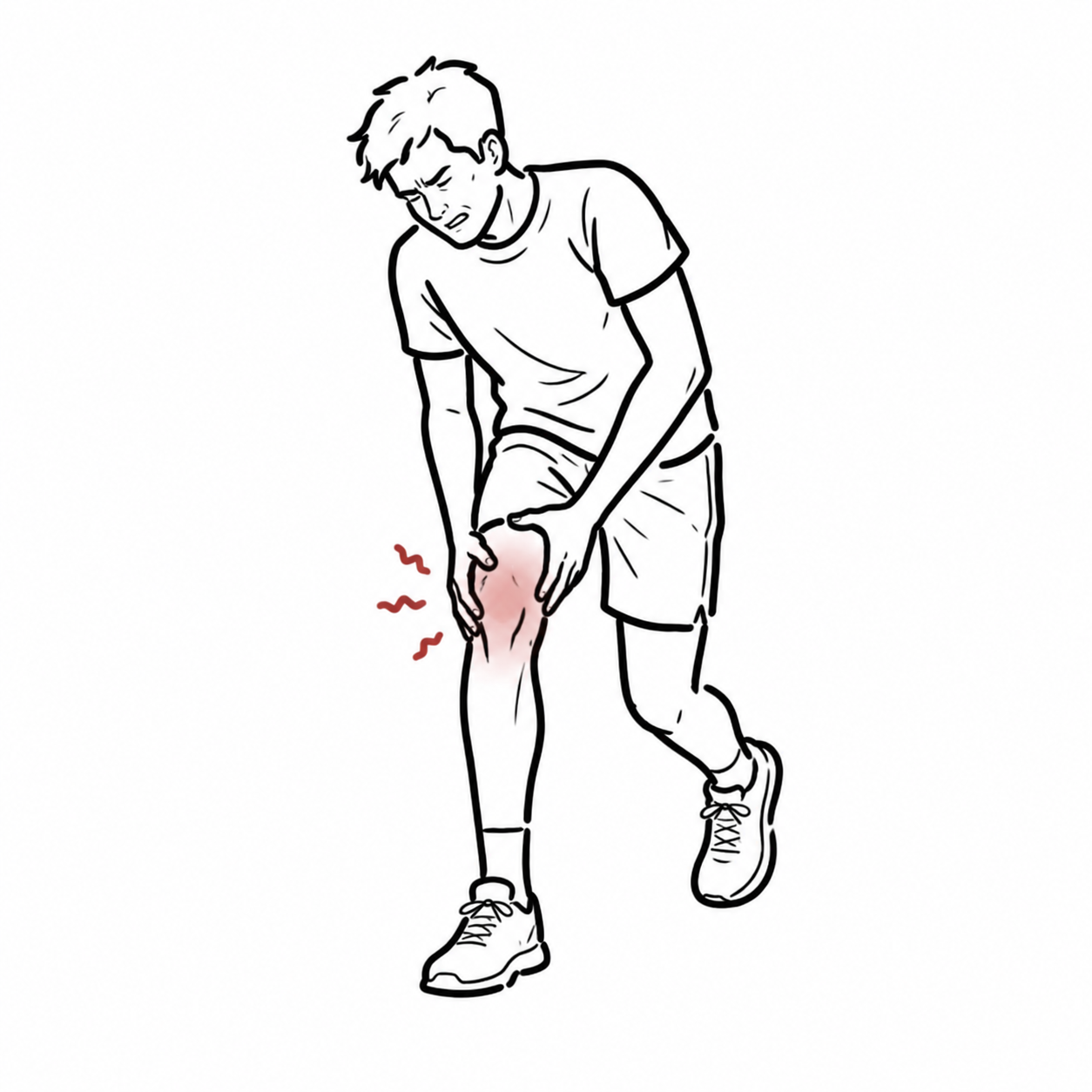
Patellar tendinopathy, commonly called jumper’s knee, is a load-related disorder of the patellar tendon. It typically causes localised anterior knee pain at the tendon’s attachment to the inferior pole of the patella.
The condition is particularly associated with sports involving repeated jumping, landing, acceleration and rapid changes of direction, such as basketball, volleyball and athletics. It may also affect recreationally active individuals following a sudden increase in training load.1
The preferred term is tendinopathy, rather than tendinitis, because established disease is characterised predominantly by collagen disorganisation, altered extracellular matrix and abnormal tendon-cell activity—not simply acute inflammation.
Definition
- Patellar tendinopathy
- A load-related disorder characterised by localised pain and impaired function of the patellar tendon.
- Tendon capacity
- The amount and type of mechanical load that a tendon can tolerate without developing an adverse symptomatic response.
- Energy-storage load
- Rapid loading in which the tendon stores and releases elastic energy, as occurs during jumping, landing and sprinting.
- Tendinopathy
- A clinical syndrome involving tendon pain, reduced function and impaired tolerance of mechanical loading.
- Kinetic chain
- The linked contribution of the trunk, hip, knee and ankle to the production and absorption of force during movement.
Anatomy & Physiology
The patellar tendon is a strong fibrous structure extending from the inferior pole of the patella to the tibial tuberosity. Although traditionally called a tendon, it forms part of the continuous quadriceps–patella–patellar tendon extensor mechanism.
The tendon is approximately 3–5 cm long and consists predominantly of parallel type I collagen fibres. Its proximal portion is the most frequent site of patellar tendinopathy.
The knee extensor mechanism consists of:
- The quadriceps muscles
- The quadriceps tendon
- The patella
- The patellar tendon
- The tibial tuberosity
During knee extension, quadriceps contraction transmits force through the quadriceps tendon and patella into the patellar tendon, which pulls on the tibia. The patella increases the quadriceps’ mechanical advantage by maintaining the extensor mechanism away from the knee’s axis of rotation.
During jumping and landing, the patellar tendon is exposed to high tensile and energy-storage loads. The tendon must repeatedly store and release energy while the quadriceps control knee flexion and generate knee extension.
Aetiology & Risk Factors
Aetiology
Patellar tendinopathy develops when the magnitude, frequency or rate of tendon loading repeatedly exceeds the tendon’s current capacity to recover and adapt.
A rapid increase in jumping, sprinting or resistance-training load is a common precipitant. Continued high loading without adequate recovery may produce persistent changes in tendon structure and pain processing.
The condition is therefore better understood as a load-capacity imbalance than as a single traumatic injury.
Risk Factors
- Participation in jumping sports, particularly volleyball and basketball
- Sudden increases in training volume, intensity or frequency
- High weekly exposure to jumping and landing
- Inadequate recovery between high-load sessions
- Reduced quadriceps or lower-limb strength
- Limited ankle dorsiflexion
- Altered landing mechanics
- Greater body mass or body mass index
- Previous patellar tendon symptoms
- Male sex, although individuals of any sex may be affected
- Training on hard surfaces
- Poorly coordinated return to sport following inactivity or injury
Individual physical findings should not be regarded as isolated causes. Tendinopathy commonly results from an interaction between training load, recovery, tissue capacity and the mechanics of the entire lower limb.
Pathophysiology
Mechanical overload initially stimulates a tendon-cell response with increased proteoglycan production and water content. If an appropriate recovery period follows, the tendon may adapt and become more load tolerant.
When excessive loading continues, maladaptive changes may include:
- Disorganisation and separation of collagen fibres
- Increased ground substance and tendon thickening
- Abnormal tenocyte activity
- Neovascularisation
- Altered mechanical properties
- Reduced ability to tolerate high-rate loading
- Increased local and central pain sensitivity
The tendon-continuum model describes reactive tendinopathy, tendon dysrepair and degenerative tendinopathy as overlapping tissue states.2 It is a conceptual model rather than a definitive clinical staging system; different regions of the same tendon may display different structural features.
Pain severity does not correlate reliably with the extent of structural change. Abnormal ultrasound or MRI appearances may be found in asymptomatic athletes, while substantial pain may occur without extensive imaging abnormalities.
Clinical Manifestations
The characteristic presentation is:
- Gradual-onset pain at the inferior pole of the patella
- Pain that is well localised rather than diffuse
- Pain during jumping, landing, sprinting or rapid changes of direction
- Pain during squatting, lunging or stair descent
- Symptoms associated with an increase in training load
- Stiffness or discomfort after rest
- Reduced jumping performance or exercise tolerance
Pain may initially occur only after activity. With progression, it may be present during activity, limit performance and persist afterwards.
Some patients describe a “warm-up phenomenon,” in which pain improves temporarily during exercise but returns later or is worse the following day. Temporary improvement during activity should not be interpreted as complete tendon recovery.
Clinical Examination
- Focal tenderness at the inferior pole of the patella
- Pain with a single-leg decline squat
- Pain during jumping, hopping or resisted knee extension
- Reduced quadriceps strength or muscle bulk
- Reduced lower-limb power
- Deficits in hip, knee or ankle control during landing
- Restricted ankle dorsiflexion
- Thickening of the proximal patellar tendon
The tendon should be palpated systematically. Diffuse peripatellar pain, joint-line tenderness, a large effusion or mechanical locking suggests an alternative diagnosis.
Diagnosis
Patellar tendinopathy is primarily a clinical diagnosis based on:
- Load-related pain
- Focal symptoms at the patellar tendon—usually its proximal attachment
- Reproduction of pain during tendon-loading activities
- Exclusion of competing causes of anterior knee pain
Imaging
Imaging is not routinely required when the clinical presentation is typical.
Ultrasound may demonstrate:
- Tendon thickening
- Focal hypoechoic regions
- Loss of the normal fibrillar pattern
- Increased Doppler vascularity
MRI may demonstrate:
- Increased tendon thickness
- Increased intratendinous signal
- Changes near the inferior pole of the patella
Structural abnormality on ultrasound or MRI does not establish the diagnosis by itself. Imaging findings must be interpreted alongside the patient’s pain location, loading history and functional examination.
Treatment
The foundation of treatment is education, load management and progressive exercise rehabilitation. Passive treatments alone do not restore the tendon’s ability to tolerate sporting loads.1
Education and Load Management
Management begins by identifying the activities provoking the greatest tendon load.
- Temporarily reducing the volume or frequency of jumping, sprinting and rapid direction changes
- Avoiding sudden spikes in training load
- Maintaining non-provocative physical activity where possible
- Allowing adequate recovery between high-load sessions
- Monitoring symptoms during activity and the following day
- Explaining that meaningful recovery commonly takes several months
Complete rest is generally discouraged because prolonged unloading reduces tendon and muscle capacity. The objective is to reduce excessive load while maintaining an appropriate level of mechanical stimulus.
Load management does not mean complete rest.
Reduce or temporarily remove the most provocative energy-storage activities, maintain tolerated conditioning and progressively rebuild the tendon’s capacity. Symptoms that are substantially worse the next morning usually indicate that the previous load was excessive.
Exercise Rehabilitation
A staged programme is adjusted to symptom irritability, strength and sporting requirements.
Stage 1: Pain Modulation and Isometric Loading
Isometric quadriceps exercises may provide short-term pain relief and allow loading to begin in patients with irritable symptoms.3 Examples include:
- Isometric leg-extension holds
- Spanish squats
- Isometric squat holds
Analgesic benefit is variable, so isometric exercise should not be presented as curative.
Stage 2: Progressive Isotonic Strengthening
Slow resistance exercises progressively restore quadriceps and tendon capacity. Options include:
- Leg press
- Knee extension
- Squat
- Split squat
- Hack squat
Exercises can progress from bilateral to unilateral loading and from lighter to heavier resistance. Both concentric and eccentric muscle actions should eventually be trained.
Heavy slow resistance and eccentric decline-squat programmes can improve symptoms and function. Progressive tendon-loading programmes that incorporate multiple contraction types may provide better outcomes than eccentric exercise alone.4
Stage 3: Energy-Storage Loading
Once slow resistance is well tolerated and strength is improving, faster activities are introduced:
- Skipping
- Running drills
- Bilateral jumps
- Hopping
- Bounding
- Acceleration and deceleration tasks
- Change-of-direction drills
Volume, intensity and frequency should be increased gradually. Adequate recovery is particularly important because energy-storage exercises impose high tendon loads.
Stage 4: Sport-Specific Loading and Return to Sport
Adjunctive Treatments
Adjuncts may be considered to support—not replace—progressive rehabilitation:
- Patellar straps or taping: may provide temporary symptom relief
- Simple analgesia or NSAIDs: may help short-term pain but do not restore tendon capacity
- Extracorporeal shockwave therapy: evidence is inconsistent
- Platelet-rich plasma injections: evidence is mixed and protocols vary
- Corticosteroid injection: may provide short-term improvement but has poor longer-term results and is not routinely recommended for tendon disease1
- Other injections: evidence is insufficient for routine use
Surgery
Surgical referral may be considered when symptoms remain substantially limiting after a prolonged, well-supervised rehabilitation programme and the diagnosis has been reassessed.
Procedures may include excision or debridement of abnormal tendon tissue, with or without treatment of the inferior patellar pole. Evidence supporting surgery is limited, and postoperative rehabilitation remains necessary.
A practical rehabilitation sequence is:
- 1. Settle highly irritable symptoms.
- 2. Restore slow strength and tendon-loading tolerance.
- 3. Reintroduce rapid energy-storage loading.
- 4. Rebuild sport-specific volume and intensity.
- 5. Maintain strength and monitor workload after return to sport.
Progression is guided by function and symptom response rather than imaging appearance alone.
Complications & Prognosis
Complications
- Persistent anterior knee pain
- Reduced training and competition availability
- Loss of quadriceps strength and lower-limb power
- Altered movement patterns
- Reduced physical activity
- Recurrent symptom flares
- Psychological distress or fear of loading
- Rare progression to partial or complete tendon rupture
Failure to identify the provoking load pattern, excessive reliance on passive treatment and premature return to repeated jumping can prolong symptoms.
Prognosis
Patellar tendinopathy may follow a prolonged course. Improvement often requires several months of consistent, progressive rehabilitation rather than a brief period of rest.
Many patients achieve meaningful improvements in pain and function, but not every athlete returns rapidly to their previous level of performance.
Recurrence is possible when high-volume jumping is resumed too quickly. Continued strength training and planned management of weekly tendon load are important after return to sport.
Pain relief is not the same as restored tendon capacity. An athlete may feel better before the tendon and lower limb can tolerate repeated high-rate jumping. Rehabilitation should continue until strength, energy-storage tolerance and sport-specific workload have been rebuilt.
References
- Rosen AB, Wellsandt E, Nicola M, Tao MA. Clinical management of patellar tendinopathy. J Athl Train. 2022;57(7):621–631. Available from: https://pubmed.ncbi.nlm.nih.gov/35576245/
- Cook JL, Purdam CR. Is tendon pathology a continuum? A pathology model to explain the clinical presentation of load-induced tendinopathy. Br J Sports Med. 2009;43(6):409–416. doi:10.1136/bjsm.2008.051193.
- Rio E, Kidgell D, Purdam C, et al. Isometric exercise induces analgesia and reduces inhibition in patellar tendinopathy. Br J Sports Med. 2015;49(19):1277–1283. doi:10.1136/bjsports-2014-094386.
- Breda SJ, Oei EHG, Zwerver J, et al. Effectiveness of progressive tendon-loading exercise therapy in patients with patellar tendinopathy: a randomised clinical trial. Br J Sports Med. 2021;55(9):501–509. doi:10.1136/bjsports-2020-103403.
- Malliaras P, Cook J, Purdam C, Rio E. Patellar tendinopathy: clinical diagnosis, load management, and advice for challenging case presentations. J Orthop Sports Phys Ther. 2015;45(11):887–898. doi:10.2519/jospt.2015.5987.






Members only discussions coming soon…