

Chronic Peripheral Arterial Disease: Intermittent Claudication
Overview
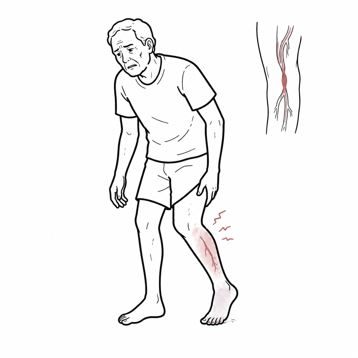
Peripheral arterial disease (PAD) is an atherosclerotic disorder that reduces blood flow to the limbs, most often the legs. Intermittent claudication is exertional muscle discomfort caused by a mismatch between oxygen supply and demand; it is reproducibly triggered by walking and usually resolves within about 10 minutes of rest.
Although claudication is a limb symptom, PAD is a marker of systemic atherosclerosis and increased risk of myocardial infarction, stroke and cardiovascular death. Management therefore aims both to reduce cardiovascular and limb events and to improve walking ability and quality of life.
Pain at rest, non-healing ulceration, gangrene, or a suddenly cold, pale, painful or neurologically impaired limb is not uncomplicated intermittent claudication and requires urgent vascular assessment.
Definition
- Peripheral arterial disease
- Atherosclerotic narrowing or occlusion of arteries supplying the limbs.
- Intermittent claudication
- Reproducible exertional limb-muscle discomfort caused by ischaemia and relieved by rest.
- Ankle-brachial index
- Ratio of ankle systolic pressure to brachial systolic pressure, used to identify haemodynamically significant lower-limb arterial disease.
- Chronic limb-threatening ischaemia
- Chronic ischaemic rest pain, non-healing wound or gangrene attributable to PAD.
Anatomy & Physiology
The abdominal aorta divides into the common iliac arteries. Blood reaches each lower limb through the external iliac and common femoral arteries, then the profunda femoris and superficial femoral-popliteal pathways, before passing into the anterior tibial, posterior tibial and peroneal arteries.
At rest, collateral flow may meet tissue demand despite an arterial stenosis. During exercise, skeletal-muscle oxygen demand rises and flow across a fixed obstruction cannot increase sufficiently. The muscle group that becomes painful often lies distal to the principal lesion.
Aetiology & Risk Factors
Aetiology
Atherosclerosis is the usual cause of chronic lower-limb PAD. Less common causes of exertional arterial symptoms include arterial entrapment, cystic adventitial disease, vasculitis, thromboangiitis obliterans, fibromuscular dysplasia and prior radiation or trauma; these should be considered when the presentation is atypical or occurs in a younger person.
Risk Factors
- Cigarette smoking, including cumulative exposure.
- Diabetes mellitus.
- Increasing age.
- Hypertension and dyslipidaemia.
- Chronic kidney disease.
- Established atherosclerotic disease in another vascular bed, including coronary or cerebrovascular disease.
- Family history and other cardiovascular risk factors.
Pathophysiology
Endothelial injury and lipid deposition drive plaque formation, inflammation, calcification and progressive arterial stenosis. Distal perfusion pressure falls and collateral vessels develop, but flow reserve remains limited. Exercise increases metabolic demand, producing reversible muscle ischaemia, metabolite accumulation and pain. Symptoms resolve when rest lowers demand and restores the supply-demand balance.
Disease progression or plaque thrombosis may further reduce perfusion and lead to chronic limb-threatening ischaemia or acute limb ischaemia. PAD also reflects a systemic atherosclerotic burden, explaining the associated cardiovascular risk.
A patient may have a near-normal resting examination because collateral flow is adequate at rest. Reproducible exertional symptoms with a normal or borderline resting ABI should prompt exercise ABI testing rather than exclusion of PAD.
Clinical Manifestations
Classical claudication is fatigue, aching, cramping, tightness or pain in a muscle group during walking. Features include:
- Cramp-like pain in a muscle group (usually calf, thigh, or buttock) triggered by exertion (e.g. walking) and relieved by rest.
- Pain consistently occurs after walking a specific distance
- Relieved within minutes of stopping activity.
- Location of pain localises disease level:
- Calf claudication (most common) → femoropopliteal disease.
- Thigh claudication → superficial femoral or common femoral disease.
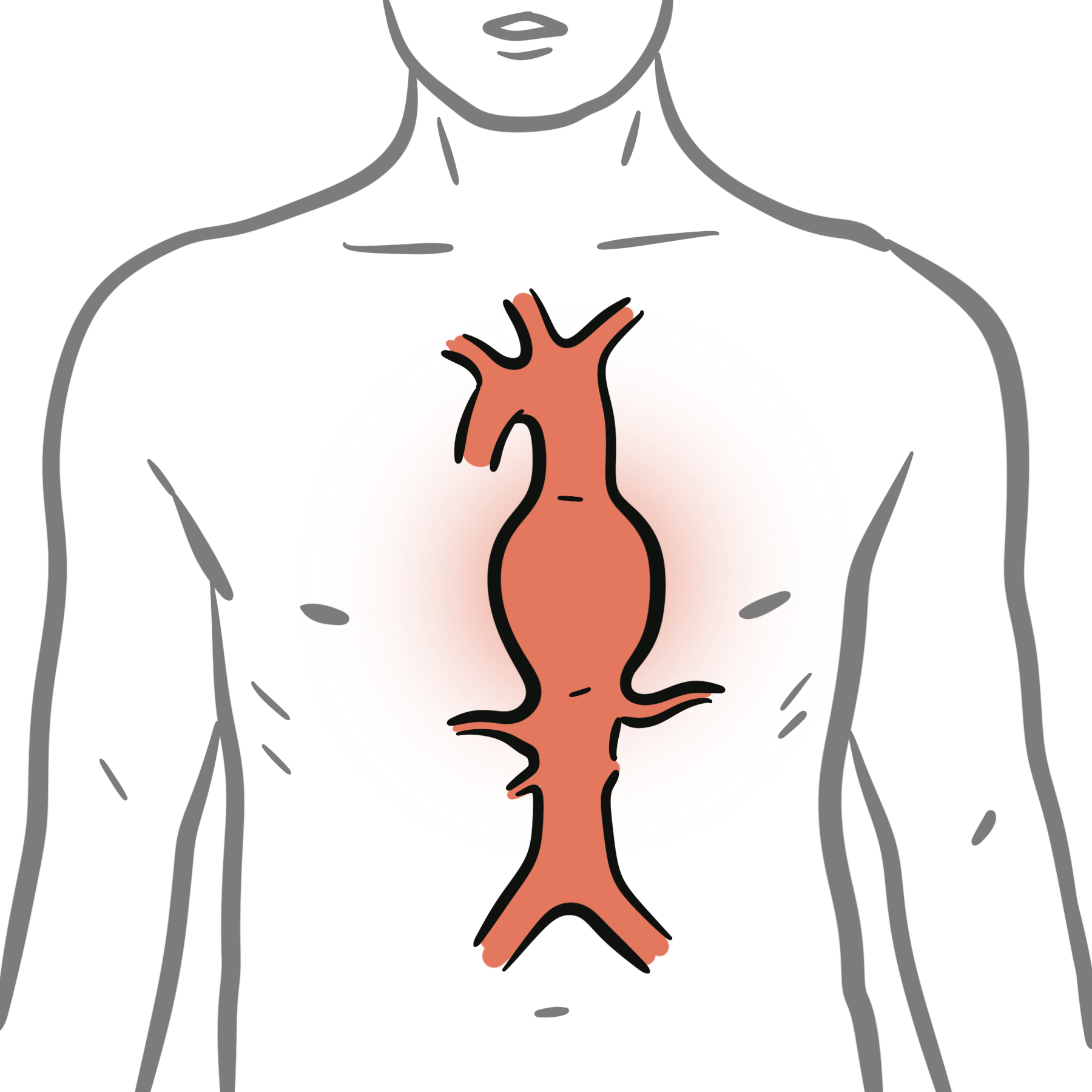
- Buttock/hip claudication ± impotence → aortoiliac disease (Leriche syndrome).
| Predominant symptom site | Common anatomical implication |
|---|---|
| Buttock or hip | Aorto-iliac disease |
| Thigh | Aorto-iliac or common femoral disease |
| Calf | Femoropopliteal disease; the most common pattern |
| Foot | Tibial or pedal disease, often with multilevel disease |
Clinical Examination
- Arterial pulses: weak or absent pulses distally to diseased artery
- Skin changes: dryness, thinning, or hair loss, and possibly signs of poor circulation such as slow-healing wounds.
- Buerger’s test positive: pallor on elevation, rubor on dependency.
Erectile dysfunction → think aortoiliac disease (Leriche syndrome).
Atypical symptoms are common. Neurogenic claudication, osteoarthritis, radiculopathy, chronic venous disease and musculoskeletal pain are important mimics.
New rest pain, tissue loss or gangrene suggests chronic limb-threatening ischaemia. Sudden severe limb pain with pallor, pulselessness, paraesthesia, paralysis or poikilothermia suggests acute limb ischaemia and requires emergency vascular assessment.
Diagnosis
Diagnosis begins with a compatible history, vascular examination and a resting ankle-brachial index (ABI). Resting ABI values are reported as abnormal (≤0.90), borderline (0.91–0.99), normal (1.00–1.40) or non-compressible (>1.40).
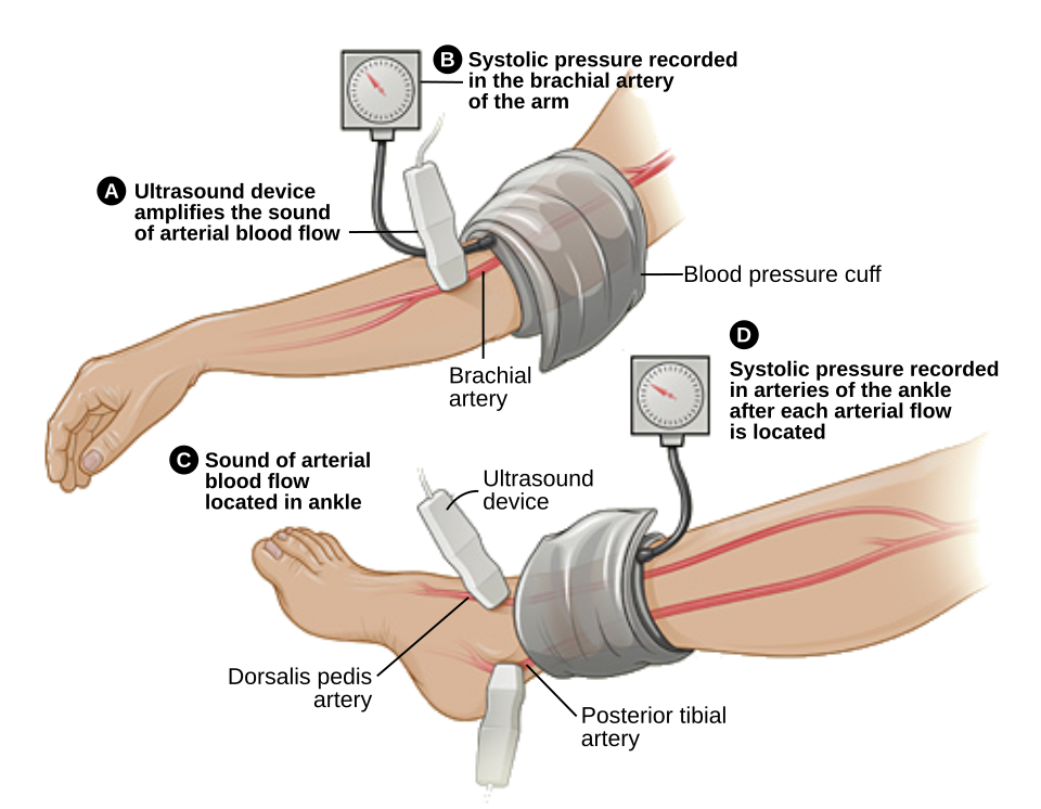
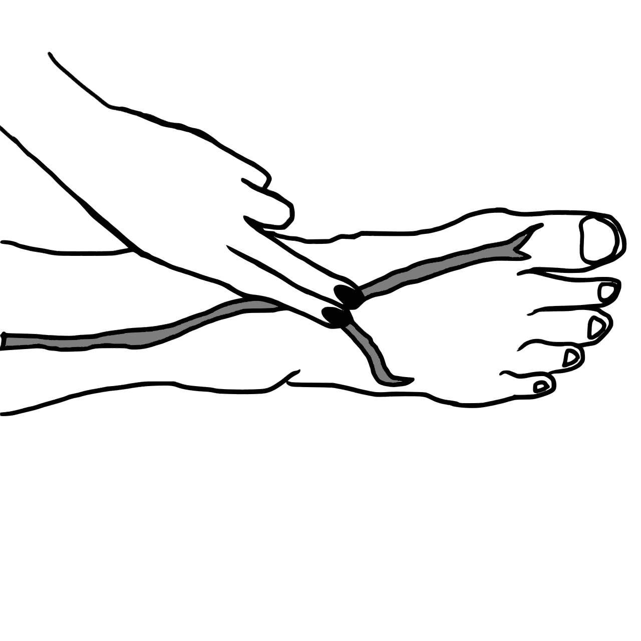
- Exercise ABI: Recommended when exertional non-joint leg symptoms suggest PAD but the resting ABI is normal or borderline.
- Toe-brachial index: Useful when the ABI is >1.40 because calcified ankle arteries may be non-compressible; a value ≤0.70 is abnormal.
- Segmental pressures and pulse-volume recordings: Help localise the level of haemodynamic disease.
- Duplex ultrasound: Defines arterial anatomy and haemodynamics without contrast.
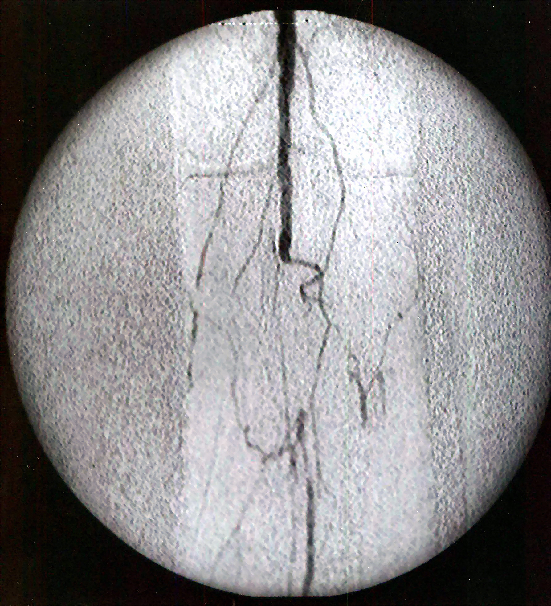
- CT angiography or MR angiography: Used for anatomical planning when revascularisation is being considered. Catheter angiography is generally reserved for cases proceeding toward intervention or when non-invasive imaging is inadequate.
- Cardiovascular assessment: Blood pressure, fasting lipids, glycaemic status, renal function and assessment for coronary, cerebrovascular and other arterial disease.
Important differentials include lumbar spinal stenosis, peripheral neuropathy, radiculopathy, hip or knee osteoarthritis, exertional compartment syndrome, chronic venous obstruction and popliteal artery entrapment. Unlike vascular claudication, neurogenic symptoms may depend more on posture, improve with spinal flexion and take longer to settle.
An ABI may be falsely high in diabetes or chronic kidney disease because medial arterial calcification prevents cuff compression. In this setting, toe pressures and waveform assessment are more informative.
Classification
Intermittent claudication corresponds to Fontaine stage II and Rutherford categories 1–3. Classification communicates symptom severity but does not replace anatomical assessment or evaluation of functional limitation.
| Fontaine | Rutherford | Clinical description |
|---|---|---|
| I | 0 | Asymptomatic PAD |
| IIa | 1 | Mild claudication; Fontaine traditionally uses a walking distance >200 m |
| IIb | 2–3 | Moderate-to-severe claudication; Fontaine traditionally uses a walking distance <200 m |
| III | 4 | Ischaemic rest pain |
| IV | 5–6 | Minor or major tissue loss |
Treatment
Treatment combines guideline-directed cardiovascular risk reduction with therapies that improve walking. Management should be individualised according to symptom burden, comorbidity, bleeding risk, anatomy and patient goals.
Cardiovascular and limb risk reduction
- Smoking cessation
- Statin therapy
- Use single antiplatelet therapy for symptomatic PAD, commonly clopidogrel or aspirin, according to the relevant guideline and patient factors.
- Low-dose rivaroxaban (2.5 mg twice daily) plus low-dose aspirin can reduce major cardiovascular and limb events in selected symptomatic patients, but increases bleeding and requires individual assessment of indication, renal function, interactions and bleeding risk.
- Provide preventive foot care, particularly for people with diabetes, neuropathy or previous ulceration.
Improving walking and symptoms
- Offer supervised exercise therapy as first-line treatment. Structured walking sessions at least three times weekly for at least 12 weeks are recommended, with continuation thereafter.
- If supervised exercise is unavailable or unsuitable, use a structured home- or community-based programme with goal setting, monitoring and behavioural support.
- Cilostazol can improve walking distance and symptoms, but must not be used in heart failure. Availability and prescribing practice vary by jurisdiction.
- Pentoxifylline is not recommended for claudication in the 2024 ACC/AHA guideline.
Revascularisation
Revascularisation is considered for functionally or lifestyle-limiting claudication that persists despite guideline-directed medical therapy and a structured exercise programme. The goal is symptom and functional improvement, not prevention of progression in an otherwise stable, mildly symptomatic limb.1,3,4
Duplex ultrasound, CT angiography, MR angiography or catheter angiography is used to define anatomy before intervention. Endovascular therapy or surgical bypass is selected through shared decision-making based on lesion location and complexity, operative risk, expected durability, conduit availability and patient preference. Infrapopliteal intervention for claudication alone is generally discouraged because benefit is uncertain and harm is possible.
Do not proceed directly from an arterial stenosis on imaging to intervention. For stable claudication, cardiovascular risk reduction and structured exercise are first-line; revascularisation is reserved for persistent, meaningful functional limitation after shared decision-making.
Complications & Prognosis
Complications
- Progression to chronic limb-threatening ischaemia, with rest pain, ulceration or gangrene.
- Acute limb ischaemia due to thrombosis or embolism.
- Reduced mobility, loss of independence and impaired quality of life.
- Myocardial infarction, stroke and cardiovascular death due to systemic atherosclerosis.
- Following revascularisation: bleeding, contrast-associated kidney injury, embolisation, restenosis, graft or stent occlusion, wound complications and need for repeat intervention.
Prognosis
Many patients with intermittent claudication have a stable limb course, but cardiovascular morbidity and mortality remain the dominant prognostic concern. Smoking, diabetes, chronic kidney disease, polyvascular disease and poor adherence to preventive therapy are associated with worse outcomes.1,3
Structured exercise improves walking performance and quality of life. Revascularisation can provide additional symptom relief in appropriately selected patients, but durability varies and ongoing exercise and risk-factor management remain necessary.
References
- Gornik HL, Aronow HD, Goodney PP, et al. 2024 ACC/AHA/AACVPR/APMA/ABC/SCAI/SVM/SVN/SVS/SIR/VESS guideline for the management of lower extremity peripheral artery disease. Circulation. 2024;149(24):e1313–e1410. doi:10.1161/CIR.0000000000001251
- National Institute for Health and Care Excellence. Peripheral arterial disease: diagnosis and management [Internet]. London: NICE; 2012 [updated 2020 Dec 11; cited 2026 Sep 7]. Available from: https://www.nice.org.uk/guidance/cg147
- Nordanstig J, Behrendt CA, Baumgartner I, et al. Editor’s Choice – European Society for Vascular Surgery (ESVS) 2024 clinical practice guidelines on the management of asymptomatic lower limb peripheral arterial disease and intermittent claudication. Eur J Vasc Endovasc Surg. 2024;67(1):9–96. doi:10.1016/j.ejvs.2023.08.067
- Conte MS, Farber A, Kinlay S, et al. Society for Vascular Surgery clinical practice guideline on the management of intermittent claudication: focused update. J Vasc Surg. 2025;82:303–326.e11. doi:10.1016/j.jvs.2025.04.016



Members only discussions coming soon…