

Catecholaminergic Polymorphic Ventricular Tachycardia (CPVT)
Overview
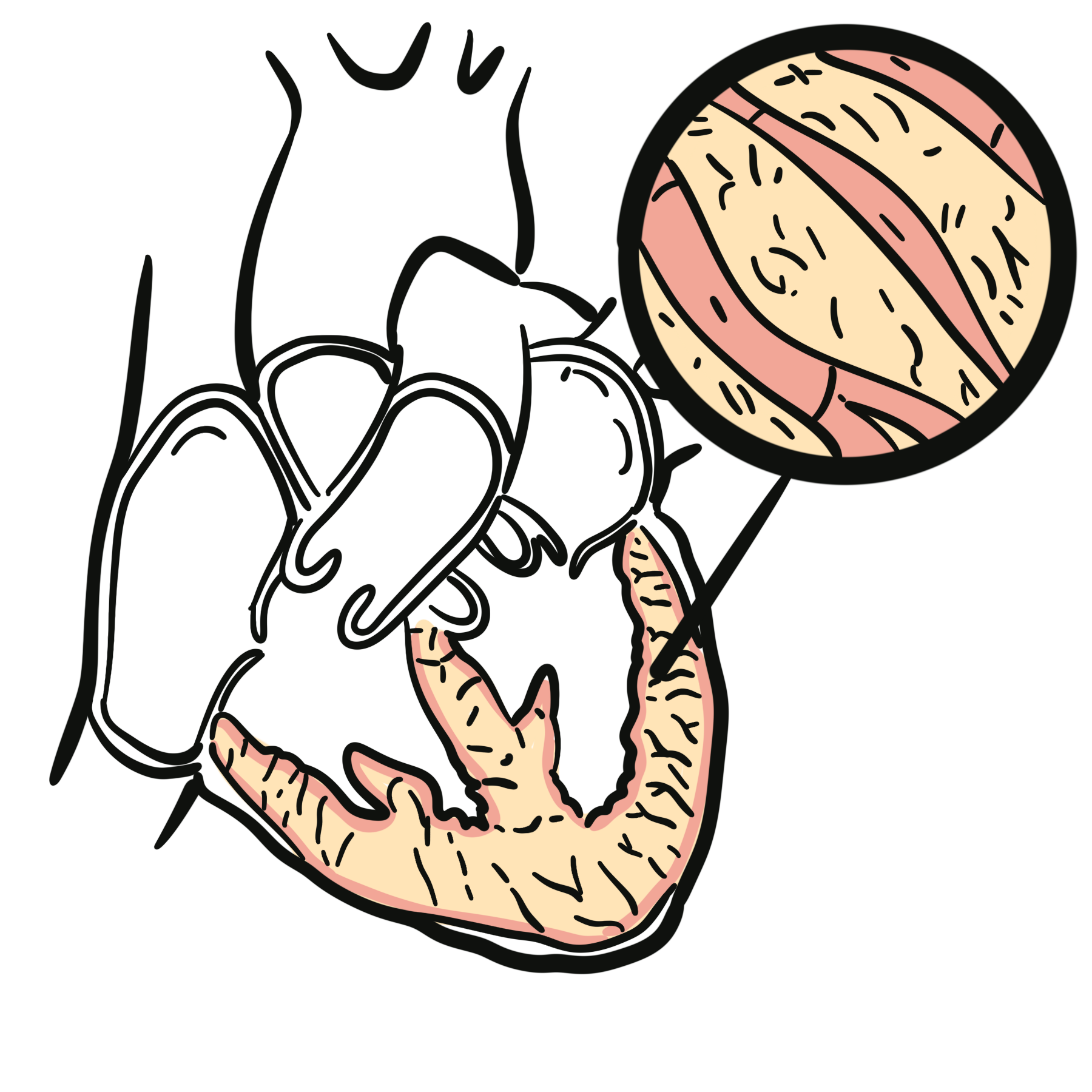
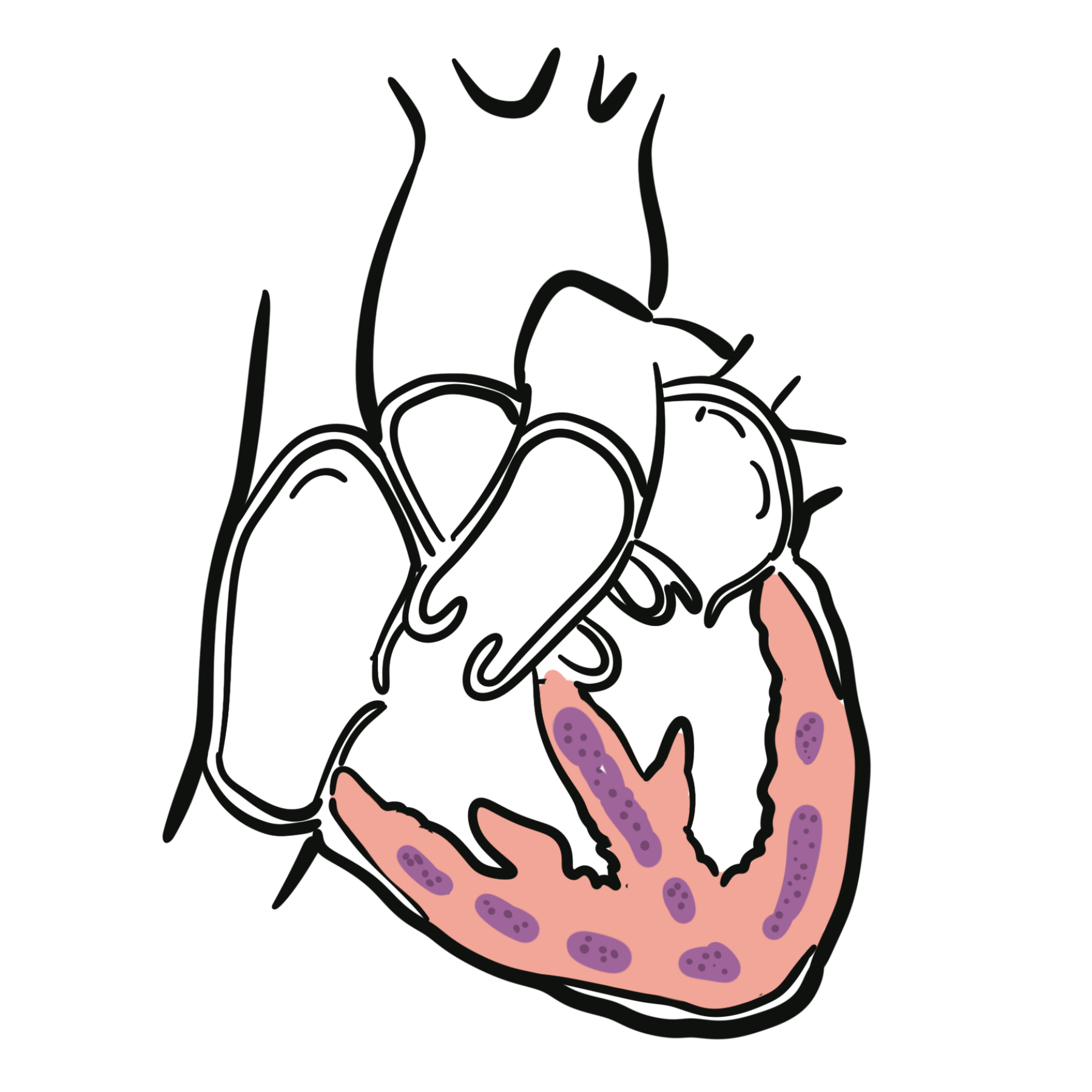
Catecholaminergic polymorphic ventricular tachycardia (CPVT) is an inherited cardiac channelopathy in which physical exertion or acute emotion provokes ventricular arrhythmias. The heart is usually structurally normal and the resting electrocardiogram (ECG) is often normal. During adrenergic stress, ventricular ectopy may progress to bidirectional or polymorphic ventricular tachycardia (VT), ventricular fibrillation and sudden cardiac death.1
CPVT commonly presents during childhood or adolescence, although onset in adulthood is recognised. The characteristic history is exertional or emotion-triggered syncope, seizure-like activity or cardiac arrest. Misdiagnosis as vasovagal syncope or epilepsy is common because routine examination, resting ECG and echocardiography may be unremarkable.2
CPVT is treatable but potentially lethal. Prompt recognition, exercise-based assessment, family screening and lifelong treatment are essential.
Definition
- Catecholaminergic polymorphic ventricular tachycardia
- An inherited arrhythmia syndrome characterised by adrenergically induced bidirectional or polymorphic ventricular tachycardia in a usually structurally normal heart.
- Bidirectional ventricular tachycardia
- Ventricular tachycardia in which the frontal QRS axis or morphology alternates from beat to beat.
- Polymorphic ventricular tachycardia
- Ventricular tachycardia with continuously changing QRS morphology.
- Channelopathy
- A disorder caused by abnormal function of an ion channel or an associated regulatory protein.
Anatomy & Physiology
Cardiac contraction depends on tightly regulated movement of calcium between the cardiomyocyte cytoplasm and the sarcoplasmic reticulum.
During each action potential:
- Calcium enters the cardiomyocyte through L-type calcium channels.
- This activates cardiac ryanodine receptor type 2 channels (RyR2) in the sarcoplasmic reticulum.
- RyR2 releases a larger amount of stored calcium into the cytoplasm.
- Cytoplasmic calcium binds to the contractile apparatus and produces contraction.
- Calcium is subsequently returned to the sarcoplasmic reticulum, where it is stored partly through binding to calsequestrin-2.
Adrenergic stimulation during exercise or emotion increases heart rate, calcium entry and sarcoplasmic-reticulum calcium loading. In a healthy heart this produces a stronger, faster contraction without spontaneous calcium release.
Aetiology & Risk Factors
Aetiology
CPVT is most commonly caused by pathogenic variants affecting proteins responsible for intracellular calcium handling.2,4
| Gene | Usual inheritance | Functional effect |
|---|---|---|
| RYR2 | Autosomal dominant | Abnormal cardiac ryanodine-receptor function and inappropriate calcium release |
| CASQ2 | Usually autosomal recessive | Impaired calcium storage within the sarcoplasmic reticulum |
| CALM1, CALM2 and CALM3 | Usually autosomal dominant | Abnormal calmodulin-dependent regulation of calcium handling |
| TRDN | Autosomal recessive | Abnormal triadin function and sarcoplasmic-reticulum calcium regulation |
| TECRL | Autosomal recessive | Overlapping CPVT and long-QT-like phenotype |
A pathogenic variant is not identified in every clinically diagnosed patient. Conversely, an uncertain genetic variant does not establish CPVT without compatible clinical and phenotypic evidence.
Risk Factors
- Strenuous physical exercise
- Sudden bursts of exertion
- Acute emotional stress, fear or excitement
- Poor adherence to medication
- Inadequate beta-blocker dosing
- Use of sympathomimetic drugs or stimulants
- Failure to recognise affected relatives
- Previous cardiac arrest
- Recurrent syncope or ventricular arrhythmia despite treatment
CPVT often occurs in families, but an apparently negative family history does not exclude it. Pathogenic RYR2 variants may arise de novo, and relatives with the same variant may have different clinical severity.
Pathophysiology
Pathogenic variants affecting RyR2 or its regulatory proteins destabilise calcium handling within the sarcoplasmic reticulum.
During adrenergic stimulation:
- Intracellular cyclic AMP and protein-kinase activity increase.
- Sarcoplasmic-reticulum calcium loading rises.
- Abnormal channels release calcium spontaneously during diastole.
- The sodium–calcium exchanger removes excess intracellular calcium in exchange for sodium.
- This produces a transient inward current and a delayed afterdepolarisation.
- If the afterdepolarisation reaches threshold, it triggers a premature ventricular complex.
- Repeated triggered beats progress from isolated ectopy to bigeminy, couplets, bidirectional VT and polymorphic VT.
This explains why the resting ECG can appear normal: the electrical instability becomes apparent when adrenergic stimulation increases intracellular calcium loading.
Why is exercise testing so important when the resting ECG is normal? CPVT is not primarily a disorder of baseline ventricular depolarisation or repolarisation. It is a disorder of intracellular calcium handling exposed by adrenergic stress. Increasing the heart rate recreates the physiological setting in which spontaneous calcium release and triggered activity occur.
Clinical Manifestations
Typical Presentation
The classic presentation is syncope during:
- Running, swimming or competitive sport
- Sudden exertion
- Acute fear, anger or excitement
- Emotional stress
Other presentations include:
- Palpitations
- Presyncope
- Seizure-like movements caused by cerebral hypoperfusion
- Aborted sudden cardiac arrest
- Sudden unexplained death
- Detection during family screening
Symptoms often begin in childhood or adolescence. Adult presentations occur and should not be dismissed solely because of age.2
Clinical Clues
- Exertional or emotion-triggered syncope
- Recurrent “seizures” with normal neurological investigations
- Syncope while swimming
- A family history of exertional syncope, unexplained drowning or sudden death
- A structurally normal heart despite a serious ventricular arrhythmia
- A normal resting QT interval
- Exercise-induced ventricular ectopy that becomes progressively more complex as heart rate rises
Arrhythmia Pattern
During exercise, the ECG may show a characteristic progression:
- Isolated premature ventricular complexes
- Ventricular bigeminy
- Couplets
- Bidirectional VT
- Polymorphic VT
- Ventricular fibrillation
Arrhythmia commonly reduces as the heart rate falls during recovery.
Exertional syncope in a child or young adult with a normal resting ECG is not automatically benign. CPVT must be considered, particularly when episodes occur during running, swimming or acute emotion.
Diagnosis
CPVT is diagnosed from the combination of a compatible clinical history and adrenergically induced ventricular arrhythmia in the absence of structural heart disease or another explanation. International consensus criteria recognise CPVT when an otherwise unexplained exercise- or catecholamine-induced bidirectional or polymorphic VT occurs in a structurally normal heart with a normal resting ECG. A pathogenic variant in an established CPVT-associated gene can also establish the diagnosis in an appropriate setting.3
Resting ECG
The resting 12-lead ECG is usually normal and is primarily used to identify alternatives such as:
- Long QT syndrome
- Brugada syndrome
- Pre-excitation
- Conduction disease
- Baseline ventricular ectopy
- Andersen–Tawil syndrome features, including prominent U waves
Sinus bradycardia may occur but is not diagnostic. A normal resting ECG does not exclude CPVT.
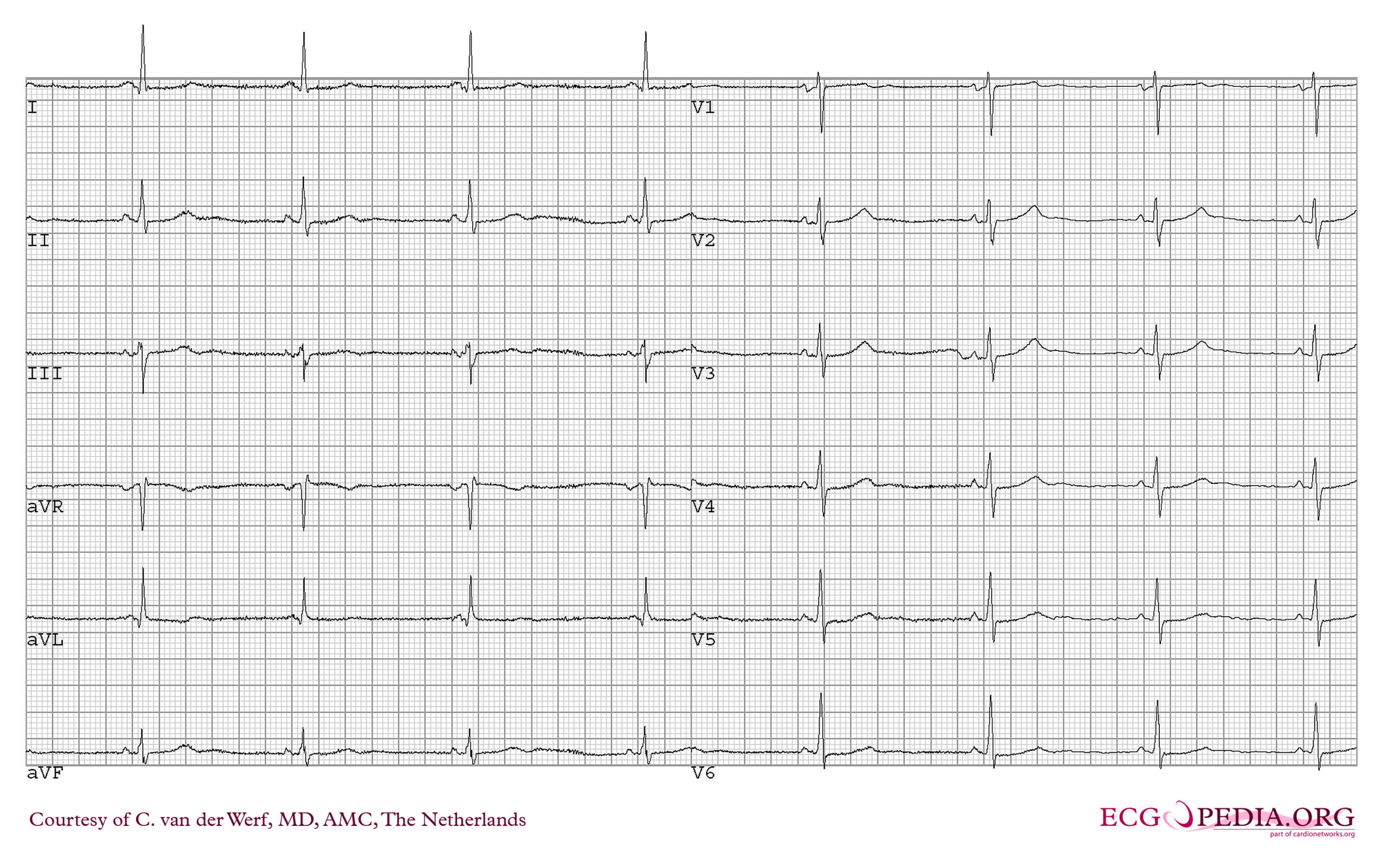
Resting ECG from a patient with CPVT. CardioNetworks ECGpedia. Licensed under CC BY-SA 3.0, via Wikimedia Commons.
What to notice
Detailed image description
Source: https://commons.wikimedia.org/wiki/File:ECG000032_(CardioNetworks_ECGpedia).jpg Licence: Creative Commons Attribution-ShareAlike 3.0 Unported (https://creativecommons.org/licenses/by-sa/3.0/). No modifications.
Exercise Stress Test
A symptom-limited exercise ECG is the principal diagnostic investigation in a patient able to exercise.
- Heart rate at the onset of ventricular ectopy
- Increasing ectopic burden as workload rises
- Progression from isolated premature ventricular complexes to bigeminy, couplets or VT
- Bidirectional or polymorphic morphology
- Reproduction of symptoms
- Reduction of arrhythmia during recovery
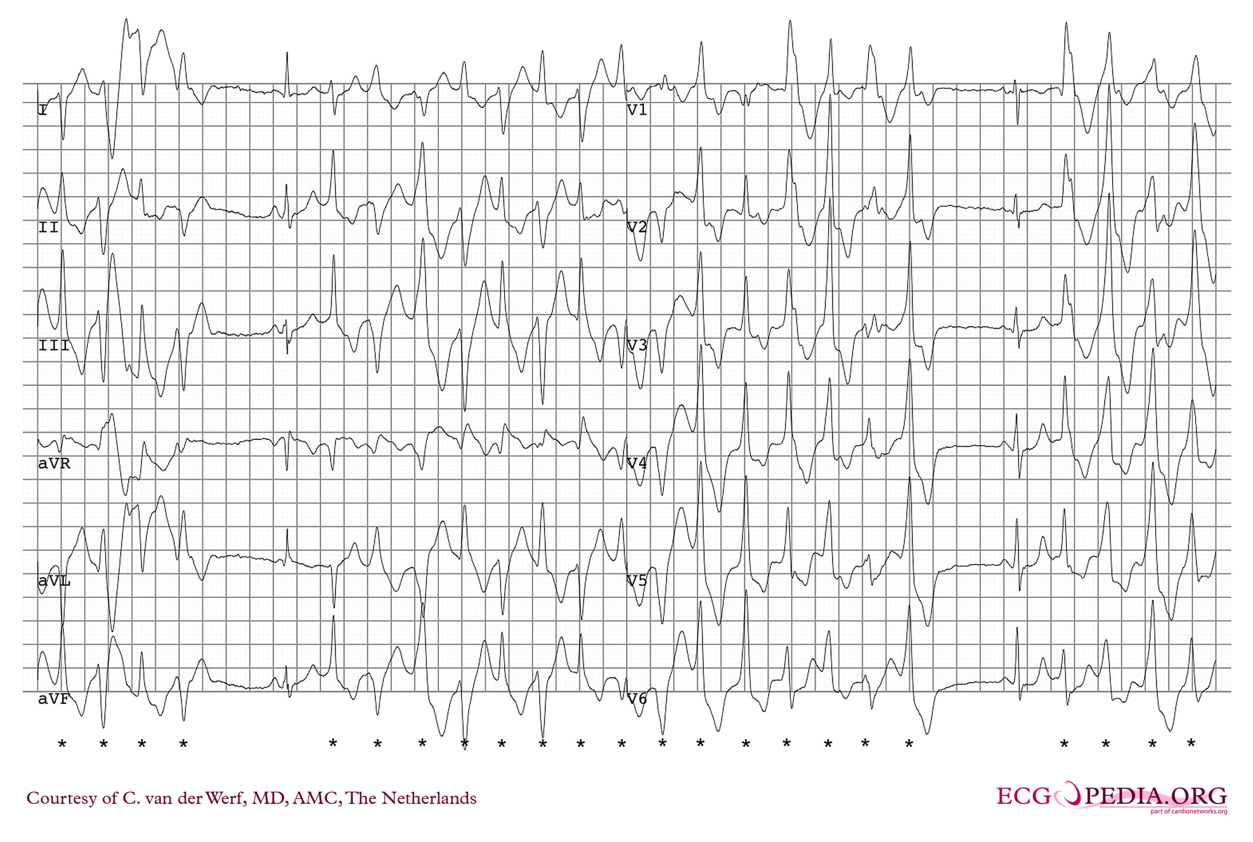
Exercise ECG from the same patient showing polymorphic ventricular ectopy. CardioNetworks ECGpedia. Licensed under CC BY-SA 3.0, via Wikimedia Commons.
What to notice
Detailed image description
Source: https://commons.wikimedia.org/wiki/File:ECG000033_(CardioNetworks_ECGpedia).jpg Licence: Creative Commons Attribution-ShareAlike 3.0 Unported (https://creativecommons.org/licenses/by-sa/3.0/). No modifications.
The test should be undertaken in an appropriately supervised environment with resuscitation facilities.
A positive test with the characteristic phenotype strongly supports CPVT. A negative test does not reliably exclude it, particularly when the history or family genotype is compelling. Repeat testing or a specialist modified exercise protocol may be appropriate when suspicion remains high.2
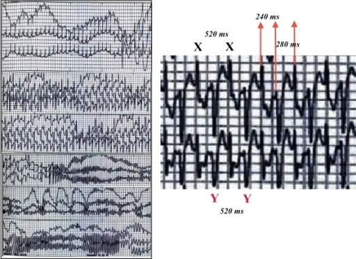
Bidirectional ventricular tachycardia in CPVT. Femenia F, Barbosa-Barros R, Sampaio SV, Arce M, Perez-Riera A, Baranchuk A. Licensed under CC BY 2.5, via Wikimedia Commons.
What to notice
Detailed image description
Source: https://commons.wikimedia.org/wiki/File:Abnormal_heart_rhythms_in_a_case_of_CPVT_(ECG_graphs).png Licence: Creative Commons Attribution 2.5 Generic (https://creativecommons.org/licenses/by/2.5/). No modifications.
Exercise testing is also used after diagnosis to:
- Assess response to beta-blockade and flecainide
- Identify breakthrough ventricular ectopy
- Guide medication titration
- Support individualised advice about physical activity
The diagnostic exercise test should not be reported simply as “negative for ischaemia”. The rhythm tracing must be examined for progressive ventricular ectopy, couplets and bidirectional or polymorphic VT.
Other investigations
- Ambulatory ECG Monitoring
- Cardiac Imaging: Echocardiography should confirm normal cardiac structure and ventricular function and exclude structural causes of ventricular arrhythmia.
- Pharmacological Adrenergic Challenge: Intravenous adrenaline or another catecholamine challenge may be considered in an expert setting when exercise testing is not feasible.
- Genetic Testing
Important Differentials
| Differential | Distinguishing features |
|---|---|
| Vasovagal syncope | Usually associated with prolonged standing, heat or prodromal nausea; not accompanied by exercise-induced ventricular arrhythmia |
| Epilepsy | Events may occur without exertional triggers; arrhythmic syncope may produce convulsive movements |
| Long QT syndrome | Prolonged or abnormal QT phenotype, genotype-specific triggers and torsades de pointes rather than typical bidirectional VT |
| Andersen–Tawil syndrome | Ventricular arrhythmia with periodic paralysis and characteristic facial or skeletal features; prominent U waves may be present |
| Digoxin toxicity | Medication exposure, gastrointestinal or neurological features and characteristic ECG abnormalities; may also produce bidirectional VT |
| Arrhythmogenic cardiomyopathy | Structural or tissue abnormalities, characteristic ECG findings and ventricular arrhythmias often arising from a consistent ventricular origin |
| Myocarditis | Acute systemic illness, chest pain, troponin elevation and myocardial abnormalities on imaging |
| Anomalous coronary artery | Exertional symptoms with an anatomical coronary abnormality rather than a primary calcium-handling disorder |
Bidirectional VT is highly distinctive but not unique to CPVT. In a young patient with exertional syncope, a normal heart and no digoxin exposure, CPVT becomes the central diagnosis; dysmorphic features or periodic paralysis should redirect attention towards Andersen–Tawil syndrome.
Genetics & Family Screening
Once a pathogenic or likely pathogenic familial variant is identified, first-degree relatives should be offered targeted cascade testing. Relatives who carry the variant require clinical assessment even if they are asymptomatic.4
When no causative variant is identified, first-degree relatives should undergo clinical screening that generally includes:
- Detailed personal and family history
- Resting ECG
- Exercise stress testing
- Additional ambulatory monitoring when clinically indicated
A single normal exercise test may not completely exclude disease in a genotype-positive relative or a person with a compelling family history. Follow-up should be directed by an inherited-arrhythmia service.
Genetic counselling should address:
- Autosomal dominant and autosomal recessive inheritance
- Variable penetrance and clinical severity
- De novo variants
- Implications for siblings, parents and children
- Reproductive options
- The limitations of variants of uncertain significance
Genetic testing is most informative when it begins with a clinically affected family member. Broad testing of an unaffected relative without a defined familial variant increases the risk of finding uncertain results that do not clarify management.
Treatment
Management aims to prevent adrenergically induced ventricular arrhythmia while allowing safe, sustainable daily activity.
Lifestyle and Trigger Management
- Avoiding competitive sport and strenuous exercise
- Avoiding sudden bursts of intense activity
- Reducing exposure to extreme emotional stress where practicable
- Avoiding sympathomimetic stimulants and recreational drugs
- Checking over-the-counter cold, flu and weight-loss preparations
- Maintaining strict medication adherence
- Developing an emergency plan for schools, workplaces and sporting environments
Exercise advice should be individualised using treatment response, exercise-test findings and shared decision-making with an inherited-arrhythmia specialist.1
Beta-Blockers
A non-selective beta-blocker without intrinsic sympathomimetic activity is first-line therapy. Nadolol is generally preferred when available; propranolol is an alternative.1,2
Flecainide
Flecainide is added when ventricular arrhythmia persists during exercise despite an adequately dosed beta-blocker. It may also reduce recurrent arrhythmia and ICD shocks.1
Flecainide is usually used with, rather than instead of, beta-blockade. Follow-up should assess ECG intervals, medication tolerance and exercise-induced arrhythmia.
The effectiveness of CPVT treatment cannot be judged only by the absence of symptoms. Exercise testing is needed to determine whether ventricular ectopy has been adequately suppressed.
Left Cardiac Sympathetic Denervation
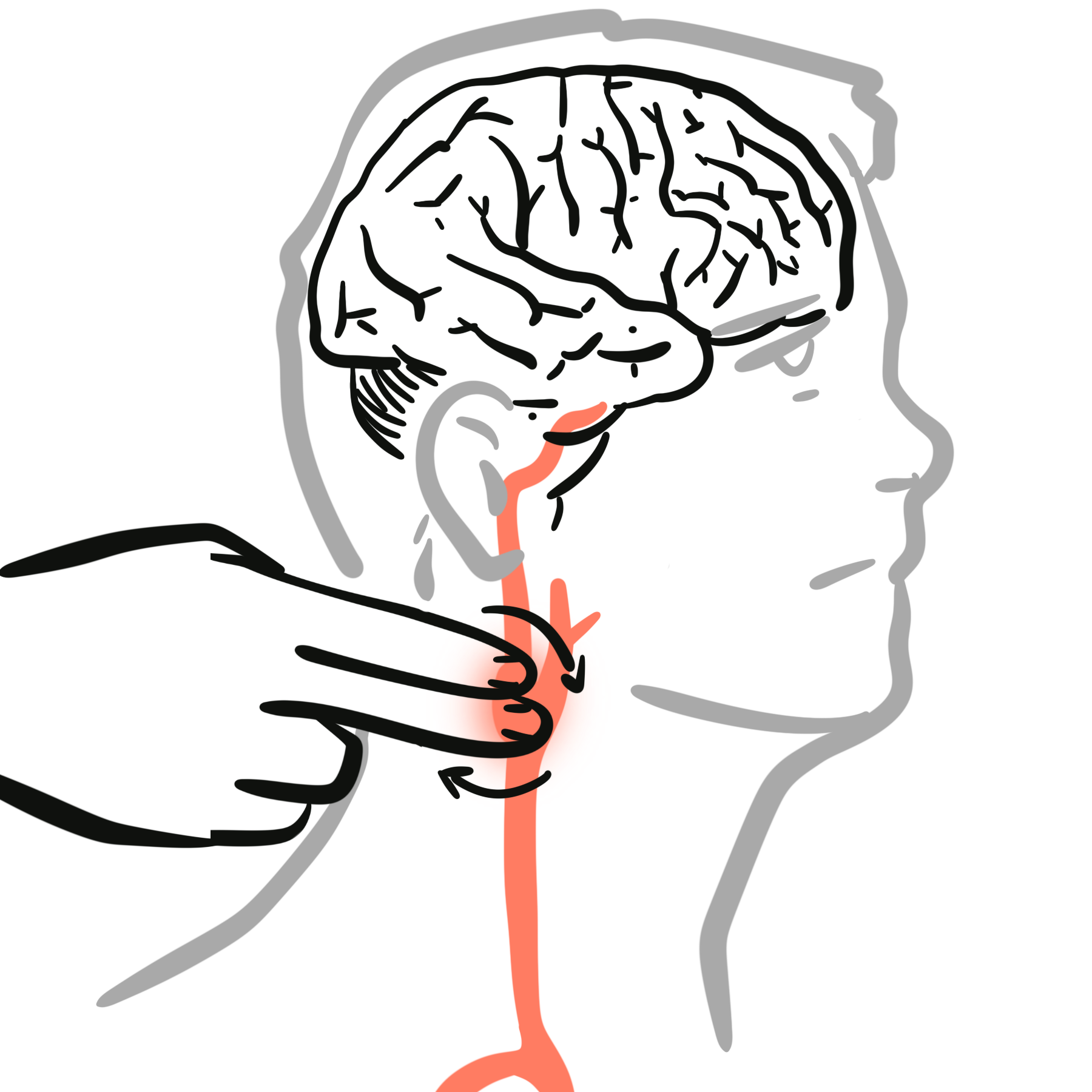
Left cardiac sympathetic denervation reduces adrenergic input to the heart and may be considered when ventricular arrhythmia persists despite maximally tolerated beta-blocker and flecainide, medication is not tolerated, recurrent appropriate ICD shocks occur, or an ICD is unsuitable or cannot provide adequate control.
Denervation reduces risk but does not necessarily eliminate the need for medication.
Implantable Cardioverter-Defibrillator
An ICD may be indicated after cardiac arrest or in patients with recurrent syncope or sustained ventricular arrhythmia despite optimal medical therapy.1
- A painful shock may trigger further catecholamine release
- This may intensify ventricular arrhythmia and produce an electrical storm
- Shocks may be ineffective against some bidirectional or polymorphic episodes
- Inappropriate shocks may occur
- Device complications are especially important in young patients
An ICD should not be used as a substitute for beta-blockade, flecainide, trigger control or consideration of sympathetic denervation. Device programming should be directed by an electrophysiologist experienced in inherited arrhythmia syndromes.
Why can an ICD shock worsen CPVT? The shock treats the rhythm but is also painful and frightening. The resulting adrenergic surge can recreate the mechanism that initiated the arrhythmia, producing recurrent ventricular tachycardia or ventricular fibrillation unless the underlying catecholamine-driven instability is medically controlled.
Complications & Prognosis
Complications
- Recurrent exertional syncope
- Traumatic injury during syncope
- Seizure-like episodes and misdiagnosis as epilepsy
- Sustained bidirectional or polymorphic VT
- Ventricular fibrillation
- Sudden cardiac arrest or death
- Recurrent ICD shocks
- Electrical storm
- Psychological distress and activity restriction
- Device- and procedure-related complications
Prognosis
Untreated CPVT carries a substantial risk of recurrent syncope, cardiac arrest and sudden death.2 Outcomes improve considerably with early diagnosis, strict adherence to non-selective beta-blockade, addition of flecainide when required, trigger modification and specialist follow-up.1
Higher-risk clinical features include:
- Previous cardiac arrest
- Diagnosis at a younger age
- Exercise-induced complex ventricular arrhythmia
- Recurrent syncope during treatment
- Persistent arrhythmia despite adequate therapy
- Poor medication adherence
- Failure to restrict unsafe high-intensity activity
Breakthrough events frequently reflect missed medication or inadequate dosing. Regular review should therefore include adherence, interval symptoms, exercise-test response, growth-related dose adjustment and screening of relatives.
References
- Zeppenfeld K, Tfelt-Hansen J, de Riva M, Winkel BG, Behr ER, Blom NA, et al. 2022 ESC Guidelines for the management of patients with ventricular arrhythmias and the prevention of sudden cardiac death. Eur Heart J. 2022;43(40):3997–4126. doi:10.1093/eurheartj/ehac262.
- Napolitano C, Mazzanti A, Bloise R, Priori SG. Catecholaminergic polymorphic ventricular tachycardia. In: Adam MP, Feldman J, Mirzaa GM, Pagon RA, Wallace SE, Amemiya A, editors. GeneReviews® [Internet]. Seattle: University of Washington, Seattle; 2004 [updated 2022 Jun 23; cited 2026 Aug 26]. Available from: https://www.ncbi.nlm.nih.gov/books/NBK1289/
- Priori SG, Wilde AA, Horie M, Cho Y, Behr ER, Berul C, et al. HRS/EHRA/APHRS expert consensus statement on the diagnosis and management of patients with inherited primary arrhythmia syndromes. Heart Rhythm. 2013;10(12):1932–1963. doi:10.1016/j.hrthm.2013.05.014.
- Wilde AAM, Semsarian C, Márquez MF, Sepehri Shamloo A, Ackerman MJ, Ashley EA, et al. European Heart Rhythm Association/Heart Rhythm Society/Asia Pacific Heart Rhythm Society/Latin American Heart Rhythm Society expert consensus statement on the state of genetic testing for cardiac diseases. Europace. 2022;24(8):1307–1367. doi:10.1093/europace/euac030.
- Femenia F, Barbosa-Barros R, Sampaio SV, Arce M, Perez-Riera A, Baranchuk A. Bidirectional ventricular tachycardia: a hallmark of catecholaminergic polymorphic ventricular tachycardia. Indian Pacing Electrophysiol J. 2012;12(2):65–68.


Members only discussions coming soon…