

Brugada Syndrome
Overview
Brugada syndrome is an inherited cardiac arrhythmia syndrome characterised by a distinctive type 1 Brugada electrocardiogram (ECG) pattern and susceptibility to polymorphic ventricular tachycardia (VT), ventricular fibrillation (VF) and sudden cardiac death, usually without overt structural heart disease. The ECG phenotype is dynamic and may be concealed until it is unmasked by fever, sodium-channel-blocking drugs or increased vagal tone.1–3
It occurs worldwide but is more prevalent in Southeast Asia. Clinical events occur more often in males and commonly arise during rest, sleep or fever rather than exercise.2,3
The crucial clinical task is not merely to recognise a Brugada-like ECG: it is to confirm that the pattern is genuine, exclude reversible phenocopies and determine whether the person has sufficient arrhythmic risk to justify an implantable cardioverter-defibrillator (ICD).1,3
Definition
- Brugada syndrome
- An inherited arrhythmia syndrome defined by a diagnostic type 1 Brugada ECG pattern and an increased risk of ventricular arrhythmia and sudden cardiac death.
- Type 1 Brugada ECG pattern
- Coved ST-segment elevation of at least 2 mm followed by a negative T wave in at least one right precordial lead positioned over the right ventricular outflow tract.
- Brugada phenocopy
- A reversible Brugada-like ECG pattern caused by another condition rather than true Brugada syndrome.
Anatomy & Physiology
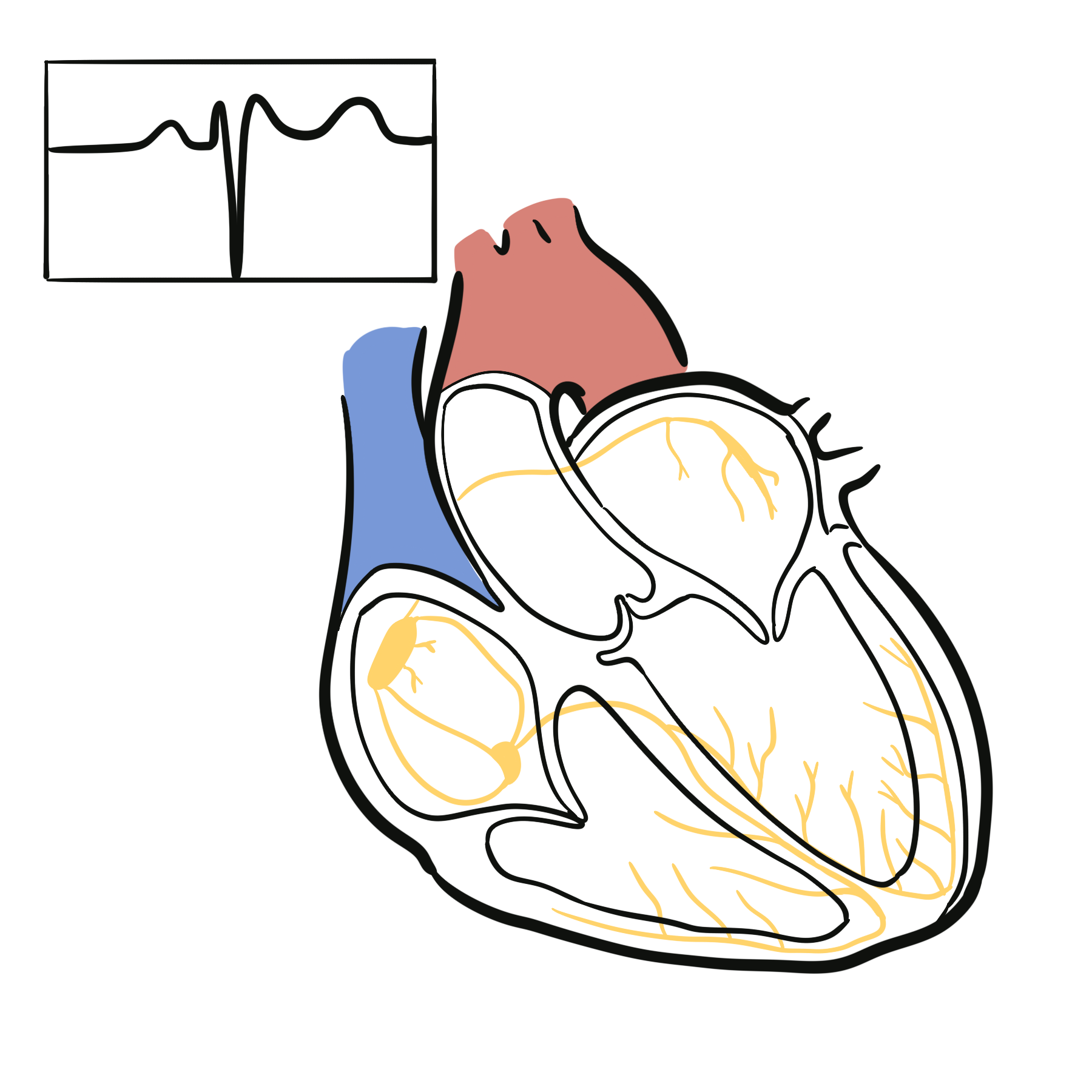
The right ventricular outflow tract (RVOT), particularly its epicardial surface, is central to the Brugada ECG phenotype and arrhythmogenic substrate. Leads V1 and V2 overlie this region; moving them from the conventional fourth intercostal space to the second or third intercostal space may improve detection of a concealed type 1 pattern.1,3
Normal cardiac depolarisation depends on rapid inward sodium current through the Naᵥ1.5 channel during phase 0 of the ventricular action potential. Balanced inward sodium and calcium currents and outward potassium currents allow orderly propagation and recovery. Disturbance of this balance can create delayed or heterogeneous activation in the RVOT and facilitate re-entry and VF.2,3
Aetiology & Risk Factors
Aetiology
Brugada syndrome is genetically heterogeneous. It is usually inherited in an autosomal dominant pattern with reduced penetrance and variable expression. Pathogenic loss-of-function variants in SCN5A, which encodes the cardiac Naᵥ1.5 sodium channel, are the best-established monogenic cause and are identified in only a minority of clinically diagnosed cases. A negative genetic test therefore does not exclude Brugada syndrome.2,3
The genetic architecture is often complex: common susceptibility variants and environmental modifiers influence whether and how the phenotype manifests. Some individuals with an SCN5A variant develop overlapping conduction disease, sinus-node dysfunction, atrial arrhythmias or other SCN5A-related phenotypes.2,3
Risk Factors
Factors associated with expression or unmasking of the Brugada phenotype include:
- Fever, particularly when high or persistent.
- Sodium-channel-blocking medicines and other drugs known to aggravate Brugada syndrome.
- Cocaine and excessive alcohol intake.
- Electrolyte or metabolic disturbances.
- Increased vagal tone, bradycardia, rest and sleep.
- A family history of Brugada syndrome or otherwise unexplained sudden death, especially at a young age.
Male sex strongly influences clinical expression and arrhythmic risk, although the genetic disorder affects all sexes.1–3
“Bangungut” in the Philippines and sudden unexpected nocturnal death syndrome described elsewhere in Southeast Asia are historically associated with the Brugada phenotype.2
Pathophysiology
Reduced inward sodium current impairs myocardial excitability and conduction. In the RVOT, where electrophysiological properties are particularly vulnerable, this can produce conduction delay and marked spatial heterogeneity between adjacent regions. Competing depolarisation and repolarisation hypotheses have been proposed; both converge on a vulnerable RVOT substrate capable of generating phase-2 re-entry, premature ventricular complexes and VF.2,3
Fever further reduces function of some mutant sodium channels, whereas bradycardia and increased vagal tone can increase the imbalance between inward and outward currents. This explains why the ECG pattern and ventricular arrhythmias may emerge during febrile illness, rest or sleep.2,3
A normal ECG when the patient is well does not exclude Brugada syndrome. The phenotype is dynamic: repeat the ECG during fever and consider high-right-precordial leads when clinical suspicion remains high.
Clinical Manifestations
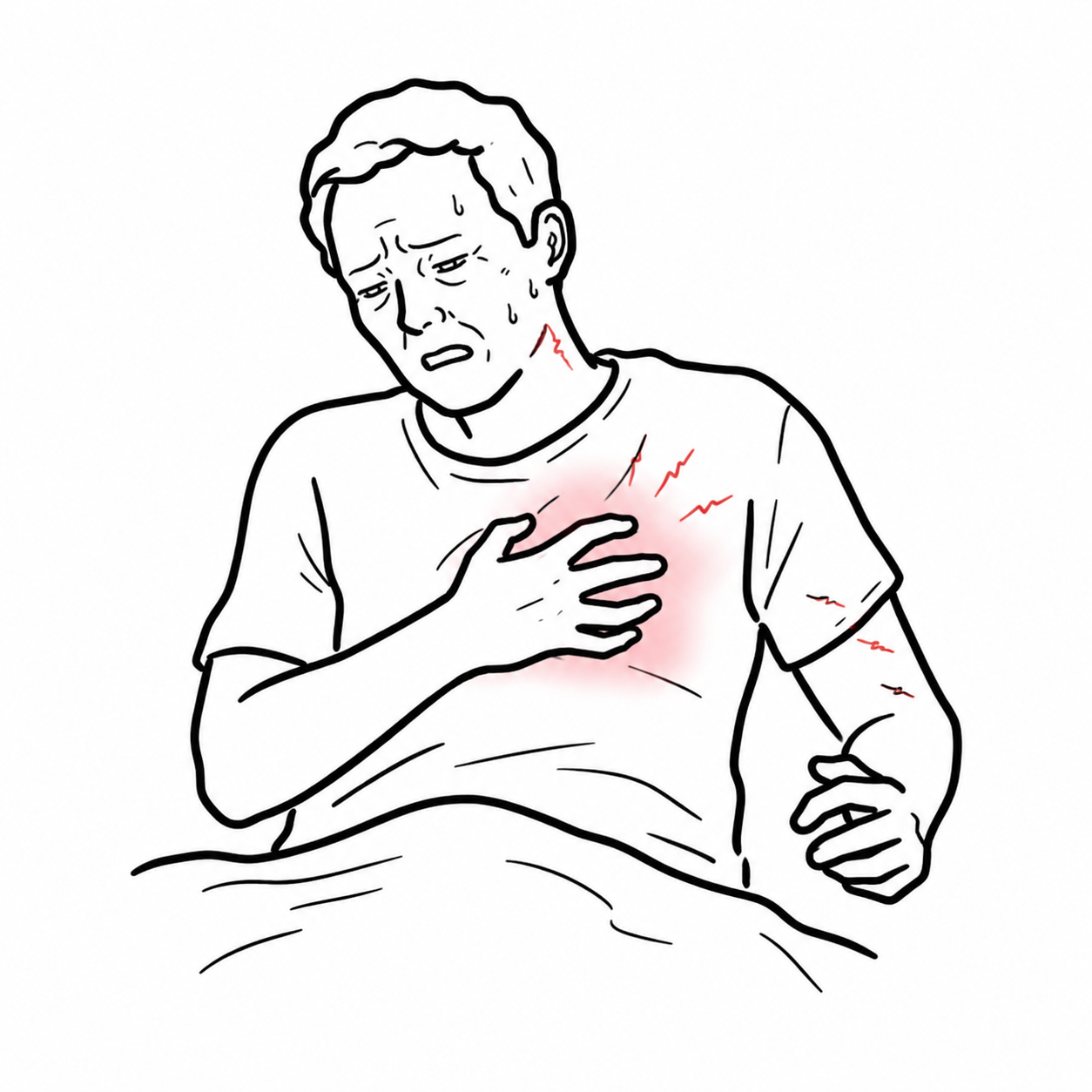
Many affected individuals are asymptomatic and are identified through an incidental ECG, family screening or investigation after a relative’s sudden death. When symptoms occur, they reflect self-terminating or sustained ventricular arrhythmia:
- Sudden cardiac arrest due to VF or polymorphic VT.
- Unexplained syncope, often without a prolonged prodrome.
- Nocturnal agonal respiration or gasping during sleep.
- Palpitations, seizure-like activity or brief nocturnal episodes caused by cerebral hypoperfusion.
- Sudden unexpected death, typically at rest or during sleep.
Atrial fibrillation, sinus-node dysfunction and conduction disease may coexist, especially with SCN5A-related disease.2,3
Features favouring arrhythmic syncope include occurrence at rest or during sleep, absent or very brief prodrome, injury from abrupt loss of consciousness, nocturnal agonal breathing, and a spontaneous type 1 ECG. A clear vasovagal trigger and typical autonomic prodrome favour reflex syncope, but diagnostic uncertainty should prompt specialist assessment.1,3
In Brugada syndrome, “seizure” may be the manifestation of transient cerebral hypoperfusion from ventricular arrhythmia. Always review the ECG and circumstances before diagnosing epilepsy in isolation.
Diagnosis
Diagnostic ECG pattern
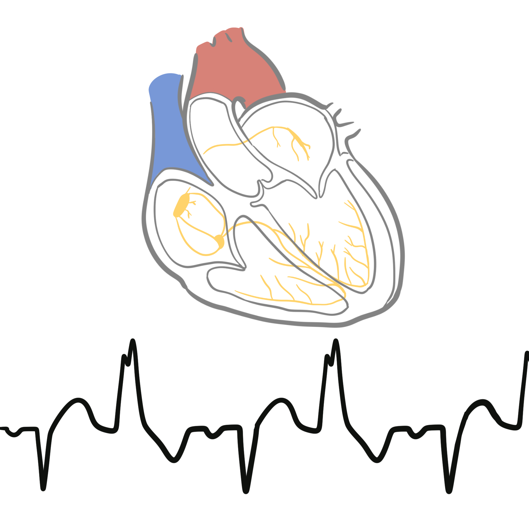
The diagnostic ECG finding is a type 1 Brugada pattern, occurring spontaneously or induced by fever or a supervised sodium-channel blocker challenge. It consists of coved ST-segment elevation of at least 2 mm in at least one right precordial lead (V1 or V2) placed in the second, third or fourth intercostal space, followed by a negative T wave. Type 2 and type 3 saddleback patterns are suspicious but are not independently diagnostic.1,3,4
| ECG pattern | Key morphology | Interpretation |
|---|---|---|
| Type 1 | Coved ST elevation ≥2 mm with descending ST segment and negative T wave in a right precordial lead | Diagnostic ECG pattern after exclusion of mimics |
| Type 2 | Saddleback ST elevation with a positive or biphasic T wave | Suggestive only; consider further evaluation if clinical suspicion is meaningful |
| Type 3 | Coved or saddleback appearance with less ST elevation | Non-diagnostic |
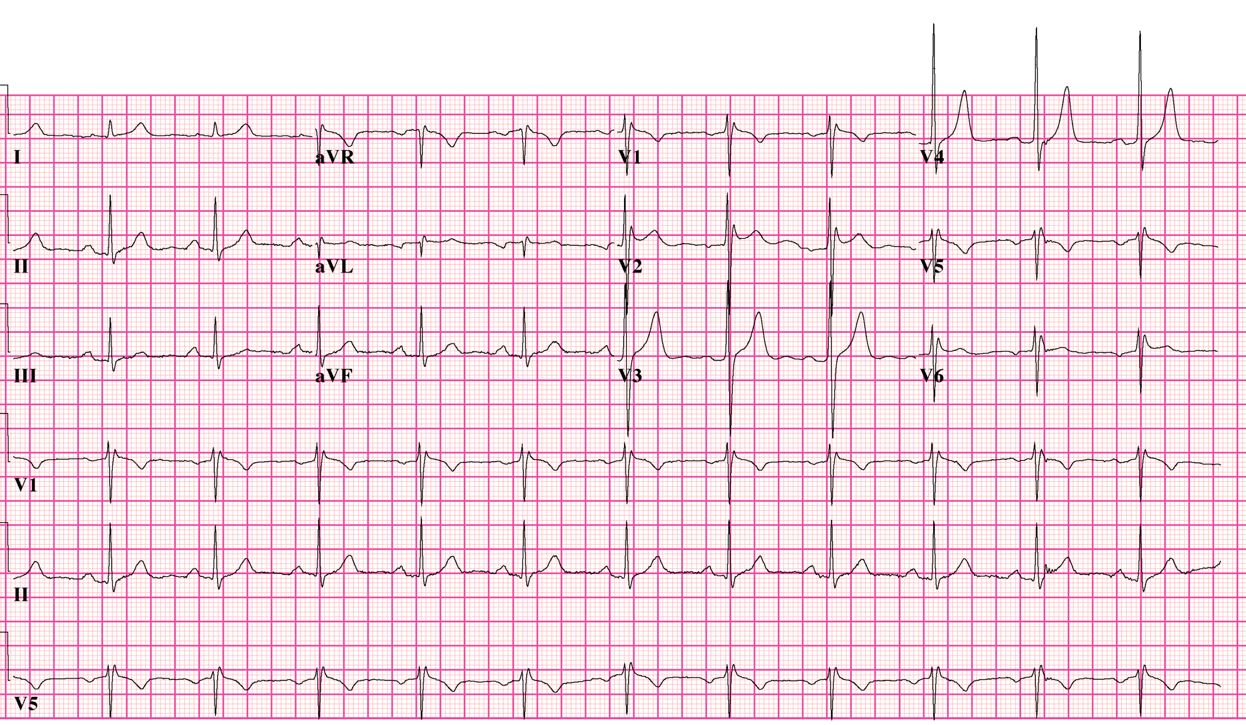
What to notice
Detailed image description
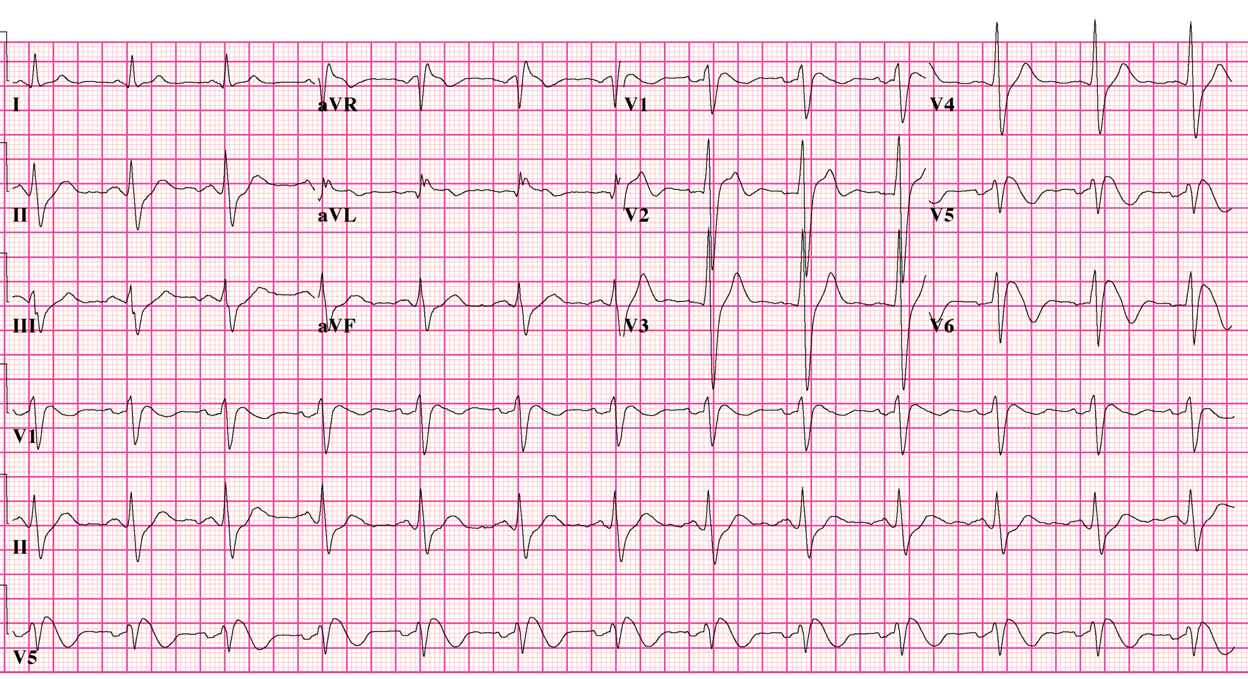
What to notice
Detailed image description
Only the type 1 ECG pattern is diagnostic. A “saddleback” type 2 or type 3 pattern alone is not Brugada syndrome.
Focused Diagnostic Assessment
- Repeat the 12-lead ECG: compare previous tracings and record V1–V2 in both standard and high right precordial positions. Obtain an ECG during fever whenever possible.
- Clarify the phenotype: document syncope characteristics, nocturnal agonal breathing, resuscitated arrest, documented VT/VF, provoking drugs and a three-generation family history of Brugada syndrome or unexplained sudden death.
- Exclude Brugada phenocopies and structural disease: direct testing toward the clinical context. Important mimics include acute myocardial ischaemia, pericarditis, pulmonary embolism, hyperkalaemia, hypothermia, sodium-channel-blocker toxicity and mechanical RVOT compression. Echocardiography is generally appropriate; cardiac magnetic resonance imaging is reserved for suspected cardiomyopathy or structural pathology rather than performed indiscriminately.1,3
- Sodium-channel blocker challenge: consider in an experienced centre when clinical suspicion persists without a spontaneous type 1 pattern. Ajmaline, flecainide or procainamide may be used according to local protocols, with continuous ECG and resuscitation capability because malignant ventricular arrhythmias can be provoked. A drug-induced type 1 pattern must be interpreted in the clinical context because overdiagnosis and false-positive results occur.4
- Rhythm documentation: ambulatory monitoring or an implantable loop recorder may be useful when recurrent unexplained syncope remains diagnostically uncertain, but a negative short recording does not exclude intermittent VF.
- Genetic testing: offer targeted testing with genetic counselling after a clinical diagnosis, primarily to enable cascade testing when a pathogenic or likely pathogenic familial variant is identified. Genetic testing is not the first-line diagnostic test for an isolated non-diagnostic ECG, and a negative result does not exclude the syndrome.1–3
Electrophysiological study with programmed ventricular stimulation is not a diagnostic test. It has a limited and debated role in risk stratification, mainly in selected asymptomatic patients with a spontaneous type 1 pattern when the result would change management.1,3
The ECG label and the clinical syndrome are not interchangeable. Before diagnosing Brugada syndrome from a drug-induced pattern, ask whether the history, family context and exclusion of phenocopies make the result clinically credible.
Classification
Brugada ECG patterns are classified morphologically as type 1, type 2 or type 3. Only type 1 is diagnostic; types 2 and 3 are non-diagnostic patterns that may justify specialist evaluation when supported by symptoms or family history.1,3
For risk assessment, the more useful clinical distinction is:
- Spontaneous type 1 pattern: present without pharmacological provocation; carries greater arrhythmic significance.
- Fever-induced type 1 pattern: appears during febrile illness.
- Drug-induced type 1 pattern: appears only during sodium-channel blocker challenge; usually confers lower risk in an asymptomatic person than a spontaneous pattern.
Treatment
Immediate Management
- Treat cardiac arrest and unstable ventricular arrhythmia according to advanced life support principles, including prompt defibrillation.
- Admit and monitor patients with a type 1 pattern during significant fever, arrhythmic syncope or ventricular arrhythmia.
- Treat fever promptly with antipyretics and management of its cause; maintain hydration and correct electrolyte abnormalities.
- For recurrent VF or electrical storm, intravenous isoproterenol is used acutely in a monitored specialist setting. Quinidine may suppress recurrent ventricular arrhythmia and can be used as adjunctive therapy.1,2
Long-Term Prevention
All patients should receive education to:
- Treat fever early and seek urgent assessment when fever is persistent or associated with syncope, palpitations or a type 1 ECG.
- Avoid medicines known to provoke Brugada ECG changes or ventricular arrhythmias; check a current specialist drug-safety list before prescribing.
- Avoid cocaine and excessive alcohol intake.
- Inform clinicians of the diagnosis before anaesthesia or new medication is given.
An ICD is the only established treatment that terminates potentially fatal VT/VF. It is indicated for survivors of cardiac arrest and patients with documented spontaneous sustained ventricular arrhythmia. ICD implantation should be considered for arrhythmic syncope with a spontaneous type 1 pattern after careful assessment. The benefit is uncertain in asymptomatic patients, and inappropriate shocks, lead complications and device infection are important harms, particularly in young people.1–3
Quinidine may be considered when an ICD is contraindicated or declined, for recurrent ICD shocks or electrical storm, and in selected specialist-managed cases. Epicardial catheter ablation targeting the abnormal RVOT substrate may be considered for recurrent electrical storm or repeated appropriate ICD shocks despite medical therapy; it is not routine treatment for an asymptomatic ECG pattern.1,3
First-degree relatives should receive clinical assessment and ECG screening. If a pathogenic familial variant is identified, cascade genetic testing can distinguish relatives who require ongoing surveillance from non-carriers.2
Fever can unmask the type 1 pattern and trigger ventricular arrhythmia. Treat it promptly, record an ECG and escalate urgently if arrhythmic symptoms or a diagnostic pattern occurs.
Complications & Prognosis
Complications
- Polymorphic VT, VF and sudden cardiac death.
- Recurrent syncope, injury or seizure-like episodes from transient cerebral hypoperfusion.
- Atrial fibrillation and progressive conduction disease.
- ICD-related inappropriate shocks, lead failure, infection and psychological burden.
- Misdiagnosis and unnecessary treatment after an incorrectly interpreted Brugada-like ECG or provocative test.
Prognosis
Prognosis varies widely. Previous cardiac arrest or sustained ventricular arrhythmia predicts the greatest recurrence risk. Arrhythmic syncope and a spontaneous type 1 ECG also identify higher-risk patients. In contrast, most asymptomatic individuals with only a drug-induced type 1 pattern have a substantially lower event rate, so the potential harms of prophylactic ICD implantation may outweigh benefit.1,3
The ECG and clinical phenotype can change over time. Long-term follow-up should therefore focus on new syncope or nocturnal symptoms, fever-related ECG change, interval arrhythmia, medication exposure and evolving family history rather than relying on a single reassuring ECG.1–3
References
- Zeppenfeld K, Tfelt-Hansen J, de Riva M, et al. 2022 ESC Guidelines for the management of patients with ventricular arrhythmias and the prevention of sudden cardiac death. Eur Heart J. 2022;43(40):3997–4126. doi:10.1093/eurheartj/ehac262
- Brugada R, Campuzano O, Sarquella-Brugada G, Brugada J, Brugada P. Brugada syndrome. In: Adam MP, Feldman J, Mirzaa GM, et al., editors. GeneReviews® [Internet]. Seattle (WA): University of Washington, Seattle; 2005 [updated 2022 Aug 25; cited 2026 Aug 23]. Available from: https://www.ncbi.nlm.nih.gov/books/NBK1517/
- Brugada J, Campuzano O, Arbelo E, Sarquella-Brugada G, Brugada R. Present status of Brugada syndrome: JACC state-of-the-art review. J Am Coll Cardiol. 2018;72(9):1046–1059. doi:10.1016/j.jacc.2018.06.037
- Viskin S, Chorin E, Rosso R, Amin AS, Wilde AAM. Diagnosis of Brugada syndrome with a sodium-channel-blocker test: who should be tested? Who should not? Circulation. 2024;150(8):642–650. doi:10.1161/CIRCULATIONAHA.124.069138
- Honarbakhsh S, Providencia R, Garcia-Hernandez J, et al. A primary prevention clinical risk score model for patients with Brugada syndrome (BRUGADA-RISK). JACC Clin Electrophysiol. 2021;7(2):210–222. doi:10.1016/j.jacep.2020.08.032


Members only discussions coming soon…