

Infertility
OVERVIEW
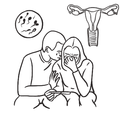
Infertility is a disease of the reproductive system characterised by failure to achieve a pregnancy after 12 months or more of regular, unprotected sexual intercourse. It may arise from female factors, male factors, combined factors, or remain unexplained after standard evaluation.
When to Begin Evaluation:
- Female partner younger than 35 years: Evaluate after 12 months of regular unprotected intercourse.
- Female partner aged 35 years or older: Evaluate after 6 months because fertility declines more rapidly with age.
- Female partner older than 40 years: Consider immediate evaluation and treatment.
- Evaluate without delay at any age when there is oligomenorrhoea, amenorrhoea or suspected anovulation; known or suspected endometriosis, tubal disease, pelvic inflammatory disease or uterine abnormality; previous pelvic surgery, chemotherapy, radiotherapy or gonadotoxic treatment; suspected diminished ovarian reserve or premature ovarian insufficiency; known male-factor infertility, sexual dysfunction or abnormal genital examination; or a need for donor gametes, fertility preservation or assisted reproduction independent of intercourse.
Classification:
- Primary infertility: No previous pregnancy has occurred.
- Secondary infertility: Difficulty conceiving after a previous pregnancy, irrespective of its outcome.
- Unexplained infertility: No cause identified after confirmation of ovulation, adequate semen parameters, and tubal patency with assessment of the uterine cavity.
Clinical Significance: Female and male factors contribute with similar frequency, and several factors may coexist. Both partners should therefore be assessed concurrently rather than completing the female evaluation before investigating the male partner.
Infertility is a couple or individual reproductive disorder, not solely a female diagnosis. Begin semen analysis and female reproductive assessment at the same time to prevent avoidable delays.
APPROACH
Step 1: Determine Whether Immediate Referral Is Required
- Advanced reproductive age: Expedite specialist referral for women aged 35 years or older. Do not delay evaluation in women older than 40 years.
- Amenorrhoea or marked menstrual irregularity: Investigate pregnancy, hypothalamic dysfunction, PCOS, hyperprolactinaemia, thyroid disease, and premature ovarian insufficiency.
- Possible premature ovarian insufficiency: Consider in patients younger than 40 years with amenorrhoea or irregular cycles and menopausal symptoms.
- Known tubal or pelvic disease: Previous pelvic inflammatory disease, ectopic pregnancy, endometriosis, pelvic surgery, or sterilisation warrants earlier assessment.
- Severe male-factor features: Azoospermia, severe oligozoospermia, small testes, testicular mass, cryptorchidism, previous testicular torsion, genital surgery, chemotherapy, or ejaculatory dysfunction.
- Before gonadotoxic treatment: Arrange urgent fertility-preservation referral before chemotherapy, pelvic radiotherapy, gonadectomy, or other treatment likely to impair fertility.
- Possible pregnancy complication: Pelvic pain, vaginal bleeding, dizziness, or syncope following fertility treatment or a positive pregnancy test requires urgent assessment for ectopic pregnancy.
- Possible ovarian hyperstimulation syndrome: Abdominal distension, severe pain, vomiting, oliguria, dyspnoea, rapid weight gain, or thromboembolic symptoms after ovarian stimulation requires urgent specialist review.
Step 2: Establish Reproductive and Sexual History
Female or Egg-Producing Partner
- Duration of attempts to conceive and frequency and timing of intercourse or insemination.
- Menstrual cycle length, regularity, amenorrhoea, intermenstrual bleeding, dysmenorrhoea, and features of ovulation.
- Previous pregnancies, miscarriages, ectopic pregnancy, and obstetric complications.
- Pelvic inflammatory disease, sexually transmitted infections, endometriosis, pelvic surgery, uterine procedures, or cervical treatment.
- Symptoms of hyperandrogenism, galactorrhoea, thyroid disease, premature ovarian insufficiency, or hypothalamic dysfunction.
- Previous chemotherapy, radiotherapy, ovarian surgery, or gonadotoxic medication.
- Family history of early menopause, genetic disease, infertility, or recurrent pregnancy loss.
Male or Sperm-Producing Partner
- Pubertal development, cryptorchidism, testicular torsion, orchitis, trauma, genital or inguinal surgery, and varicocele.
- Previous fertility or pregnancies with another partner.
- Erectile function, ejaculation, libido, frequency of intercourse, and use of lubricants.
- Febrile illness, occupational heat exposure, anabolic steroids, testosterone therapy, chemotherapy, radiotherapy, and recreational drugs.
- Family history of infertility, cystic fibrosis, or genetic disease.
Both Partners
- Smoking, vaping, alcohol, cannabis, anabolic steroids, and other recreational substances.
- Current medicines and supplements.
- Weight, nutrition, exercise, chronic disease, vaccination status, and mental health.
- Relationship context, reproductive goals, cultural preferences, donor-conception needs, and treatment expectations.
Step 3: Decide Which Reproductive Component Is Impaired
Assess four essential components:
- Ovulation and ovarian function
- Tubal patency and pelvic anatomy
- Uterine cavity and implantation environment
- Sperm production and delivery
A defect in more than one component is common.
Step 4: Perform Focused Examination
Female Examination
- BMI, blood pressure, and signs of nutritional deficiency or excessive exercise.
- Hirsutism, acne, androgenic alopecia, or acanthosis nigricans.
- Thyroid enlargement, galactorrhoea, or visual-field abnormality.
- Features of oestrogen deficiency or Cushing syndrome.
- Abdominal or pelvic mass and endometriosis-related tenderness where examination is indicated.
Male Examination
- Secondary sexual characteristics and gynaecomastia.
- Testicular size, consistency, and position.
- Epididymis and presence of each vas deferens.
- Varicocele assessed while standing and performing Valsalva.
- Penile abnormality, phimosis, hypospadias, or features preventing semen deposition.
- Digital rectal examination only when clinically indicated.
Regular menstrual cycles usually indicate ovulation, but they do not assess ovarian reserve, tubal patency, uterine anatomy, sperm quality, or the age-related decline in oocyte competence.
DIFFERENTIAL DIAGNOSIS
Ovulatory and Endocrine Causes
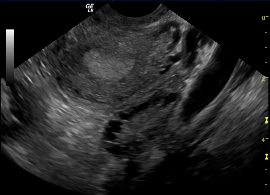
- Polycystic ovary syndrome: Oligo-ovulation or anovulation with clinical or biochemical hyperandrogenism. May be associated with acne, hirsutism, obesity, insulin resistance, and acanthosis nigricans.
- Functional hypothalamic amenorrhoea: Energy deficiency, low body weight, excessive exercise, psychological stress, or chronic illness. Typically associated with low or inappropriately normal gonadotropins and low oestradiol.
- Premature ovarian insufficiency: Loss of ovarian activity before age 40, presenting with irregular cycles or amenorrhoea, elevated FSH, and symptoms of oestrogen deficiency.
- Age-related diminished ovarian reserve: Declining oocyte number and quality, especially from the mid-30s onward.
- Hyperprolactinaemia: Oligomenorrhoea, amenorrhoea, galactorrhoea, reduced libido, headache, or visual disturbance.
- Thyroid dysfunction: Hyperthyroidism or hypothyroidism may disrupt ovulation and menstruation.
- Congenital adrenal hyperplasia or severe androgen excess: Consider with marked hyperandrogenism, rapid virilisation, or relevant family history.
- Iatrogenic ovarian dysfunction: Chemotherapy, pelvic radiotherapy, bilateral ovarian surgery, or gonadotoxic medication.
Tubal and Peritoneal Causes
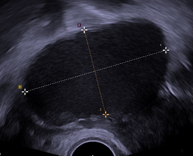
- Previous pelvic inflammatory disease: Tubal scarring or occlusion following chlamydial or gonococcal infection.
- Endometriosis: Dysmenorrhoea, deep dyspareunia, chronic pelvic pain, endometrioma, or distorted pelvic anatomy.
- Previous ectopic pregnancy or tubal surgery: May reduce tubal patency and increase recurrent ectopic pregnancy risk.
- Postoperative pelvic adhesions: Following pelvic, abdominal, or bowel surgery.
- Genital tuberculosis: Consider in patients from higher-prevalence regions or with relevant exposure.
- Hydrosalpinx: Distal tubal obstruction associated with reduced implantation and IVF success.
Uterine and Cervical Causes
- Submucosal fibroid: Distorts the endometrial cavity and may impair implantation.
- Endometrial polyp: May cause intermenstrual bleeding and interfere with implantation.
- Congenital uterine anomaly: Septate, bicornuate, unicornuate, or other Müllerian anomaly.
- Intrauterine adhesions: Asherman syndrome following uterine surgery, curettage, infection, or postpartum haemorrhage.
- Adenomyosis: Heavy painful menstruation, enlarged tender uterus, and possible impairment of fertility.
- Cervical stenosis or mucus abnormality: Uncommon as an isolated cause but may impair sperm transport or insemination access.
Male Causes
Pre-Testicular and Endocrine
- Hypogonadotropic hypogonadism: Pituitary or hypothalamic disease, congenital GnRH deficiency, hyperprolactinaemia, severe systemic illness, or anabolic-steroid use.
- Exogenous testosterone or anabolic steroids: Suppress gonadotropins and intratesticular testosterone, potentially causing severe oligozoospermia or azoospermia.
- Thyroid or other systemic endocrine disease.
Primary Testicular Failure
- Klinefelter syndrome or another chromosomal disorder.
- Y-chromosome microdeletion.
- Cryptorchidism, testicular torsion, trauma, orchitis, chemotherapy, or radiotherapy.
- Sertoli-cell-only syndrome or maturation arrest.
- Clinically significant varicocele.
- Testicular malignancy.
Post-Testicular and Sperm-Delivery Disorders
- Congenital bilateral absence of the vas deferens: Strongly associated with pathogenic CFTR variants.
- Ejaculatory-duct obstruction or acquired reproductive-tract obstruction.
- Retrograde ejaculation or anejaculation: May follow diabetes, neurological disease, pelvic surgery, or medication.
- Erectile dysfunction, premature ejaculation, severe hypospadias, or infrequent intercourse.
- Previous vasectomy.
Combined and Unexplained Infertility
- Combined infertility: More than one female or male factor is present.
- Unexplained infertility: Standard assessment shows ovulation, patent tubes, no important uterine abnormality, and adequate semen parameters, but conception has not occurred.
- Reduced fecundability associated with reproductive ageing: May exist despite apparently normal investigations.
Exogenous testosterone is a contraceptive, not a fertility treatment. It suppresses LH and FSH and may cause azoospermia; do not prescribe testosterone to a man actively seeking fertility.
INVESTIGATIONS
Concurrent First-Line Assessment
Semen Analysis
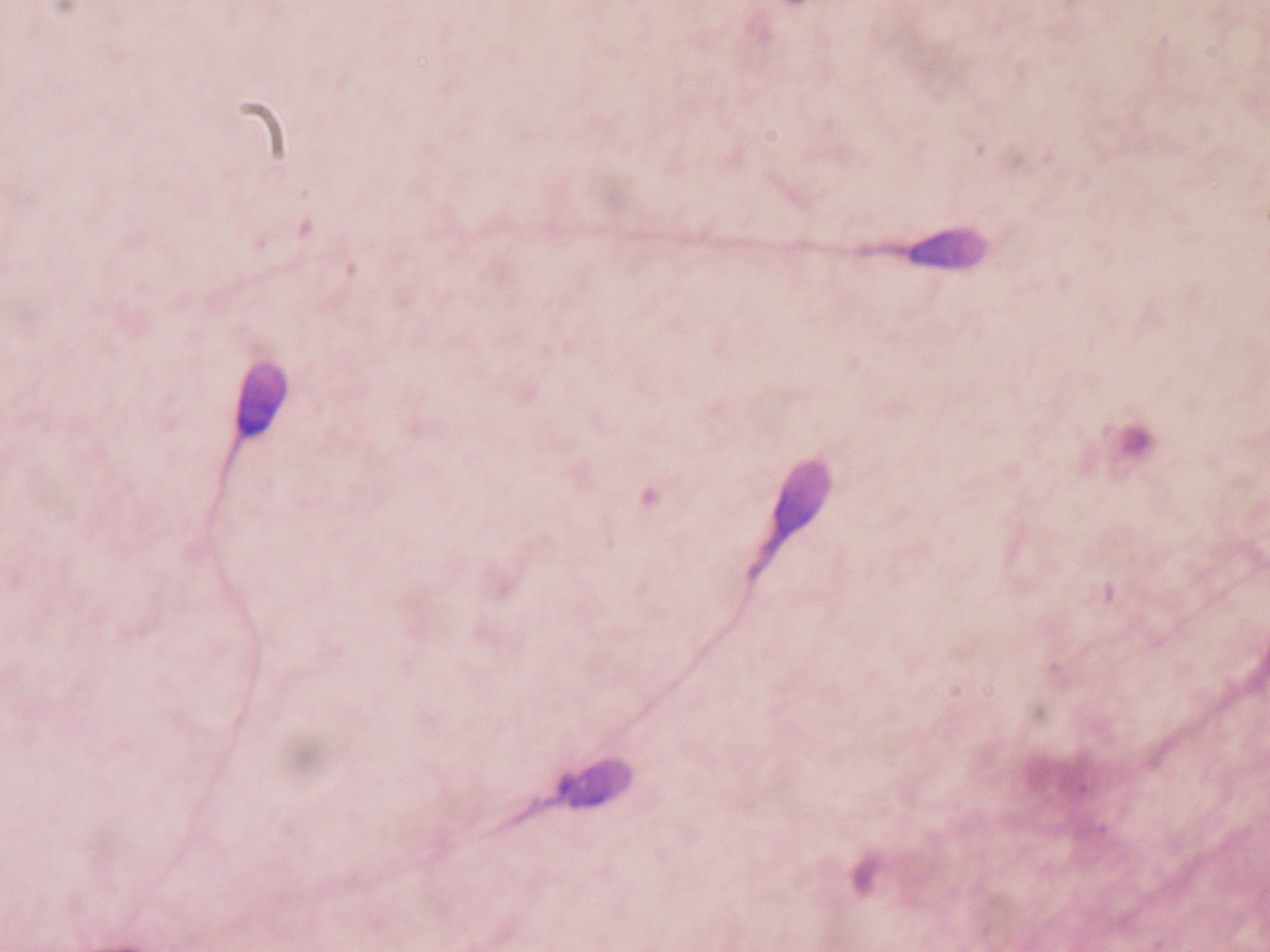
- Obtain at least one semen analysis early in the evaluation.
- Collect the complete ejaculate after the laboratory’s recommended period of abstinence and transport it under appropriate conditions.
- Assess semen volume, sperm concentration, total sperm number, motility, morphology, and other laboratory parameters against current WHO reference standards.
- Because semen results vary biologically, repeat an abnormal analysis—usually after an interval determined by the degree of abnormality and clinical circumstances.
- Severe abnormalities or azoospermia require specialist male-reproductive evaluation.
Assessment of Ovulation
- A menstrual history demonstrating regular cycles of approximately 21–35 days usually supports ovulation.
- If ovulation is uncertain, measure serum progesterone approximately one week before the expected next menstruation rather than automatically on “day 21.”
- Ovulation-prediction kits or ultrasound monitoring may assist in selected patients.
- Do not routinely perform endometrial biopsy to confirm ovulation.
Pelvic Ultrasound
- Transvaginal ultrasound assesses uterine anatomy, fibroids, polyps, adenomyosis, ovarian morphology, antral follicles, endometrioma, adnexal masses, and structural causes of pelvic pain or abnormal bleeding.
- Polycystic ovarian morphology alone does not diagnose PCOS.
Ovarian Reserve Testing
- Anti-Müllerian hormone and antral follicle count: Estimate expected ovarian response to stimulation and help guide medication dosing and assisted-reproduction counselling, but do not independently predict spontaneous conception or oocyte quality.
- Early-follicular FSH and oestradiol: May assist when ovarian reserve is uncertain.
- Interpret ovarian reserve in the context of age, menstrual history, prior ovarian treatment, and planned therapy.
- Do not use ovarian-reserve testing as a general fertility screening test in people who do not meet criteria for infertility.
Tubal Patency and Uterine Cavity
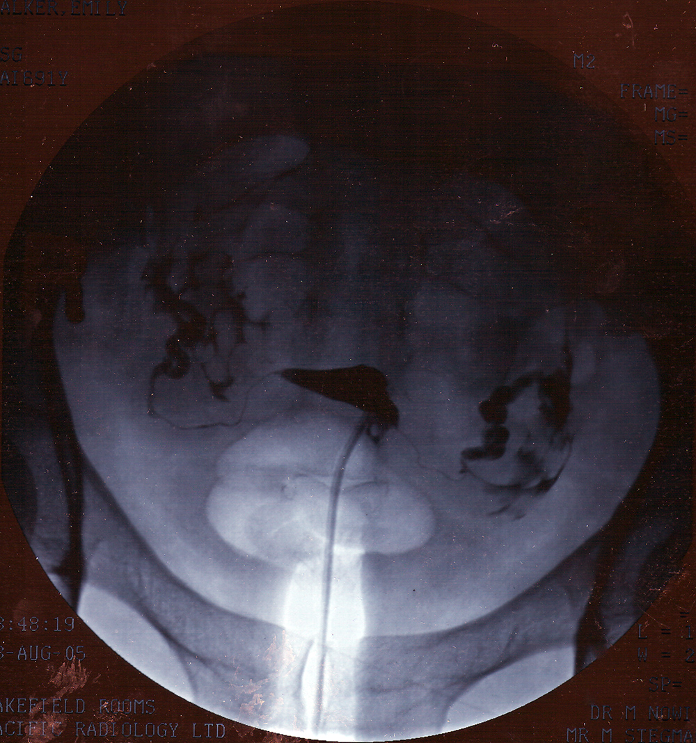
- Hysterosalpingography: Assesses tubal patency and outlines the uterine cavity.
- Hysterosalpingo-contrast sonography: Ultrasound-based alternative where expertise is available.
- Saline-infusion sonography: More accurately defines intracavitary polyps, submucosal fibroids, and adhesions.
- Hysteroscopy: Definitive evaluation and treatment of suspected intracavitary pathology.
- Laparoscopy with chromopertubation: Not a routine first-line investigation; consider when endometriosis, significant pelvic disease, or surgically treatable tubal pathology is suspected.
Targeted Female Hormonal Investigation
- Pregnancy test: In amenorrhoea or before treatment and imaging where relevant.
- TSH: With ovulatory dysfunction, menstrual disturbance, or thyroid symptoms.
- Prolactin: With galactorrhoea, oligomenorrhoea, amenorrhoea, headache, or visual symptoms.
- FSH and oestradiol: When amenorrhoea or premature ovarian insufficiency is suspected.
- Total testosterone, SHBG, and other androgen testing: With hirsutism, acne, virilisation, or suspected PCOS.
- 17-hydroxyprogesterone: When non-classic congenital adrenal hyperplasia is suspected.
Targeted Male Investigation
- FSH and total testosterone: Indicated with oligozoospermia, azoospermia, reduced libido, erectile dysfunction, testicular atrophy, or other evidence of endocrine disease.
- LH and prolactin: Add according to testosterone findings and clinical features.
- Scrotal ultrasound: Not routine for every patient; use for a testicular mass, examination uncertainty, or selected pathology.
- Post-ejaculatory urine testing: Consider when retrograde ejaculation is suspected.
- Genetic testing: Karyotype and Y-chromosome microdeletion testing in appropriately selected patients with azoospermia or severe oligozoospermia; CFTR testing when congenital absence of the vas deferens or obstructive azoospermia suggests a CFTR-related disorder. Offer genetic counselling before and after clinically significant testing.
Tests Not Recommended Routinely
- Postcoital cervical-mucus testing.
- Endometrial biopsy for luteal-phase dating.
- Diagnostic laparoscopy without a clinical indication.
- Routine sperm DNA-fragmentation testing in the initial evaluation.
- Routine antisperm-antibody testing.
- Routine thrombophilia, immunological, natural-killer-cell, or broad infection panels without a specific indication.
- Routine prolactin testing in an ovulatory patient without suggestive symptoms.
AMH measures ovarian quantity and expected response to stimulation, not egg quality and not the chance of natural conception by itself. Female age remains the most important predictor of oocyte competence and treatment success.
CRITICAL MANAGEMENT
Urgent Fertility Preservation
- Refer before gonadotoxic treatment whenever clinically possible.
- Options may include sperm cryopreservation, oocyte cryopreservation, embryo cryopreservation, ovarian-tissue cryopreservation in selected patients, and testicular-tissue preservation in specialised paediatric or research settings.
- Do not delay urgent cancer treatment without multidisciplinary agreement.
- Discuss future use, storage, consent, genetic risk, and the possibility that preserved material may not result in a live birth.
Preconception and Natural-Conception Optimisation
- Recommend intercourse every one to two days during the fertile window; rigid scheduling is unnecessary if it creates distress.
- Prescribe folic acid before conception, with dose adjusted for individual risk.
- Optimise diabetes, thyroid disease, hypertension, epilepsy, and other chronic conditions.
- Review medicines for teratogenicity and reproductive effects.
- Encourage smoking and recreational-drug cessation.
- Avoid testosterone and anabolic steroids in men seeking fertility.
- Address harmful alcohol use and excessive occupational heat or toxin exposure.
- Offer weight-related support when relevant, while avoiding unnecessary treatment delays—particularly with advancing reproductive age.
- Provide rubella, varicella, hepatitis, STI, cervical-screening, and genetic-carrier assessment according to clinical risk and local guidance.
Ovulatory Infertility
- PCOS-related anovulation: Letrozole is generally first-line pharmacological ovulation induction when no other major infertility factor exists. Alternatives include clomifene citrate or gonadotropins in selected patients. Metformin is primarily indicated for metabolic features and may support ovulation in selected PCOS phenotypes. Monitor treatment to reduce multiple-pregnancy and ovarian-hyperstimulation risks.
- Hyperprolactinaemia: Treat the underlying cause; a dopamine agonist such as cabergoline is commonly used for prolactinoma.
- Hypothyroidism: Restore euthyroidism before conception.
- Functional hypothalamic amenorrhoea: Correct energy deficiency, excessive exercise, psychological stress, and low bone-health risk before specialist ovulation induction.
- Hypogonadotropic hypogonadism: Specialist pulsatile GnRH or gonadotropin therapy may restore ovulation.
- Premature ovarian insufficiency: Spontaneous ovulation may occasionally occur, but donor-oocyte IVF offers the most effective established route to pregnancy.
Tubal, Uterine and Endometriosis-Related Infertility
- Proximal tubal obstruction: Confirm possible spasm or technical occlusion before definitive treatment.
- Distal tubal disease or hydrosalpinx: IVF is frequently preferred for severe bilateral disease. Salpingectomy or proximal tubal occlusion before IVF may improve outcomes when hydrosalpinx is present.
- Intracavitary pathology: Hysteroscopic removal of a submucosal fibroid, polyp, or adhesions may be appropriate when cavity distortion is clinically important.
- Endometriosis: Individualise expectant management, surgery, ovarian stimulation with intrauterine insemination, or IVF according to age, pain, ovarian reserve, disease severity, tubal anatomy, semen findings, and previous surgery. Avoid repeated ovarian surgery that may unnecessarily reduce ovarian reserve.
Male-Factor Infertility
- Stop testosterone and anabolic steroids.
- Treat infection only when clinically demonstrated.
- Correct endocrine causes: gonadotropin therapy may restore spermatogenesis in hypogonadotropic hypogonadism; aromatase inhibitors, selective oestrogen-receptor modulators, or hCG-based treatment should be specialist-directed.
- Consider microsurgical varicocele repair for a palpable varicocele, infertility, and abnormal semen parameters in an appropriate couple.
- Treat ejaculatory dysfunction or obstruction where reversible.
- Use surgical sperm retrieval with IVF–ICSI for selected obstructive or non-obstructive azoospermia.
- Offer genetic counselling when a heritable cause or use of surgically retrieved sperm is relevant.
Unexplained Infertility
- Management depends principally on female age, duration of infertility, previous treatment, and prognosis.
- Short expectant management may be reasonable when prognosis for natural conception is favourable.
- A common active-treatment pathway is a limited course of ovarian stimulation using an oral agent with intrauterine insemination.
- Proceed to IVF when initial treatment fails, prognosis is poor, infertility is prolonged, or reproductive age makes further delay inappropriate.
- Avoid unproven immune therapies and other costly “add-ons” without evidence of benefit.
Assisted Reproductive Technology
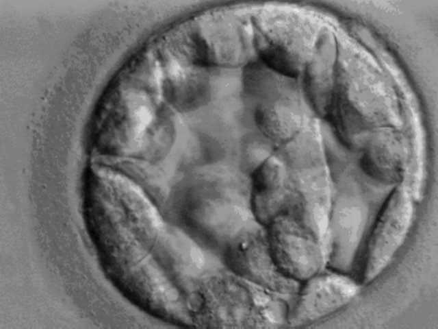
- Intrauterine insemination: Useful in selected unexplained, mild male-factor, cervical, or ovulatory infertility when at least one tube is patent.
- In vitro fertilisation: Used for severe tubal disease, prolonged unexplained infertility, unsuccessful simpler treatment, or other indications.
- Intracytoplasmic sperm injection: Primarily indicated for important male-factor infertility or previous fertilisation failure; it is not automatically required for every IVF cycle.
- Prefer single-embryo transfer when clinically appropriate to reduce multiple pregnancy.
- Counsel regarding success rates, ovarian hyperstimulation, procedure complications, ectopic pregnancy, multiple pregnancy, emotional burden, financial cost, embryo storage, and cumulative live-birth probability.
Psychosocial Care
- Recognise infertility as a potentially major psychological and relationship stressor.
- Offer counselling before, during, and after treatment.
- Use inclusive language and provide equitable fertility assessment to single people, same-sex couples, transgender and gender-diverse people, and those requiring donor gametes or gestational-carrier arrangements.
Fertility treatment should follow the least invasive effective pathway without losing time that matters biologically. Age, duration of infertility, ovarian reserve, tubal anatomy, semen findings, and patient goals determine when to move from expectant management to IUI or IVF.
REFERENCES
- World Health Organization. Guideline for the prevention, diagnosis and treatment of infertility [Internet]. Geneva: WHO; 2025. Available from: https://www.who.int/publications/i/item/9789240115774
- National Institute for Health and Care Excellence. Fertility problems: assessment and treatment. NICE guideline NG257 [Internet]. London: NICE; 2026. Available from: https://www.nice.org.uk/guidance/ng257
- Practice Committee of the American Society for Reproductive Medicine. Fertility evaluation of infertile women: a committee opinion. Fertil Steril. 2021;116(5):1255–1265. Available from: ASRM guidance
- American Urological Association; American Society for Reproductive Medicine. Diagnosis and treatment of infertility in men: AUA/ASRM guideline [Internet]. Published 2020; amended 2024. Available from: AUA/ASRM guideline
- World Health Organization. WHO laboratory manual for the examination and processing of human semen. 6th ed. Geneva: WHO; 2021.
- Practice Committee of the American Society for Reproductive Medicine. Optimizing natural fertility: a committee opinion. Fertil Steril. 2022;117(1):53–63. Available from: ASRM guidance
- Teede HJ, Tay CT, Laven JJE, et al. Recommendations from the 2023 international evidence-based guideline for the assessment and management of polycystic ovary syndrome. Fertil Steril. 2023;120(4):767–793.
- European Society of Human Reproduction and Embryology. ESHRE guideline: endometriosis [Internet]. Grimbergen: ESHRE; 2022. Available from: https://www.eshre.eu/Guideline/Endometriosis


Members only discussions coming soon…