

Headache
OVERVIEW
Headache is pain perceived in the head, face, or upper neck. It may represent a primary headache disorder, in which headache itself is the condition, or a secondary headache, caused by another underlying disease.
Most headaches are benign primary disorders such as migraine or tension-type headache. The immediate clinical priority is to identify secondary causes requiring urgent treatment, including subarachnoid haemorrhage, meningitis, raised intracranial pressure, cerebral venous sinus thrombosis, arterial dissection, acute angle-closure glaucoma, hypertensive emergency, pituitary apoplexy, and giant cell arteritis.
Primary Headache Patterns:
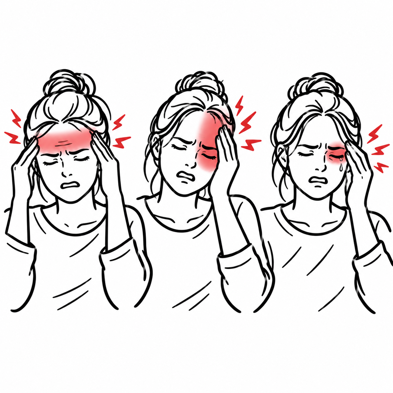
- Migraine: Recurrent attacks lasting 4–72 hours, typically unilateral, pulsating, moderate to severe, aggravated by routine activity, and associated with nausea and/or photophobia and phonophobia.
- Tension-type headache: Bilateral, pressing or tightening, mild to moderate pain that is not aggravated by routine activity and is usually not associated with prominent nausea.
- Cluster headache: Excruciating strictly unilateral orbital, supraorbital, or temporal pain lasting 15–180 minutes, accompanied by ipsilateral autonomic features and marked restlessness.
- Medication-overuse headache: Headache occurring on at least 15 days per month in a patient regularly overusing acute headache medication for more than three months.
A familiar migraine phenotype does not automatically exclude secondary pathology. A first, sudden, severe, progressive, or substantially changed headache requires reassessment for an underlying cause.
APPROACH
Step 1: Identify Emergency Red Flags
Use a systematic red-flag screen such as SNOOP10:
- Systemic illness or symptoms: Fever, weight loss, night sweats, rash, malignancy, immunosuppression, or pregnancy.
- Neurological abnormality: Focal deficit, confusion, reduced consciousness, seizure, ataxia, or papilloedema.
- Onset sudden: Thunderclap headache reaching maximum intensity within one minute.
- Older age at onset: New headache after approximately 50 years of age.
- Pattern change or progression: Increasing frequency or severity, new phenotype, persistent daily headache, or failure of usual treatment.
- Precipitated by exertion or Valsalva: Coughing, sneezing, exercise, or sexual activity.
- Positional headache: Markedly worse upright or supine.
- Pregnancy or puerperium: Increased risk of pre-eclampsia, cerebral venous sinus thrombosis, posterior reversible encephalopathy syndrome, and pituitary apoplexy.
- Painful eye with autonomic or visual disturbance: Consider acute angle-closure glaucoma or ocular inflammation.
- Post-traumatic onset or anticoagulation: Consider intracranial haemorrhage.
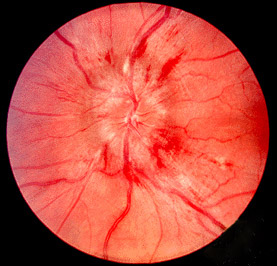
- Papilloedema or pulsatile tinnitus: Suggests raised intracranial pressure.
- Immunocompromise or cancer: Consider opportunistic infection or intracranial malignancy.
Step 2: Recognise Time-Critical Presentations
- Thunderclap headache: Suspect subarachnoid haemorrhage until excluded
- Headache with fever, neck stiffness, photophobia, rash, or altered mental state: Suspect meningitis or encephalitis.
- Headache with focal neurological deficit, seizure, or reduced consciousness: Urgently evaluate for stroke, haemorrhage, cerebral venous sinus thrombosis, intracranial mass, infection, or posterior reversible encephalopathy syndrome.
- New headache with papilloedema, vomiting, or sixth-nerve palsy: Suspect raised intracranial pressure.
- New headache in pregnancy or the puerperium: Check blood pressure and urine protein and evaluate urgently for pre-eclampsia and cerebrovascular causes.
- New headache after age 50 with scalp tenderness, jaw claudication, constitutional symptoms, or visual disturbance: Suspect giant cell arteritis.
- Painful red eye, blurred vision, halos, nausea, or a fixed mid-dilated pupil: Suspect acute angle-closure glaucoma.
- Sudden headache with neck pain, partial Horner syndrome, pulsatile tinnitus, or posterior-circulation symptoms: Suspect carotid or vertebral artery dissection.
Step 3: Define the Headache Phenotype
- Onset: Sudden or gradual; first-ever or recurrent.
- Timing: Duration, frequency, time to peak intensity, morning or nocturnal pattern, and continuous versus episodic.
- Location: Unilateral, bilateral, orbital, occipital, facial, or diffuse.
- Character: Pulsating, pressure-like, stabbing, electric-shock-like, or explosive.
- Severity and disability: Interference with routine activity, work, sleep, or mobility.
- Associated features: Nausea, vomiting, photophobia, phonophobia, aura, autonomic symptoms, fever, neurological symptoms, or visual loss.
- Triggers: Menstruation, sleep deprivation, dehydration, missed meals, alcohol, exertion, coughing, posture, or medication use.
- Medication exposure: Document the number of days per month each acute medication is used.
- Family history: Migraine and other primary headache disorders frequently cluster in families.
Step 4: Perform a Focused Examination
- Record temperature, blood pressure, pulse, oxygen saturation, and level of consciousness.
- Perform a complete neurological examination, including cranial nerves, visual fields, power, sensation, coordination, gait, and reflexes.
- Examine pupils, visual acuity, ocular movements, and the optic discs for papilloedema.
- Assess for meningism and rash.
- Palpate the temporal arteries when giant cell arteritis is possible.
- Measure intraocular pressure when acute angle-closure glaucoma is suspected.
- Examine the head and neck for trauma, sinus disease, cervical tenderness, Horner syndrome, and signs of infection.
A thunderclap headache is defined by speed of onset, not simply severity. Any headache reaching maximum intensity within one minute requires urgent investigation even if the pain later improves or the neurological examination is normal.
DIFFERENTIAL DIAGNOSIS
Primary Headache Disorders
- Migraine without aura: Recurrent attacks lasting 4–72 hours. Typically unilateral and pulsating, moderate to severe, aggravated by routine activity, and associated with nausea and/or photophobia and phonophobia.
- Migraine with aura: Fully reversible visual, sensory, speech/language, motor, brainstem, or retinal symptoms that usually develop gradually. Typical aura evolves over at least five minutes, lasts 5–60 minutes, and is followed by or accompanied by headache. Abrupt negative neurological symptoms should be treated as possible transient ischaemic attack or stroke until assessed.
- Tension-type headache: Bilateral pressing or tightening pain, mild to moderate intensity, without prominent nausea or activity limitation.
- Cluster headache: Severe or excruciating unilateral orbital, supraorbital, or temporal pain lasting 15–180 minutes. Associated with ipsilateral lacrimation, conjunctival injection, nasal congestion, rhinorrhoea, eyelid oedema, ptosis, miosis, facial sweating, and/or marked agitation.
- Other trigeminal autonomic cephalalgias: Paroxysmal hemicrania, hemicrania continua, and short-lasting unilateral neuralgiform headache attacks. Indomethacin responsiveness is characteristic of paroxysmal hemicrania and hemicrania continua.
- Primary stabbing, cough, exertional, or sexual-activity headache: These diagnoses require exclusion of secondary vascular or structural causes at first presentation.
Vascular and Haemorrhagic Causes
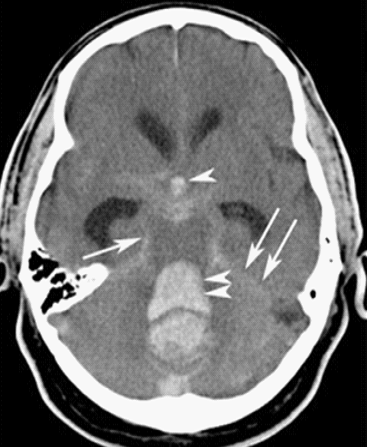
- Subarachnoid haemorrhage: Thunderclap headache, vomiting, neck stiffness, collapse, photophobia, altered consciousness, or focal neurological signs. Exertional onset may occur; neurological examination can initially be normal.
- Cervical artery dissection: Unilateral head, face, or neck pain with partial Horner syndrome, pulsatile tinnitus, cranial neuropathy, or ischaemic neurological symptoms.
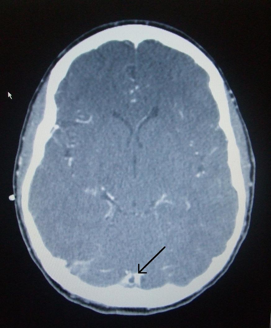
- Cerebral venous sinus thrombosis: Progressive or thunderclap headache, papilloedema, seizure, focal deficit, or encephalopathy. Risk factors include pregnancy, puerperium, oestrogen exposure, malignancy, thrombophilia, dehydration, and infection.
- Reversible cerebral vasoconstriction syndrome: Recurrent thunderclap headaches over days to weeks, often triggered by exertion, sexual activity, vasoactive drugs, or the postpartum state.
- Intracerebral, subdural, or epidural haemorrhage: Consider after trauma, in older adults, with anticoagulation, or with focal neurological deterioration.
- Hypertensive emergency / posterior reversible encephalopathy syndrome: Severe hypertension with headache, visual disturbance, seizure, confusion, or other acute organ dysfunction.
Infectious and Inflammatory Causes
- Meningitis or encephalitis: Fever, headache, neck stiffness, photophobia, rash, altered mental state, seizure, or focal neurological signs. The classic triad may be incomplete.
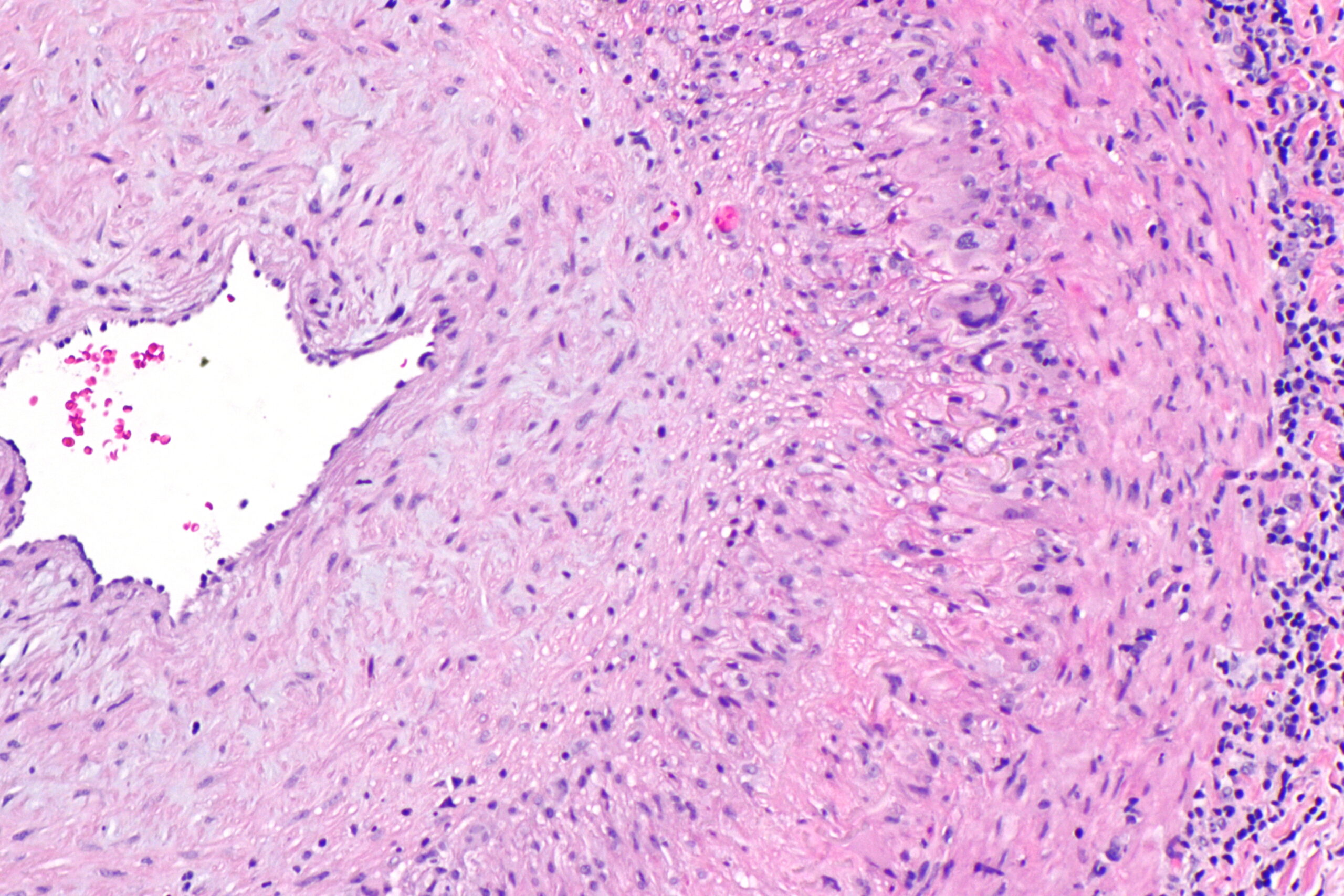
- Giant cell arteritis: New headache after age 50, scalp tenderness, jaw or tongue claudication, constitutional symptoms, polymyalgia rheumatica, temporal-artery abnormality, or transient/permanent visual loss.
- Sinusitis: Purulent nasal discharge, nasal obstruction, facial pain or pressure, reduced smell, and fever. Facial pressure alone without infective sinonasal features is commonly migraine rather than sinusitis.
- Systemic infection: Viral illness, COVID-19, influenza, or other systemic inflammatory conditions may cause headache.
Raised or Reduced Intracranial Pressure
- Intracranial mass lesion: Progressive headache, seizure, focal deficit, personality change, vomiting, papilloedema, or known malignancy.
- Idiopathic intracranial hypertension: Daily headache, transient visual obscurations, pulsatile tinnitus, diplopia from sixth-nerve palsy, and papilloedema. More common in women of reproductive age with obesity but can occur outside this phenotype.
- Spontaneous intracranial hypotension / cerebrospinal fluid leak: Orthostatic headache that worsens upright and improves when supine. May be accompanied by neck pain, nausea, hearing disturbance, or diplopia.
- Hydrocephalus: Headache, vomiting, reduced consciousness, gait disturbance, or papilloedema depending on acuity.
Ocular, Neuropathic and Other Causes
- Acute angle-closure glaucoma: Painful red eye, blurred vision, halos, vomiting, corneal haze, and fixed mid-dilated pupil.
- Trigeminal neuralgia: Brief recurrent unilateral electric-shock-like facial pain triggered by touch, chewing, talking, or tooth brushing.
- Occipital neuralgia: Paroxysmal stabbing pain in the distribution of the greater, lesser, or third occipital nerves.
- Temporomandibular disorder: Jaw pain, clicking, bruxism, and tenderness around the temporomandibular joint.
- Cervicogenic headache: Headache associated with reduced cervical movement and provocation by neck movement or pressure.
- Medication-overuse headache: Headache on at least 15 days per month with regular overuse of acute medications for more than three months. Typically associated with triptan, opioid, combination-analgesic or ergot use on at least 10 days per month, or simple analgesics/NSAIDs on at least 15 days per month.
- Carbon monoxide toxicity: Headache, dizziness, nausea, confusion, and exposure of multiple people in the same environment.
Migraine aura usually develops gradually, spreads from one modality or anatomical area to another, and produces positive symptoms such as flashing lights or tingling. Sudden-onset negative symptoms—loss of vision, sensation, speech, or power—should be managed as possible cerebral ischaemia.
INVESTIGATIONS
First-Line Assessment
- No routine investigation is required for a stable recurrent headache with a typical primary-headache phenotype, a normal neurological examination, and no red flags.
- Headache diary: Record frequency, duration, severity, associated symptoms, triggers, menstruation, disability, and all acute medications used. A diary over at least eight weeks helps confirm the phenotype and identify medication overuse.
- Pregnancy testing: Obtain when pregnancy is possible and would affect the differential diagnosis, imaging, or treatment.
- Targeted blood tests: FBC, electrolytes, renal and liver function, glucose, CRP/ESR, coagulation studies, blood cultures, or toxicology testing according to the suspected cause. In suspected giant cell arteritis, obtain ESR, CRP, FBC, platelets, and liver function tests, but do not delay glucocorticoid therapy.
Neuroimaging
- Non-contrast CT brain: First-line investigation for suspected acute intracranial haemorrhage, thunderclap headache, significant trauma, or acute neurological deterioration. Modern CT performed within six hours of thunderclap onset is highly sensitive for subarachnoid haemorrhage when interpreted under appropriate conditions, but further investigation may still be required according to timing, image quality, clinical risk, and local protocol.
- CT angiography: Evaluate suspected aneurysm, arterial dissection, reversible cerebral vasoconstriction syndrome, or other vascular pathology.
- CT venography or MR venography: Required when cerebral venous sinus thrombosis is suspected.
- MRI brain: Preferred for subacute or progressive headache, posterior-fossa pathology, suspected intracranial mass, inflammatory disease, pituitary pathology, or headache with unexplained focal neurological signs.
- MRI brain with venography: Appropriate when papilloedema or idiopathic intracranial hypertension is suspected.
- MRI brain with gadolinium and spinal imaging: Consider for suspected spontaneous intracranial hypotension or cerebrospinal fluid leak.
Lumbar Puncture
- Consider lumbar puncture after appropriate imaging when investigating suspected meningitis or encephalitis, suspected subarachnoid haemorrhage when CT has not adequately excluded it, idiopathic intracranial hypertension including accurate opening pressure measurement, or inflammatory or malignant meningeal disease.
- Do not perform lumbar puncture before imaging when there is papilloedema, focal neurological deficit, markedly reduced consciousness, new seizure, significant immunocompromise, or another concern for mass effect.
- When bacterial meningitis is suspected, obtain blood cultures and commence antibiotics promptly; do not delay treatment for imaging or lumbar puncture.
Targeted Diagnostic Procedures
- Temporal-artery ultrasound: First-line vascular imaging where expertise is available for suspected giant cell arteritis.
- Temporal-artery biopsy: Consider when imaging is unavailable, non-diagnostic, or diagnostic uncertainty remains.
- Ophthalmological assessment: Urgent slit-lamp examination, fundoscopy, visual assessment, and tonometry for suspected glaucoma or ocular pathology.
- Toxicology testing: Measure carboxyhaemoglobin when carbon monoxide exposure is possible; a normal pulse oximetry reading does not exclude toxicity.
Do not order neuroimaging solely for reassurance in a patient with a stable primary-headache phenotype, normal neurological examination, and no red flags. Conversely, a normal neurological examination does not exclude subarachnoid haemorrhage, cerebral venous sinus thrombosis, giant cell arteritis, or early meningitis.
CRITICAL MANAGEMENT
Suspected Subarachnoid Haemorrhage
- Stabilise airway, breathing, circulation, glucose, temperature, and neurological status.
- Obtain urgent non-contrast CT brain followed by vascular imaging and/or lumbar puncture according to timing and local protocol.
- Discuss immediately with neurosurgery, neurointerventional radiology, or the specialist stroke service.
- Control severe hypertension carefully according to specialist protocol while avoiding hypotension and reduced cerebral perfusion.
- Reverse anticoagulation when intracranial haemorrhage is confirmed or strongly suspected.
- Following diagnosis of aneurysmal subarachnoid haemorrhage, commence specialist-directed nimodipine and arrange early aneurysm securing by endovascular coiling or surgical clipping.
Suspected Meningitis or Encephalitis
- Obtain blood cultures and commence empirical intravenous antibiotics immediately when bacterial meningitis is suspected.
- Use local antimicrobial guidance; common adult empirical therapy includes ceftriaxone or cefotaxime plus vancomycin, with ampicillin/amoxicillin added when Listeria monocytogenes coverage is required.
- Give dexamethasone before or with the first antibiotic dose when bacterial meningitis is suspected, unless contraindicated.
- Add intravenous aciclovir when encephalitis is possible.
- Institute appropriate isolation precautions and notify public health authorities where required.
- Do not delay treatment while awaiting CT imaging or lumbar puncture.
Giant Cell Arteritis
- Commence glucocorticoids immediately when clinical suspicion is high; treatment should not wait for ultrasound or biopsy.
- Without visual symptoms, prednisone/prednisolone 40–60 mg daily is commonly used initially.
- With evolving visual loss, amaurosis fugax, or other cranial ischaemia, arrange same-day ophthalmology and rheumatology assessment and consider intravenous methylprednisolone according to specialist protocol.
- Arrange temporal-artery ultrasound and/or biopsy promptly.
- Assess for large-vessel involvement and manage cardiovascular, bone, gastric, and glucocorticoid-related risks.
Raised Intracranial Pressure
- Elevate the head of the bed, maintain oxygenation and cerebral perfusion, and treat seizures and fever.
- Obtain urgent neuroimaging and neurosurgical or neurological assessment.
- Avoid lumbar puncture until an intracranial mass causing dangerous pressure gradients has been excluded.
- In impending herniation, provide specialist-directed hyperosmolar therapy with hypertonic saline or mannitol while arranging definitive intervention.
Acute Migraine
- Treat early in the attack.
- Offer a triptan combined with an NSAID or paracetamol when not contraindicated.
- Add an antiemetic such as metoclopramide or prochlorperazine when nausea is present or oral absorption is impaired.
- Consider a non-oral triptan or antiemetic when vomiting is prominent.
- Avoid routine opioid therapy because it is less effective, promotes recurrence, and increases medication-overuse headache.
- Consider preventive therapy when attacks are frequent, prolonged, disabling, poorly responsive to acute treatment, or acute medication is being overused.
- Preventive choices may include propranolol, candesartan, topiramate, amitriptyline, sodium valproate in carefully selected patients, onabotulinumtoxinA for chronic migraine, or calcitonin gene-related peptide–targeted therapy according to patient factors and local access criteria.
- Consider pregnancy potential and teratogenic risk before prescribing preventive medication, particularly topiramate and sodium valproate.
Cluster Headache
- Give 100% oxygen at 12–15 L/min through a non-rebreather mask for approximately 15–20 minutes.
- Administer sumatriptan 6 mg subcutaneously or an appropriate intranasal triptan when not contraindicated.
- Oral analgesics and oral triptans are generally too slow for cluster attacks.
- Arrange specialist management for preventive therapy, commonly verapamil with ECG monitoring.
- A short glucocorticoid course or occipital nerve block may be used as transitional therapy while prevention becomes effective.
Medication-Overuse Headache
- Explain that frequent acute medication use perpetuates headache and reduces the effectiveness of preventive therapy.
- Withdraw the overused medication, usually abruptly for triptans and simple analgesics.
- Opioid, barbiturate, or benzodiazepine withdrawal may require gradual reduction and specialist supervision.
- Provide rescue strategies, education about expected temporary worsening, and close follow-up.
- Limit future acute treatment—generally to no more than two days per week—and consider preventive therapy for the underlying headache disorder.
Opioids should not be routine treatment for migraine or other recurrent primary headaches. They provide inferior headache-specific control, increase recurrence and dependence, and are a major driver of medication-overuse headache.
REFERENCES
- Headache Classification Committee of the International Headache Society. The International Classification of Headache Disorders. 3rd ed. Cephalalgia. 2018;38(1):1–211. Available from: https://ichd-3.org/
- National Institute for Health and Care Excellence. Headaches in over 12s: diagnosis and management. Clinical guideline CG150 [Internet]. London: NICE. Available from: https://www.nice.org.uk/guidance/cg150
- American College of Radiology. ACR Appropriateness Criteria®: Headache [Internet]. Reston (VA): American College of Radiology; 2022. Available from: https://acsearch.acr.org/docs/69482/Narrative/
- Godwin SA, Cherkas DS, Panagos PD, et al. Clinical policy: critical issues in the evaluation and management of adult patients presenting to the emergency department with acute headache. Ann Emerg Med. 2019;74(4):e41–e74.
- Hoh BL, Ko NU, Amin-Hanjani S, et al. 2023 guideline for the management of patients with aneurysmal subarachnoid hemorrhage. Stroke. 2023;54(7):e314–e370.
- Robblee J, Starling AJ. SEEDS for success: lifestyle management in migraine. Cleve Clin J Med. 2019;86(11):741–749.
- European Alliance of Associations for Rheumatology. EULAR recommendations for the use of imaging in large-vessel vasculitis in clinical practice: 2023 update. Ann Rheum Dis. 2024;83(6):741–751.
- Ailani J, Burch RC, Robbins MS. The American Headache Society consensus statement: update on integrating new migraine treatments into clinical practice. Headache. 2021;61(7):1021–1039.


Members only discussions coming soon…