

Conjunctival Disease (Conjunctivitis)
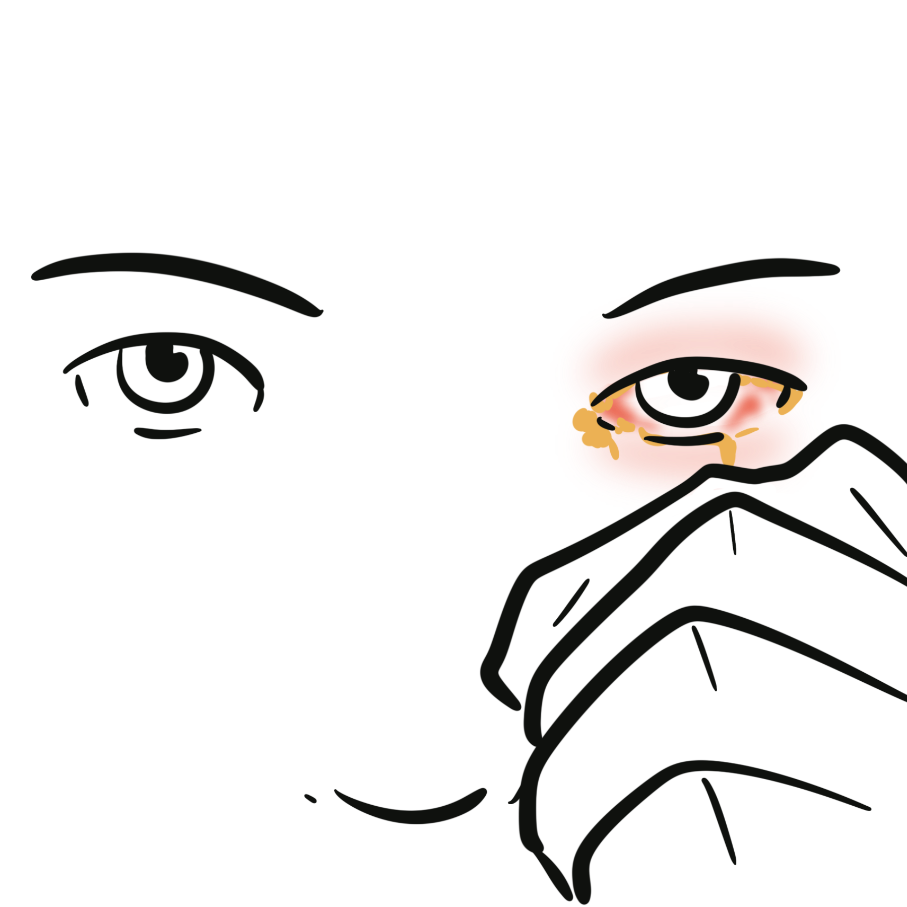
Overview
The conjunctiva is a thin, translucent mucous membrane that lines the posterior surface of the eyelids (palpebral conjunctiva), reflects over the anterior sclera (bulbar conjunctiva), and forms the superior and inferior ciliary junctions (fornices) up to the corneal limbus.
Conjunctival diseases encompass a wide spectrum of infectious, allergic, inflammatory, degenerative, and autoimmune conditions. Among these, conjunctivitis (“pink eye”) is the single most common cause of eye redness in primary care. While most cases are self-limiting, identifying key clinical features and ruling out sight-threatening “red eye” emergencies (such as bacterial keratitis, acute angle-closure glaucoma, and anterior uveitis) is a vital clinical skill.
Viral Conjunctivitis
Overview
Viral conjunctivitis is an acute, highly contagious infection of the conjunctival membrane. It is the single most common cause of infectious conjunctivitis overall, accounting for up to 75% of acute cases in primary care settings.
Definition
Epidemic Keratoconjunctivitis (EKC): A severe, highly contagious subtype caused by specific adenoviral serotypes, featuring severe conjunctival hyperemia, keratitis, and subepithelial corneal infiltrates.
Pharyngoconjunctival Fever (PCF): A clinical syndrome characterized by triad of viral conjunctivitis, fever, and acute pharyngitis.
Follicle: A small (0.5–1.5 mm) pale, avascular lymphoid aggregation on the palpebral conjunctiva with fine blood vessels encircling its base—the classic physical finding in viral conjunctivitis.
Aetiology & Risk Factors
- Aetiology
- Adenovirus: Responsible for 65% to 90% of viral cases (Serotypes 8, 19, 37 cause EKC; Serotypes 3, 7 cause PCF).
- Herpes Simplex Virus (HSV-1): Important unilateral cause, often accompanied by vesicular skin lesions.
- Enterovirus 70 / Coxsackievirus A24: Cause Acute Hemorrhagic Conjunctivitis.
- Risk Factors
- Recent upper respiratory tract infection (URI)
- Direct contact with infected individuals
- Exposure in crowded environments (schools, swimming pools)
- Contaminated ophthalmic equipment.
[Viral Inoculation onto Conjunctival Epithelium]
│
▼
[Epithelial Cell Invasion & Intracellular Replication]
│
▼
[Cell Lysis & Local Inflammatory Cytokine Release]
│
▼
[Subepithelial Lymphocytic Infiltration -> Follicle Formation]
│
▼
[Regional Lymph Node Transport -> Tender Preauricular Lymphadenopathy]
Clinical Manifestations
- Sudden onset of watery/serous discharge, burning, grittiness, and foreign body sensation
- Starts in one eye and frequently spreads to the fellow eye within 24–48 hours.
- Clinical examination
- Lids & Discharge: Eyelid edema with profuse watery (non-purulent) lacrimation.
- Conjunctiva: Diffuse conjunctival injection and lower tarsal follicles.
- Lymph Nodes: Tender preauricular lymphadenopathy (pathognomonic hallmark).
- Pseudomembranes: Coagulated fibrin exudate adhering to the palpebral conjunctiva in severe adenoviral EKC.
- Diagnosis is clinical. Laboratory testing (viral PCR or rapid adenoviral antigen immunoassay) is reserved for severe or atypical cases.
- Differential Diagnosis:
- Bacterial Conjunctivitis: Distinguished by purulent, thick discharge and matted eyelids.
- HSV Keratoconjunctivitis: Identified via fluorescein staining showing classic dendritic corneal ulcers.
- Anterior Uveitis / Acute Glaucoma: Differentiated by deep eye pain, ciliary flush, photophobia, and altered pupils.
- Supportive Care (First-Line): Cold compresses, preservative-free artificial tears, and topical OTC decongestants for symptom relief. Reassure the patient that symptoms typically peak at days 3–5 and take 2–3 weeks to resolve fully.
- Strict Infection Control: High contagiousness; instruct patients on hand hygiene, changing towels/pillowcases, and staying off work or school until discharge resolves.
- HSV Conjunctivitis: Topical Ganciclovir 0.15% gel or oral Acyclovir. Topical corticosteroids are strictly contraindicated.
- Subepithelial Infiltrates (SEIs): Immune-mediated focal corneal opacities that can persist for months after adenoviral infection, causing glare and reduced vision.
- Prognosis: Excellent; self-limiting condition that resolves without permanent visual deficits in uncomplicated cases.
Bacterial Conjunctivitis
Overview
Bacterial conjunctivitis is an acute pyogenic infection of the conjunctival mucosa. While less common than viral cases in adults, it accounts for up to 50% of conjunctivitis cases in pediatric populations.
Definition
Mucopurulent / Purulent Discharge: Thick, yellow-green inflammatory exudate composed of dead neutrophils, bacteria, and epithelial debris; classic feature of bacterial infection.
Papilla: A vascular projection with a central red capillary core surrounded by edema, forming a polygonal red dot on the palpebral conjunctiva.
Hyperacute Bacterial Conjunctivitis: Rapidly progressive, explosive infection (typically N. gonorrhoeae) carrying an immediate threat of corneal perforation.
Aetiology & Risk Factors
- Aetiology
- Staphylococcus aureus (most common cause in adults).
- Streptococcus pneumoniae and Haemophilus influenzae (most common in children).
- Pseudomonas aeruginosa (predominant pathogen in contact lens wearers).
- Neisseria gonorrhoeae (sexually transmitted or neonatally acquired).
- Risk Factors
- Contact lens wear (especially overnight wear)
- Trauma
- Compromised tear production
- Exposure to infected individuals.
Pathophysiology
[Bacterial Adherence to Conjunctival Epithelial Cells]
│
▼
[Exotoxin Release & Disruption of Epithelial Tight Junctions]
│
▼
[Acute Neutrophilic Migration & Vascular Permeability]
│
▼
[Accumulation of Purulent Exudate & Papillary Hypertrophy]
Clinical Manifestations
- Rapid onset of red eye
- Matted eyelids stuck shut upon waking
- Thick purulent discharge
- Mild globe discomfort
- Clinical examination
- Discharge: Continuous purulent or mucopurulent yellow-green discharge that reaccumulates within minutes of wiping.
- Conjunctiva: Marked conjunctival injection, chemosis, and tarsal papillae.
- Preauricular Nodes: Characteristically absent (except in hyperacute Neisseria infection).
Diagnosis
- Uncomplicated cases are diagnosed clinically.
- Microbiological Workup (Gram Stain & Culture): Mandatory in:
- Hyperacute purulent conjunctivitis (Gonococcal)
- Neonatal conjunctivitis (Ophthalmia Neonatorum).
- Contact lens wearers or treatment failures.
- Differential Diagnosis:
- Viral Conjunctivitis: Watery discharge with tender preauricular node.
- Bacterial Keratitis: Presence of a corneal focal white infiltrate or ulcer; requires immediate ophthalmology evaluation.
Treatment
- Empiric First-Line Antibiotics: Topical broad-spectrum drops for 5 to 7 days:
- Standard Cases: Chloramphenicol 0.5% drops or Tobramycin / Polymyxin B.
- Contact Lens Wearers: Must cover Pseudomonas; use topical fluoroquinolones (Ciprofloxacin or Ofloxacin).
- Hyperacute Gonococcal Infection: Medical emergency requiring single-dose parenteral Ceftriaxone 1g IM/IV plus saline eye lavage and oral Azithromycin 1g for chlamydial coverage.
Complications & Prognosis
- Corneal Ulceration & Perforation: Can occur within 24–48 hours in N. gonorrhoeae or Pseudomonas infections.
- Prognosis: Excellent with prompt antibiotic therapy; uncomplicated cases resolve completely within 5 to 7 days.
Allergic Conjunctivitis
Overview
Allergic conjunctivitis is a non-infectious, IgE-mediated inflammatory condition of the conjunctiva. It is extremely common, affecting up to 20% of the population, and is closely linked to other systemic atopic diseases.
Definition
Pruritus (Itching): The hallmark, single most specific symptom of ocular allergy.
Vernal Keratoconjunctivitis (VKC): Severe, bilateral allergic disease in young males featuring giant cobblestone papillae on the upper tarsal conjunctiva.
Horner-Trantas Dots: Transient gelatinous white clumps of eosinophils and epithelial debris at the corneal limbus in severe VKC.
Aetiology & Risk Factors
- Triggering Allergens
- Airborne environmental antigens including grass/tree pollens
- Dust mites
- Mold spores
- Animal dander
- Risk Factors
- Personal or family history of atopic disease (asthma, allergic rhinitis, atopic dermatitis).
Pathophysiology
[Allergen Cross-Linking of IgE on Conjunctival Mast Cells]
│
▼
[Immediate Mast Cell Degranulation (Histamine & Tryptase Release)]
│
▼
[H1 Receptor Activation -> Intense Pruritus & Vasodilation / Chemosis]
│
▼
[Late-Phase Recruitment of Eosinophils & Th2 Cells (4-12 Hours)]
Clinical Manifestations
- Intense itching (pruritus)
- Bilateral lacrimation
- Burning sensation
- Ropy stringy mucoid discharge.
- Clinical examination
- Laterality: Strictly bilateral presentation.
- Conjunctiva: Diffuse injection, marked chemosis (glassy, swollen conjunctiva), and palpebral papillae.
- Corneal Findings (Severe VKC): Shield ulcers or limbal Horner-Trantas dots.
Diagnosis
- Clinical Diagnosis: Based on classic history of itching, bilateral presentation, atopic history, and presence of tarsal papillae.
- Differential Diagnosis
- Infectious Conjunctivitis: Absence of significant itching; presence of purulent discharge (bacterial) or preauricular nodes (viral).
- Dry Eye Syndrome: Burning sensation without true allergic pruritus.
Treatment
- General Measures: Cold compresses, allergen avoidance, and preservative-free artificial tears (flushes out allergens).
- First-Line Pharmacotherapy: Topical dual-action Antihistamine / Mast Cell Stabilizers (e.g., Olopatadine, Ketotifen).
- Refractory / Severe Cases: Short courses of low-penetration topical corticosteroids (e.g., Loteprednol or Fluorometholone) administered exclusively under ophthalmologist supervision to monitor intraocular pressure.
Complications & Prognosis
- Complications
- Corneal Shield Ulcers: Chronic mechanical abrasion from giant papillae in VKC can cause permanent corneal scarring.
- Steroid-Induced Complications: Secondary glaucoma and cataracts from unmonitored topical steroid use.
- Prognosis: Excellent for SAC/PAC; chronic subtypes (VKC/AKC) require long-term management to protect corneal integrity.
Summary Table
| Feature | Viral Conjunctivitis | Bacterial Conjunctivitis | Allergic Conjunctivitis |
| Primary Symptom | Watery eyes, grittiness | Matted eyelids, stick shut | Intense Pruritus (Itching) |
| Discharge | Serous / Watery | Purulent / Mucopurulent | Ropy / Stringy / Mucoid |
| Laterality | Unilateral starting, then bilateral | Often bilateral | Strictly Bilateral |
| Preauricular Node | Present & Tender | Absent (except Neisseria) | Absent |
| Tarsal Findings | Follicles | Papillae | Papillae (or Cobblestone) |
| First-Line Tx | Cold compresses, supportive | Topical Chloramphenicol/Tobramycin | Topical Olopatadine / Ketotifen |
References
- Azari AA, Barney NP. Conjunctivitis: a systematic review of diagnosis and treatment. JAMA. 2013;310(16):1721-1729. doi:10.1001/jama.2013.280318
- Rubenstein JB, Viradiya P. Conjunctivitis: infectious and noninfectious. Dis Mon. 2014;60(6):269-281. doi:10.1016/j.disamonth.2014.03.008
- Leonardi A, Bogacka E, Fauquert JL, et al. Ocular allergy: recognizing and managing hypersensitivity disorders of the eye. Allergy. 2012;67(11):1327-1337. doi:10.1111/all.12009


Members only discussions coming soon…