

Anklylosing Spondylitis (AS)
Overview
Ankylosing spondylitis (AS) is a chronic, progressive inflammatory arthritis that primarily affects the axial skeleton, especially the sacroiliac joints and spine. It is a subtype of the seronegative spondyloarthropathies and is characterised by inflammatory back pain, morning stiffness, and eventual spinal fusion in severe cases. AS often presents in young adults, typically males, and is strongly associated with the HLA-B27 genetic marker. Early diagnosis and treatment are essential to prevent irreversible spinal ankylosis and improve quality of life.
Definition
Spondyloarthritis: A group of inflammatory rheumatic diseases with shared features, including axial and peripheral arthritis, enthesitis, and association with HLA-B27.
Enthesitis: Inflammation at the site of tendon or ligament insertion into bone; a hallmark feature of spondyloarthropathies.
Bamboo spine: Radiographic appearance of the spine in advanced AS, resulting from syndesmophyte formation and vertebral fusion.
HLA-B27: A genetic antigen associated with increased risk of spondyloarthritis, particularly AS; present in up to 90–95% of AS patients.
Anatomy and Physiology
The sacroiliac joints are diarthrodial joints between the sacrum and ilium, supported by strong ligaments. In AS, they are the earliest sites of inflammation. The spinal vertebrae are connected by intervertebral discs and zygapophyseal joints, allowing for flexibility and movement. Inflammation in AS leads to new bone formation and fusion.
The entheses (attachment sites of ligaments/tendons to bone) are prone to inflammation in AS, leading to pain and eventual ossification.
The key affected structures in AS are sacroiliac joints, spine, and entheses. Usually it begins in the SIJ and moves up.
Aetiology and Risk Factors
- Strong genetic association with HLA-B27 antigen
- Positive family history (first-degree relatives)
- Male sex (3:1 male-to-female ratio)
- Age of onset typically <40 years
- Environmental triggers may include preceding GI or genitourinary infections
Pathophysiology
- Chronic inflammation begins at entheses (enthesitis), especially at sacroiliac joints and spine
- Leads to bone marrow oedema, erosions, and repair with new bone formation
- Progressive ossification causes syndesmophytes and ankylosis
- Systemic inflammation may also affect eyes (uveitis), heart (aortitis), and bowel
Inflammation drives both destruction and abnormal new bone formation, leading to pain and rigidity.
Clinical Manifestations
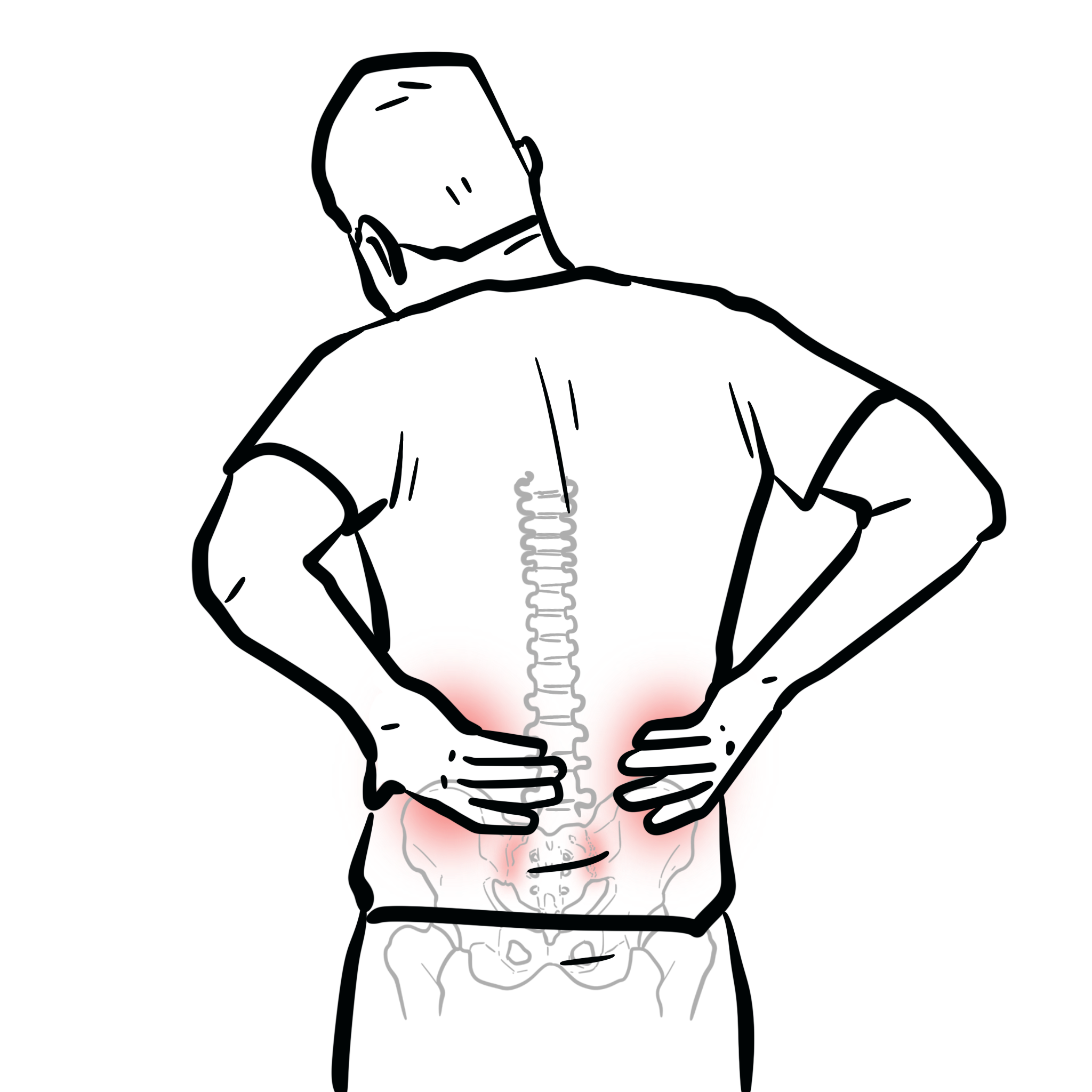
- Inflammatory back pain (insidious onset, age <40, morning stiffness >30 mins, improves with exercise)
- Alternating buttock pain (sacroiliac joint involvement)
- Limited spinal mobility (Schober test ↓, chest expansion ↓)
- Enthesitis (Achilles tendon, plantar fascia)
- Peripheral arthritis (large joints: hips, shoulders, knees)
- Extra-articular features:
- Acute anterior uveitis
- Aortic root dilatation or aortitis
- Pulmonary fibrosis (upper lobe)
- IBD association in 5–10%
Inflammatory back pain improves with exercise but not with rest — opposite of mechanical back pain.
Diagnosis
The Modified New York Criteria (1984) are the most widely used classification criteria for radiographic ankylosing spondylitis. To diagnose ankylosing spondylitis requires radiological criterion PLUS at least one clinical criterion.
- Radiological Criterion
- Bilateral sacroiliitis grade ≥2 OR unilateral sacroiliitis grade ≥3 on plain radiographs (based on the new york criteria).
- Clinical Criteria
- Low back pain and stiffness for ≥3 months, improving with exercise, but not relieved by rest.
- Limitation of lumbar spine motion in both sagittal and frontal planes.
- Reduced chest expansion compared to normal values for age and sex.
| Grade | Description |
| 0 | Normal sacroiliac joints |
| 1 | Suspicious changes: possible sclerosis or minimal joint space irregularity |
| 2 | Minimal abnormality: definite sclerosis, erosions, joint space widening or narrowing |
| 3 | Moderate abnormality: marked sclerosis, erosions, joint space narrowing, partial ankylosis |
| 4 | Severe abnormality: complete ankylosis of the sacroiliac joint |
Investigations
- ESR and CRP: often raised
- HLA-B27: positive in >90% of AS patients
- X-ray: bilateral sacroiliitis (late sign)
- MRI: detects active sacroiliac joint inflammation (bone marrow oedema) — early diagnostic tool
The association of HLA-B27 with AS is well established and occurs in 85–90% of patients. HLA-B27 is seen in 5–15% of the general population, with some variability associated with ethnic background, but only 5% of HLA-B27 positive people develop AS.
| Region | Early Findings | Late Findings |
| Sacroiliac joints | – Pseudo-widening of joint space – Subchondral erosions – Sclerosis (especially iliac side) | – Joint space narrowing – Ankylosis (fusion) – Loss of cortical margins |
| Spine | – Vertebral body squaring – Shiny corner sign (Romanus lesion) – Erosions at vertebral corners | – Marginal syndesmophytes – Bamboo spine (vertebral fusion via syndesmophytes) – Ossification of spinal ligaments (anterior longitudinal ligament) |
Differential diagnoses:
- Mechanical back pain
- Degenerative disc disease
- Psoriatic arthritis
- Reactive arthritis
- DISH (diffuse idiopathic skeletal hyperostosis)

Classification
| Feature | Ankylosing Spondylitis (AS) | Non-Radiographic Axial Spondyloarthritis (nr-axSpA) |
| Definition | Radiographic sacroiliitis (Modified New York Criteria) on X-ray | Normal X-ray; MRI evidence of active sacroiliitis and/or HLA-B27 positivity with SpA features |
| HLA-B27 positivity | ~90% | ~70–90% |
| Gender distribution | Predominantly male | More balanced or female predominant in some cohorts |
| Progression risk | May progress to spinal fusion (bamboo spine) | ~10–30% may progress to radiographic AS over time |
| Treatment approach | NSAIDs, TNF inhibitors, IL-17 inhibitors | Same treatment principles as AS; biologics used if active disease persists despite NSAIDs |
MRI allows diagnosis before structural damage occurs — critical for early treatment.
Treatment
- Exercise and physiotherapy: first-line, essential for preserving function
- NSAIDs (e.g., naproxen, indomethacin): for pain and stiffness
- Biologic therapy (TNF & IL-17 first line)
- TNF inhibitors (e.g., etanercept, adalimumab): if NSAID-refractory or high disease activity
- IL-17 inhibitors (e.g., secukinumab): second-line biologic
- JAK inhibitor
- DMARDs (e.g., sulfasalazine): for peripheral arthritis, not axial disease
- Corticosteroid injections: for enthesitis or peripheral joint flares
- Consider SIJ corticosteroid injection for if ongoing inflammatory pain
Exercise is not just adjunct — it is cornerstone therapy in AS.
Complications and Prognosis
- Progressive spinal fusion (bamboo spine) → loss of mobility, kyphosis
- Osteoporosis and vertebral fractures
- Anterior uveitis (recurs in ~40%)
- Cardiac complications: aortic regurgitation, conduction defects
- Pulmonary fibrosis (rare, upper lobes)
- Increased risk of spinal trauma due to rigidity
Prognosis improved with early biologic therapy and exercise.
References
- Braun J, Sieper J. Ankylosing spondylitis. Lancet. 2007;369(9570):1379–1390.
- Rudwaleit M, van der Heijde D, Landewe R, et al. The Assessment of SpondyloArthritis International Society (ASAS) classification criteria. Ann Rheum Dis. 2009;68(6):777–783.
- van der Heijde D, Ramiro S, Landewe R, et al. 2016 update of the ASAS-EULAR management recommendations for axial spondyloarthritis. Ann Rheum Dis. 2017;76(6):978–991.
- Baraliakos X, Haibel H, Listing J, et al. Continuous long-term anti-TNF therapy does not lead to radiographic spinal damage in AS. Arthritis Rheum. 2005;52(6):1756–1764.


Members only discussions coming soon…