Paget’s Disease of Bone

Default Category Icon

Overview

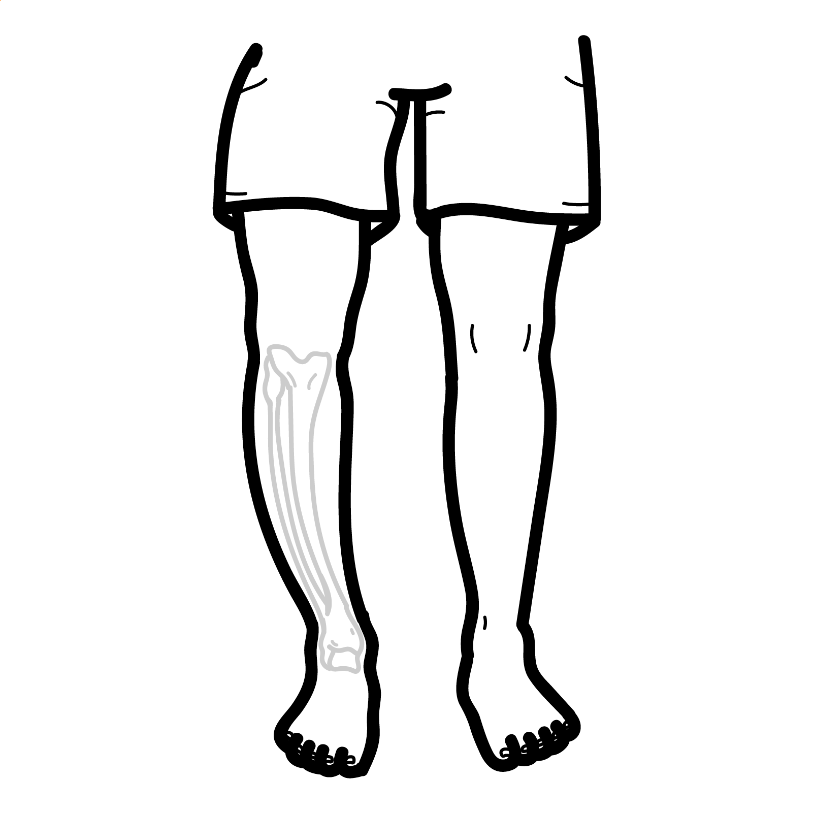
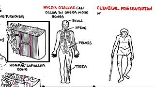
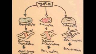
Paget’s disease of bone (PDB) is a chronic skeletal disorder characterised by focal areas of accelerated and disorganised bone remodelling. The disease begins with excessive osteoclastic bone resorption followed by a compensatory increase in osteoblastic bone formation. The result is structurally weak, enlarged, and deformed bone that is prone to fracture and complications such as arthritis or cranial nerve compression. PDB most commonly affects the pelvis, spine, skull, and long bones. It is often asymptomatic and discovered incidentally through elevated alkaline phosphatase or imaging.

Paget’s disease is the second most common metabolic bone disease after osteoporosis. It primarily affects individuals over the age of 50, with a slight male predominance. Prevalence varies by region — it is most common in people of European ancestry, particularly in the UK, New Zealand, and Australia, and is rare in Scandinavia, Asia, and Africa. The prevalence is estimated at 1–3% in adults over 55, though rates appear to be declining in some regions (1,2).

Definition

Bone remodelling: The physiological process of replacing old bone with new bone, involving both resorption (osteoclasts) and formation (osteoblasts). In PDB, this cycle is dysregulated.
Alkaline phosphatase (ALP): An enzyme produced by osteoblasts during bone formation. Elevated levels in the serum reflect increased bone turnover.
Mosaic pattern of lamellar bone: Histological hallmark of Paget’s disease, showing irregular cement lines due to disorganised bone formation
Paget’s disease of breast: Nothing related to Paget’s disease of the bone. This is a rare type of cancer of the nipple–areola complex and that is often associated with an underlying in situ or invasive carcinoma.

Anatomy & Physiology

  • Normal bone remodelling involves a tightly regulated balance between osteoclastic resorption and osteoblastic formation.
  • PDB disrupts this balance, resulting in enlarged, brittle bones prone to deformity.
  • Most commonly affects axial skeleton: pelvis, lumbar spine, femur, skull, tibia.

Aetiology & Risk Factors

Exact cause unknown — believed to involve genetic predisposition + environmental trigger
Genetic:

  • SQSTM1 gene mutation (increases osteoclast activity)
  • Family history (autosomal dominant inheritance)

Environmental:

  • Possible viral triggers (paramyxoviruses, measles virus, RSV)

Risk factors

  • Age > 50 years
  • Male sex
  • European ancestry

Pathophysiology

  • Initial osteolytic phase: Overactive, large osteoclasts cause excessive bone resorption.
  • Mixed phase: Rapid compensatory bone formation by osteoblasts leads to disorganised woven bone.
  • Sclerotic phase: Burnout stage with dense but poorly structured bone.
  • Abnormal bone is hypervascular, enlarged, and structurally weak.

Although bone is being formed, it is disorganised and weaker — explaining fractures and deformities.

Clinical Manifestations

  • Often asymptomatic (incidental diagnosis via ↑ ALP or imaging)
  • Bone pain (most common symptom) — often dull, deep, and worse at rest
  • Skeletal deformities:
    • Bowing of long bones (e.g. tibia, femur)
    • Enlargement of skull → increased hat size
    • Spinal kyphosis
      • Pathological fractures
      • Neurological complications: Hearing loss (from skull involvement, CN VIII compression)
  • Spinal stenosis or radiculopathy
    • Secondary osteoarthritis (from bone deformity altering joint mechanics

Diagnosis

Biochemistry

  • ↑ Alkaline phosphatase (isolated, with normal liver function)
  • Normal calcium and phosphate
  • ↑ Urinary hydroxyproline (optional, reflects collagen breakdown)

Imaging:

  • X-ray: Cortical thickening, bone expansion, lytic/sclerotic changes, “cotton wool” skull appearance
  • Bone scan (99mTc): To assess disease extent and activity
  • CT/MRI: For complications (e.g., spinal stenosis, fracture)

Typical Imaging Findings by Modality

ModalityFinding
X-rayCortical thickening, bone expansion, mixed lytic-sclerotic lesions
Skull X-ray“Cotton wool” appearance
Bone scanIntense uptake in involved areas
MRI/CT (if done)Structural deformity, fracture, stenosis, nerve compression

 Differential diagnoses:

  • Metastatic bone disease
  • Osteomalacia
  • Fibrous dysplasia
  • Primary bone tumors

Classification

No formal classification system by stage
Disease progression is typically described in three phases:

  1. Osteolytic phase
  2. Mixed phase
  3. Sclerotic (burnt-out) phase

Treatment

  • Indications for treatment
    • Symptomatic disease (pain, deformity, fracture)
    • Asymptomatic but involving weight-bearing bones, spine, skull
    • High ALP or bone scan activity
  • Bisphosphonates
    • First-line: Zoledronic acid (IV single dose) or Risedronate (oral)
    • Reduce bone turnover and relieve pain
  • Other medications
    • Calcitonin: For bisphosphonate-intolerant patients (less effective)
    • Analgesia for pain relief (e.g. paracetamol, NSAIDs)
    • Orthopaedic referral for fractures, deformity, joint replacement

Remember

Monitor ALP levels post-treatment as a marker of response.

Complications and Prognosis

  • Fractures (especially femur, pelvis)
  • Deformities → osteoarthritis
  • Deafness from skull involvement
  • Spinal stenosis or nerve root compression
  • High-output cardiac failure (rare, due to bone hypervascularity)
  • Osteosarcoma (very rare <1%) — aggressive, poor prognosis
  • Osteoarthritis

Prognosis is excellent in most cases with treatment.

 The increased vascularity of bone during the osteolytic and mixed phases can divert blood flow from other tissues, potentially leading to high-output cardiac failure and symptoms such as presyncope.

Remember

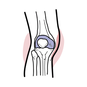
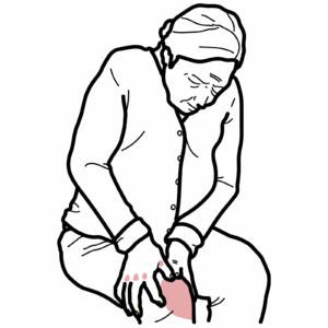
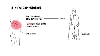
Pain in Paget’s disease should be assessed to distinguish metabolic activity from complications like osteoarthritis or other musculoskeletal conditions.

References

  1. Ralston SH, Langston AL, Reid IR. Pathogenesis and management of Paget’s disease of bone. Lancet. 2008;372(9633):155–63.
  2. Singer FR, Bone HG, Hosking DJ, et al. Paget’s disease of bone: an endocrine society clinical practice guideline. J Clin Endocrinol Metab. 2014;99(12):4408–22.
  3. Chrzanowski W, Cundy T. Paget disease of bone. Endocrinol Metab Clin North Am. 2018;47(4):865–878.
  4. Rea JA, Jameson KA, Compston JE. Effect of bisphosphonates on Paget’s disease: a systematic review. Bone. 2020;134:115243.

Related Content

Discussion

0 Comments
Most Voted
Newest Oldest
Inline Feedbacks
View all comments

Table Of Contents

The AH Community Platform is coming
Get access to member features
Early access launches soon
Armando hasudungan brain logo
Armando Hasudungan
Medical & Biology Tutorials
© 2025 Visualising Medicine. All rights reserved.
Become a member to access note taking
Orangise your medical learning
This is just one of the many AH community member perks
Become a member to access quizzes
Strengthen your medical knowledge
This is just one of the many AH community member perks
0
Would love your thoughts, please comment.x
()
x