Overview
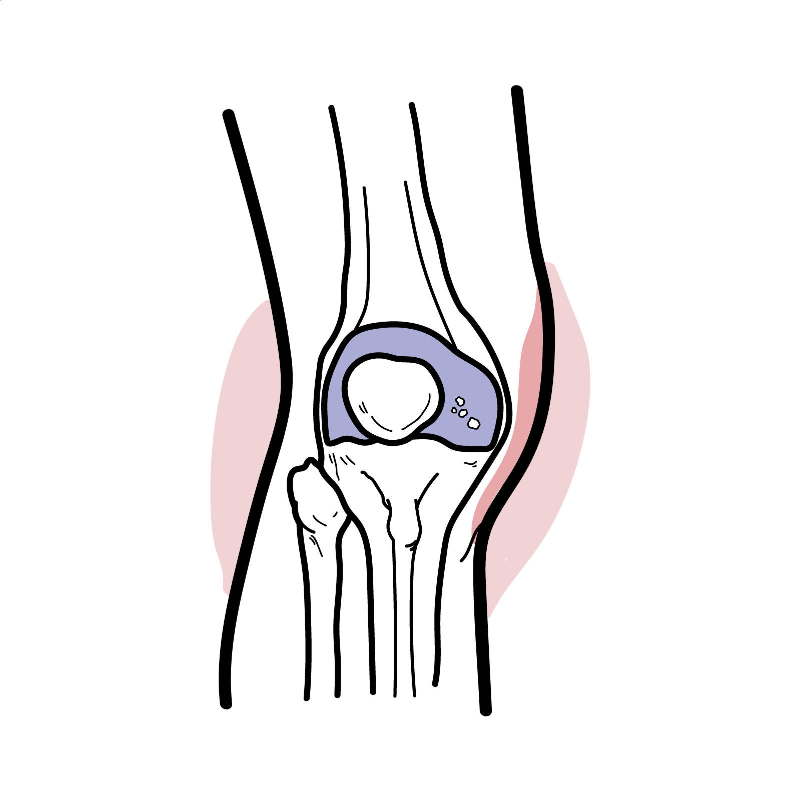
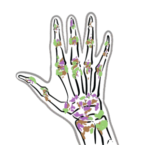
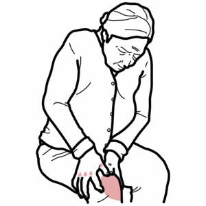
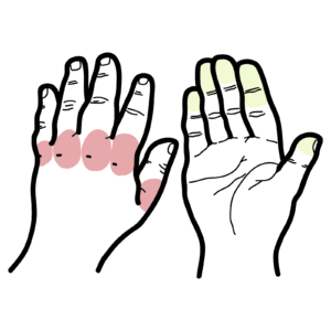
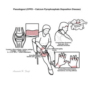
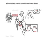
Pseudogout, more accurately known as calcium pyrophosphate deposition disease (CPPD), is a crystal arthropathy characterized by the deposition of calcium pyrophosphate dihydrate crystals in articular cartilage and soft tissues, leading to joint inflammation. It most commonly affects elderly individuals, especially over the age of 60, with a slight female predominance. Clinically, it often mimics gout or septic arthritis, and can present as acute monoarthritis or mimic chronic arthropathies like osteoarthritis or rheumatoid arthritis. The knee is the most frequently involved joint.
Definition
Calcium Pyrophosphate Deposition Disease (CPPD): A crystal arthropathy caused by the deposition of calcium pyrophosphate dihydrate crystals.
Chondrocalcinosis: Radiographic finding of cartilage calcification, often associated with CPPD.
Pseudogout: Acute inflammatory arthritis due to CPPD, mimicking gout clinically.
Crystal arthropathy: Joint disease resulting from deposition of crystals (e.g. monosodium urate in gout, CPPD in pseudogout).
Anatomy and Physiology
Aetiology and Risk Factors
Aetiology
- Abnormal production or impaired breakdown of inorganic pyrophosphate
- Enhanced crystal nucleation within cartilage matrix
- Familial forms linked to ANKH gene mutation
Risk Factors
- Advanced age (strongest risk factor)
- Joint trauma or surgery (e.g. meniscectomy)
- Osteoarthritis
- Hemochromatosis
- Hyperparathyroidism
- Hypomagnesemia
- Hypophosphatasia
- Gitelman syndrome
Remember
Metabolic causes of pseudogout (Young person with pseudogout) can be remembered by “4H”: Hemochromatosis, Hyperparathyroidism, Hypomagnesemia, Hypophosphatasia.
Pathophysiology
- Increased extracellular pyrophosphate → binds with calcium → CPPD crystal formation
- CPPD crystals deposit in fibrocartilage and hyaline cartilage (especially menisci, articular surfaces)
- Crystals are phagocytosed by synoviocytes and neutrophils → release of IL-1, IL-6, TNF-α
- Resultant neutrophilic synovitis mimics acute gout or septic arthritis
- Chronic deposition can damage cartilage → joint degeneration and secondary osteoarthritis
Think
CPPD should be considered in older adults with new-onset monoarthritis or those with “OA in unusual joints” (e.g. radiocarpal, glenohumeral joints).
Clinical Manifestations
- Acute monoarthritis (most common presentation)
- Classically affects the knee (up to 50%), also wrist, shoulder, ankle, elbow
- Sudden onset of pain, swelling, warmth, erythema
- May mimic septic arthritis
- Chronic CPPD arthropathy
- Resembles osteoarthritis but with atypical joint involvement (e.g. MCPs, wrists, shoulders)
- Pseudo-rheumatoid arthritis
- Symmetrical polyarthritis with morning stiffness
- Crowned dens syndrome
- Acute neck pain, fever, restricted movement due to crystal deposition around odontoid process
- Systemic symptoms: usually absent or mild, but fever can occur in acute flares
Triad (Crowned Dens Syndrome) Old person with acute neck pain, Fever, Restriction in cervical spine movement. Always rule out meningitis or fracture.
Diagnosis
- Synovial fluid analysis
- Rhomboid-shaped, positively birefringent CPPD crystals under polarised light microscopy
- Neutrophil-predominant fluid
- Imaging
- X-ray: chondrocalcinosis (calcified cartilage), joint space narrowing, osteophytes
- Ultrasound: hyperechoic linear deposits within cartilage
- Blood tests
- May show mild inflammatory markers elevation (ESR/CRP)
- Screen for metabolic risk factors (calcium, phosphate, magnesium, iron, PTH)
Remember
Chondrocalcinosis on imaging ≠ CPPD — it can be asymptomatic.
Gout vs Pseudogout | ||
Feature | Gout | Pseudogout |
Crystal | Monosodium urate | Calcium pyrophosphate |
Shape | Needle | Rhomboid |
Birefringence | Negative | Positive |
Most common joint | 1st MTP | Knee or wrist |
Age | 30–50 | >60 |
Remember
Always consider hemochromatosis in young patients with pseudogout.
Remember
Always rule out septic arthritis before confirming pseudogout.
Treatment
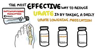
- Acute Attack:
– NSAIDs (first-line if renal function intact)
– Colchicine (low-dose, especially in recurrent attacks)
– Intra-articular corticosteroids if monoarticular
– Systemic corticosteroids (e.g. prednisone 10–30 mg/day) for polyarthritis or NSAID intolerance - Chronic/Recurrent
- Prophylactic colchicine (0.5 mg once/twice daily): no real evidence
- Address underlying metabolic conditions (e.g. treat hypomagnesemia)
- Joint protection and physiotherapy
Remember
No agents currently dissolve CPPD crystals, unlike Allopurinol which reduces uric acid in gout.
Complications and Prognosis
- Recurrent acute attacks
- Progressive joint destruction and secondary OA
- Functional impairment in chronic cases
- Crowned dens syndrome may mimic meningitis or PMR
Prognosis generally good with treatment, though no cure exists.
References
- Zhang W, et al. EULAR recommendations for the management of calcium pyrophosphate deposition. Ann Rheum Dis. 2011;70(4):563–570.
- Rosenthal AK, Ryan LM. Calcium pyrophosphate deposition disease. N Engl J Med. 2016;374(26):2575–2584.
- Abhishek A, Doherty M. Pathophysiology of articular chondrocalcinosis — role of ANKH. Nat Rev Rheumatol. 2011;7(2):96–104.
- McCarty DJ. Calcium pyrophosphate dihydrate crystal deposition disease — pseudogout: update. Rheum Dis Clin North Am. 2006;32(2):401–412.
- Pascual E, et al. Synovial fluid analysis for the diagnosis of joint disease. Best Pract Res Clin Rheumatol. 2005;19(3):371–386.
Discussion