

Rhinosinusitis
Overview
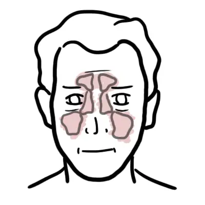
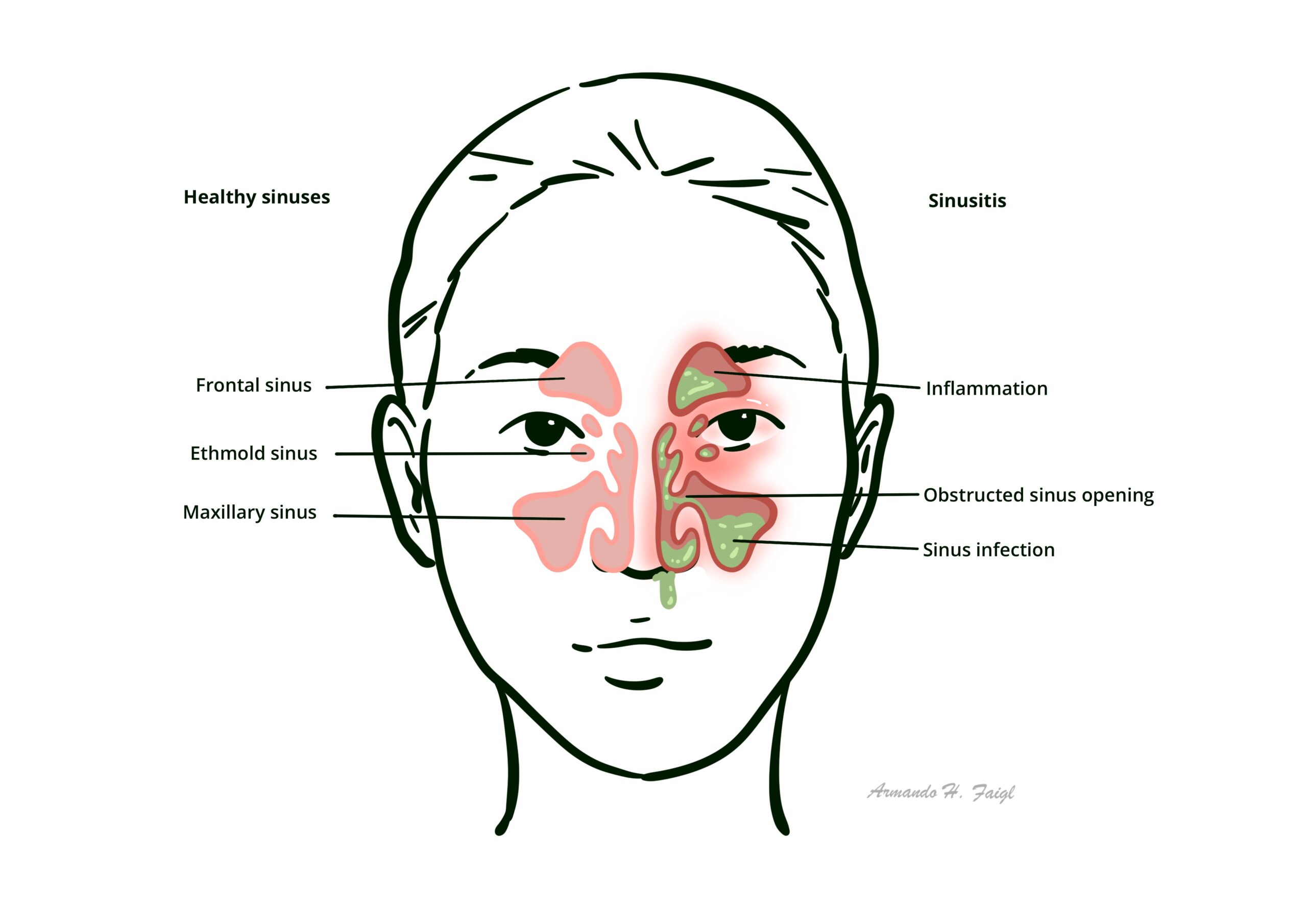
Rhinosinusitis is an inflammatory condition affecting the mucous membranes of both the nasal cavity and the paranasal sinuses (maxillary, ethmoid, frontal, and sphenoid). The clinical term “rhinosinusitis” is preferred over “sinusitis” because inflammation of the paranasal sinuses almost never occurs without concurrent inflammation of the contiguous nasal mucosa. It is one of the most common conditions encountered in outpatient medical practice, presenting across a broad spectrum ranging from acute, self-limiting viral upper respiratory infections to acute bacterial superinfections, invasive fungal emergencies, and chronic inflammatory disease.
The primary diagnostic challenge in managing acute rhinosinusitis is distinguishing between self-limiting Acute Viral Rhinosinusitis (AVRS) and Acute Bacterial Rhinosinusitis (ABRS). Over 90% of acute cases are viral in etiology, making inappropriate antibiotic prescribing a major driver of global antimicrobial resistance.
Management focuses on symptomatic relief for viral cases, reserving targeted antibiotic therapy or surgical evaluation for secondary bacterial infections, refractory chronic disease, or red-flag orbital and intracranial complications resulting from direct anatomical extension.
Definition
Acute Rhinosinusitis (ARS): Sudden onset of sinonasal symptoms lasting less than 4 weeks, with complete resolution of symptoms.
Chronic Rhinosinusitis (CRS): Persistent sinonasal inflammation lasting for 12 weeks or more without complete resolution of symptoms.
Double Sickening (Biphasic Illness): Acute worsening of fever, nasal discharge, or facial pain after initial improvement from a typical 5–6 day viral upper respiratory infection; classic sign of secondary bacterial infection.
Rhinitis Medicamentosa: Rebound nasal congestion caused by prolonged use (> 3–5 days) of topical nasal decongestants (e.g., Oxymetazoline) leading to down-regulation of alpha-adrenergic receptors.
Pott Puffy Tumor: Forehead swelling caused by frontal bone osteomyelitis and subperiosteal abscess formation secondary to complicated frontal sinusitis.
What to notice
Detailed image description
Classification
By Clinical Duration
- Acute Rhinosinusitis (ARS): Symptoms lasting < 4 weeks.
- Subacute Rhinosinusitis: Symptoms lasting 4 to 12 weeks.
- Chronic Rhinosinusitis (CRS): Symptoms lasting >= 12 weeks.
- Recurrent Acute Rhinosinusitis: >= 4 episodes of ARS per year, with complete resolution of symptoms between episodes.
By Aetiology
- Acute Viral Rhinosinusitis (AVRS): Accounts for > 90–98% of acute cases; typically resolves within 7–10 days.
- Acute Bacterial Rhinosinusitis (ABRS): Accounts for only 0.5–2% of acute cases; characterized by persistent (> 10 days), severe, or biphasic symptoms.
- Chronic Rhinosinusitis Subtypes: Classified with or without Nasal Polyposis (CRSwNP vs. CRSsNP) or as Allergic Fungal Rhinosinusitis (AFRS).
Over 90% of acute rhinosinusitis cases are viral. A diagnosis of bacterial rhinosinusitis should not be made unless symptoms persist for > 10 days without improvement or show a classic “double sickening” pattern.
Aetiology & Risk Factors
Viral Pathogens (Most Common)
- Primary Viruses: Rhinovirus, Influenza, Parainfluenza, Coronavirus, Adenovirus, Respiratory Syncytial Virus (RSV).
Bacterial Pathogens
- Acute Bacterial Community Pathogens: Streptococcus pneumoniae (~30–40%), non-typeable Haemophilus influenzae (~30–40%), and Moraxella catarrhalis (~10–15%).
- Chronic / Hospital-Acquired Pathogens: Staphylococcus aureus, Pseudomonas aeruginosa, and anaerobic species (frequently associated with odontogenic maxillary sinusitis).
Invasive Fungal Pathogens
- Mucorales (Rhizopus, Mucor) & Aspergillus: Causes fulminant, lethal invasive fungal rhinosinusitis in immunocompromised hosts (e.g., poorly controlled diabetes mellitus, neutropenia, organ transplant).
Predisposing Risk Factors
- Anatomic Obstruction: Septal deviation, concha bullosa, nasal polyps, or adenoid hypertrophy.
- Allergic & Environmental: Allergic rhinitis, active or passive cigarette smoke exposure, swim/diving barotrauma.
- Systemic Diseases: Cystic Fibrosis (impaired mucociliary clearance), Primary Ciliary Dyskinesia (Kartagener Syndrome), and Granulomatosis with Polyangiitis (GPA).
Black necrotic nasal mucosa in an immunocompromised or diabetic ketoacidosis (DKA) patient is a medical emergency indicating Invasive Mucormycosis.
Pathophysiology
- Ostial Blockade: Inflammation causes swelling of the narrow sinus ostia (particularly the ostiomeatal complex), preventing normal sinus drainage.
- Mucociliary Dysfunction: Viral toxins or allergic responses damage ciliated columnar epithelial cells, slowing mucus transport.
- Stasis & Hypoxia: Secretion retention causes reduced intra-sinus oxygen tension, creating an ideal environment for secondary bacterial proliferation.
Clinical Manifestations
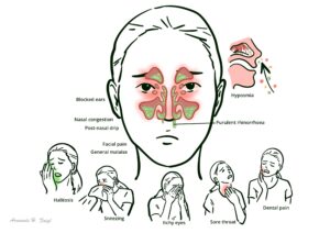
Major Clinical Features
- Facial Pain / Pressure / Fullness
- Nasal Obstruction / Congestion
- Purulent Nasal Discharge
- Hyposmia / Anosmia
Minor Clinical Features
- Headache
- Maxillary Dental Pain
- Fever
- Halitosis
- Fatigue
- Otic fullness
- Chronic cough (especially in pediatric patients due to post-nasal drip).
Clinical Patterns Suggestive of Bacterial Etiology (ABRS)
- Persistent Symptoms: Symptoms lasting >= 10 days without any clinical improvement.
- Severe Onset: High fever (>= 39.0°C / 102.2°F) accompanied by purulent nasal discharge or facial pain for at least 3–4 consecutive days at the start of illness.
- “Double Sickening”: Worsening symptoms (new fever, increased discharge, severe headache) following an initial mild viral phase lasting 5–6 days.
Pain in the upper teeth (maxillary dentition) combined with purulent nasal discharge has a high positive predictive value for Maxillary Rhinosinusitis.
Diagnosis
Clinical Diagnosis
- Uncomplicated ARS is diagnosed purely on clinical grounds based on symptom duration and presentation. Routine imaging is not recommended for uncomplicated acute cases.
Clinical examination
- Anterior Rhinoscopy
- Reveals mucosal hyperemia
- Turbinate hypertrophy
- Turulent discharge in the middle meatus.
- Diagnostic Nasal Endoscopy
- Gold standard outpatient procedure to visualize ostiomeatal complex patency, purulent drainage, mucosal edema, and nasal polyps.
Imaging Studies
- Non-Contrast CT Scan of Paranasal Sinuses
- Gold standard imaging modality.
- Shows sinus opacification, air-fluid levels, mucosal thickening, and bony erosion.
- Indicated for Chronic Rhinosinusitis, treatment failure, or suspected orbital/intracranial complications.
- Plain Sinus Radiographs (X-rays): Outdated and not recommended due to low sensitivity and high false-positive rates.
Plain X-rays of the sinuses are no longer recommended for evaluating rhinosinusitis. Non-contrast CT is the imaging modality of choice when imaging is indicated.
Treatment
Supportive & Symptomatic Care (First-Line for All Cases)
- Analgesia & Antipyretics: Paracetamol (Acetaminophen) or Ibuprofen for pain and fever relief.
- Intranasal Corticosteroids (INCS): Fluticasone, Mometasone, or Budesonide sprays reduce mucosal inflammation and ostial swelling; cornerstone of management for both ABRS and CRS.
- Saline Nasal Irrigation: Isotonic or hypertonic saline sprays/rinses enhance mucociliary clearance and remove encrusted secretions.
- Topical Decongestants: Oxymetazoline or Xylometazoline spray provides rapid relief but MUST be limited to <= 3–5 consecutive days to prevent rhinitis medicamentosa.
Antibiotic Therapy (For Confirmed ABRS)
- First-Line Regimen: Amoxicillin-clavulanate (Augmentin) oral for 5–7 days in adults (or 10–14 days in children).
- Alternative First-Line (Uncomplicated): High-dose Amoxicillin oral in areas with low penicillin-resistant S. pneumoniae.
- Penicillin Allergy (Adults): Doxycycline oral OR a Respiratory Fluoroquinolone (Levofloxacin or Moxifloxacin).
Macrolides (Azithromycin) and Trimethoprim-sulfamethoxazole (TMP-SMX) are no longer recommended first-line due to high bacterial resistance rates (> 30–40%).
Surgical Management
- Functional Endoscopic Sinus Surgery (FESS)
- Indicated for Chronic Rhinosinusitis refractory to medical therapyomplicated acute rhinosinusitis, or anatomical obstructive lesions (e.g., massive polyposis).
- Restores natural sinus ventilation and drainage pathways.
Limit topical decongestant sprays (e.g., Oxymetazoline) to a maximum of 3 to 5 days. Prolonged use causes rebound vasodilation and severe rhinitis medicamentosa.
Complications & Prognosis
Orbital Complications (Chandler Classification)
- Group I – Preseptal Cellulitis
- Group II – Orbital Cellulitis
- Group III – Subperiosteal Abscess
- Group IV – Orbital Abscess
- Group V – Cavernous Sinus Thrombosis
Intracranial Complications
- Epidural / Subdural Abscess & Brain Abscess: Secondary to direct spread through the posterior wall of the frontal or sphenoid sinus.
- Meningitis: Severe headache, photophobia, nuchal rigidity, and altered mental state.
- Pott Puffy Tumor: Frontal bone osteomyelitis with subperiosteal abscess presenting as a doughy swelling over the forehead.
Pain with eye movement, proptosis, or double vision in a patient with sinusitis indicates Orbital Cellulitis or abscess and requires urgent contrast-enhanced CT scan and emergency ENT/Ophthalmology evaluation.
- Acute Viral Rhinosinusitis: Excellent; over 90% resolve within 7–10 days without active medical intervention.
- Acute Bacterial Rhinosinusitis: Cure rates exceed 85–90% with appropriate antibiotic and supportive treatment.
References
- Fokkens WJ, Lund VJ, Hopkins C, Hellings PW, Kern R, Reitsma S, et al. European Position Paper on Rhinosinusitis and Nasal Polyps 2020 (EPOS2020). Rhinology. 2020;58(Suppl S29):1-464. doi:10.4193/Rhin20.600
- Chow AW, Benninger MS, Brook I, Brozek JL, Goldstein EJ, Hicks LA, et al. IDSA clinical practice guideline for acute bacterial rhinosinusitis in children and adults. Clin Infect Dis. 2012;54(8):e72-e112. doi:10.1093/cid/cir1043
- Rosenfeld RM, Piccirillo JF, Chandrasekhar SS, Brook I, Ashok Kumar K, Kramper M, et al. Clinical practice guideline (update): Adult sinusitis. Otolaryngol Head Neck Surg. 2015;152(2 Suppl):S1-S39. doi:10.1177/0194599815572097
Members only discussions coming soon…