Otitis Externa

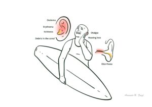
Acute otitis externa (AOE) is a common ear condition seen in the primary health care setting. Also known as ‘swimmer’s ear’ or ‘tropical ear’, it is prevalent in hot humid climates. AOE is an inflammatory condition of the ear canal skin. The causes are infective or non-infective.
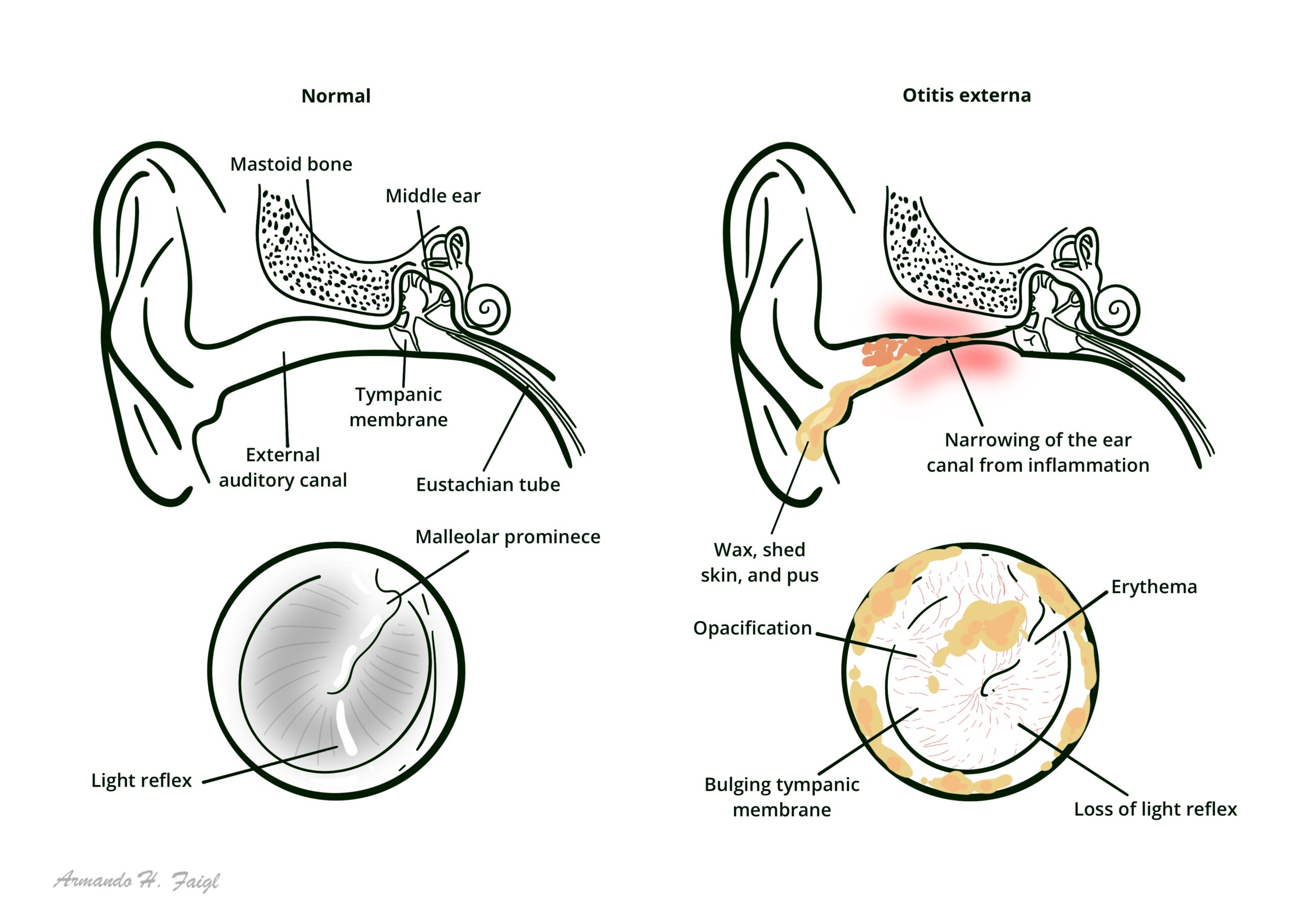
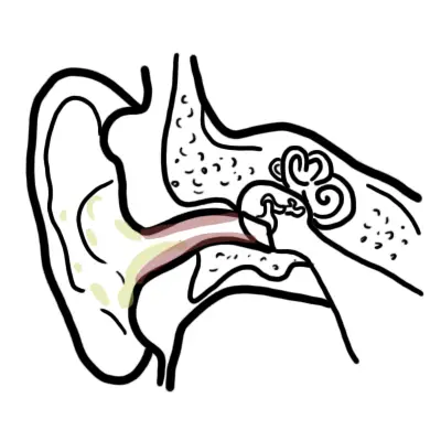
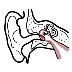
Otitis externa: Inflammation of the outer ear canal, often due to bacterial or fungal/ yeast infection. Otitis externa may occur in isolation, or can be associated with AOM and perforation of the tympanic membrane.
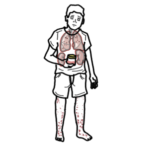
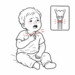
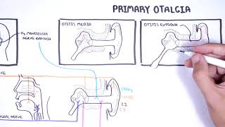
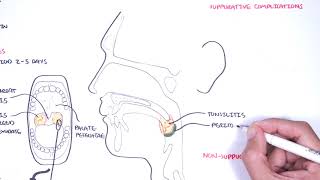
Acute Otitis Media Acute inflammation of the middle ear characterised by otalgia, bulging tympanic membrane and erythema +/- perforation and otorrhoea
Otitis Media with effusion (Glue Ear): Also known as serous otitis media characterised by fluid in the middle with without symptoms or sign of acute inflammation of ear.
AOE usually results from excessive exposure to heat and moisture and is usually very painful. Treatment involves cleaning, keeping area dry and topical antibiotics.
| Nerve | Supply |
| Great auricular (C2, C3) | Cranial surface of auricle and the lower part of the lateral surface of auricle |
| Auriculotemporal branch of the trigeminal nerve | Upper part of the lateral surface and most of the meatal skin |
| Facial nerve | Posterior–inferior portion of the external ear canal and adjacent tympanic membrane |
| Glossopharyngeal nerve tympanic branch | Mucosal surface of tympanic membrane |
| Vagus – auricular branch ‘Arnold’s nerve’ | Supplies small areas of skin on the cranial auricular surface, posterior wall and floor of the meatus and adjoining part of the tympanic membrane |
| Classification of otitis externa | ||
| Infective | Bacterial | Acute diffuse |
| Acute localised furunolosis | ||
| Chronic | ||
| Fungal | Otomycosis | |
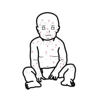
| Viral | Herpes zoster oticus | |
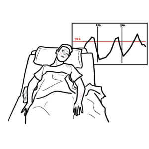
| Necrotising (Bacterial/fungal) | Malignant | |
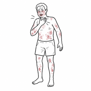
| Non-infective | Dermatitis | Seborrhoeic |
| Atopic-eczematous | ||
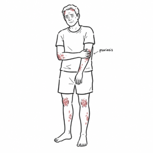
| Psoriasis | ||
AOE is an inflammatory condition of the ear canal skin. The causes are infective or non-infective.
Infective
Non – Infective
Clinical presentation
Ramsay hunt Syndrome Herpetic ulcers in canal; may have facial numbness/paralysis, severe pain, loss of taste (caused by herpes zoster – shingles).
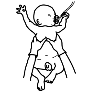
Pneumatic otoscopy
Prevention


Discussion