

Diabetes Mellitus type I
Overview
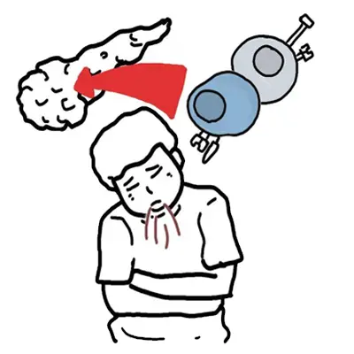
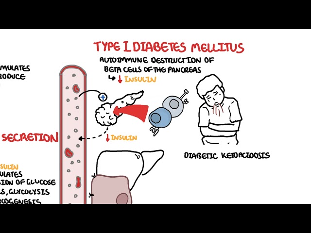
Type 1 Diabetes Mellitus (T1DM) is a chronic, autoimmune metabolic disorder characterized by the progressive, T-cell-mediated destruction of insulin-producing pancreatic beta-cells within the Islets of Langerhans. This severe loss of functional beta-cell mass results in absolute insulin deficiency, leaving affected individuals entirely dependent on exogenous insulin for survival.
Without sufficient insulin, tissues cannot take up circulating glucose, leading to severe intracellular starvation, uninhibited hepatic gluconeogenesis, glycogenolysis, and accelerated lipolysis. The disease primarily presents in children and young adults, though it can manifest at any age. If undiagnosed or improperly managed, T1DM rapidly progresses to life-threatening Diabetic Ketoacidosis (DKA) or long-term microvascular and macrovascular end-organ complications.
Definition
Absolute Insulin Deficiency: Total or near-total absence of endogenous insulin secretion due to complete autoimmune destruction of pancreatic beta-cells.
C-Peptide: A cleavage byproduct produced in a 1:1 molar ratio with endogenous insulin during proinsulin processing; serves as a direct biomarker for residual endogenous beta-cell function.
Honeymoon Phase: A transient clinical remission period shortly after initiating insulin therapy, where surviving beta-cells temporarily recover partial function, reducing exogenous insulin requirements.
Latent Autoimmune Diabetes in Adults (LADA): A slow-onset variant of Type 1 diabetes occurring in adults (> 30 years) that is initially misdiagnosed as Type 2 diabetes but is characterized by islet autoantibodies and progressive insulin dependence.
Islet Autoantibodies: Immune proteins directed against pancreatic beta-cell antigens (e.g., GAD65, IA-2, ZnT8, IAA) that serve as serological markers of autoimmune insulitis.
Classification
Type 1 Diabetes is classified under primary insulin-deficient diabetes syndromes.
1. By Etiological Subtype
- Type 1A (Immune-Mediated): Accounts for ~95% of cases. Characterized by autoimmune destruction of beta-cells with detectable serum autoantibodies and strong HLA associations.
- Type 1B (Idiopathic): Accounts for ~5% of cases. Characterized by absolute insulin deficiency and episodic ketoacidosis without detectable islet autoantibodies or HLA associations (most common in individuals of African or Asian descent).
2. By Age and Onset Pattern
- Juvenile-Onset T1DM: Classic acute/subacute presentation in children and adolescents, often presenting as initial DKA.
- Latent Autoimmune Diabetes in Adults (LADA / “Type 1.5”): Slowly progressive autoimmune beta-cell destruction presenting in adults without acute ketoacidosis at onset.
Serum C-peptide levels are extremely low or undetectable in long-standing Type 1 Diabetes, whereas they are normal or elevated in early Type 2 Diabetes.
Aetiology & Risk Factors
Genetic Susceptibility
- HLA Gene Complex: Accounts for ~50% of familial genetic risk. Strongly associated with specific Human Leukocyte Antigen (HLA) Class II alleles:
- HLA-DR3-DQ2 and HLA-DR4-DQ8 (heterozygosity for DR3/DR4 carries the highest risk of developing T1DM).
- Non-HLA Genes: Polymorphisms in the INS (insulin gene promoter), CTLA4, PTPN22, and IL2RA genes.
Environmental Triggers
- Viral Infections: Molecular mimicry triggered by Enteroviruses (especially Coxsackie B virus), Congenital Rubella, Cytomegalovirus, or Mumps.
- Dietary & Environmental Factors: Early exposure to cow’s milk protein/gluten (debated), low Vitamin D levels, and cold climate/northern latitudes.
- Hygiene Hypothesis: Reduced childhood exposure to infectious agents alters immune regulation, predisposing to autoimmunity.
Carrying both HLA-DR3 and HLA-DR4 alleles dramatically increases the lifetime risk of developing Type 1A Diabetes.
Pathophysiology
- Autoimmune Activation: Antigen-presenting cells display beta-cell peptides to CD4+ T-helper cells, which activate CD8+ cytotoxic T-cells and B-cells.
- Chronic Insulitis: Lymphocytic infiltration of the pancreatic Islets of Langerhans causes chronic inflammation, free radical release, and targeted beta-cell apoptosis.
- Loss of Glycemic Control:
- Early Phase: Destruction of 50–70% of beta-cells leads to loss of first-phase insulin response during glucose challenge.
- Symptomatic Phase: When > 80–90% of beta-cells are destroyed, overt clinical hyperglycemia and metabolic decompensation occur.
- Metabolic Derangement: Absolute lack of insulin combined with excess glucagon accelerates hepatic glycogenolysis, gluconeogenesis, and lipolysis (releasing free fatty acids that liver mitochondria convert into ketoacids: beta-hydroxybutyrate and acetoacetate).
Clinical symptoms of Type 1 Diabetes only appear after more than 80-90% of functional pancreatic beta-cells have been irreversibly destroyed.
Clinical Manifestations
Clinical features reflect acute osmotic diuresis, intracellular starvation, and severe acid-base derangements.
- Classic Osmotic & Catabolic Symptoms
- Polyuria: Excessive urination due to osmotic diuresis occurring when plasma glucose exceeds the renal threshold (~180 mg/dL or 10 mmol/L).
- Polydipsia: Excessive thirst triggered by intracellular dehydration and hyperosmolality.
- Polyphagia: Excessive hunger caused by intracellular glucose deprivation and inability to signal satiety centers.
- Unexplained Weight Loss: Rapid breakdown of adipose tissue and muscle protein to feed gluconeogenesis.
- Fatigue & Lethargy: Inability of muscle cells to utilize glucose for ATP generation.
- Diabetic Ketoacidosis (DKA) Presentation (Presenting Feature in ~30% of Children)
- Gastrointestinal: Nausea, persistent vomiting, diffuse abdominal pain (simulating an acute abdomen).
- Respiratory: Kussmaul breathing (deep, rapid hyperventilation to blow off CO2 and compensate for metabolic acidosis) and “fruity” acetone odor on breath.
- Neurological: Lethargy, confusion, altered mental status, and eventual coma due to hyperosmolality and cerebral edema risk.
Clinical Examination
- Dehydration Signs: Dry mucous membranes, sunken eyes, poor skin turgor, tachycardia, and orthostatic hypotension.
- Recurrent Infections: Vaginal candidiasis (vulvovaginitis) or cutaneous fungal infections due to chronic glucosuria/hyperglycemia.
Abdominal pain in DKA can mimic a surgical acute abdomen (e.g., appendicitis). Always check blood glucose and ketones in any child or young adult presenting with unexplained vomiting and abdominal pain.
Diagnosis
1. Diagnostic Criteria for Diabetes Mellitus
Must meet any one of the following four criteria (in the absence of unequivocal hyperglycemia, requires confirmation by repeat testing):
- Fasting Plasma Glucose (FPG): >= 126 mg/dL (7.0 mmol/L) after at least 8 hours of fasting.
- Random Plasma Glucose: >= 200 mg/dL (11.1 mmol/L) in a patient with classic osmotic symptoms or hyperosmolar crisis.
- 2-Hour Plasma Glucose (75g OGTT): >= 200 mg/dL (11.1 mmol/L).
- Glycated Hemoglobin (HbA1c): >= 6.5% (48 mmol/mol).
2. Autoantibody Screening (To Confirm Subtype 1A)
Presence of one or more autoantibodies confirms the diagnosis of autoimmune diabetes:
- Anti-GAD65: Glutamic Acid Decarboxylase 65 autoantibodies (most sensitive long-term marker).
- Anti-IA-2: Islet Antigen-2 autoantibodies.
- Anti-ZnT8: Zinc Transporter 8 autoantibodies.
- Anti-Insulin Autoantibodies (IAA): Common in young children prior to initiating exogenous insulin.
3. C-Peptide & Metabolic Evaluation
- Serum C-Peptide: Low or undetectable (< 0.2 nmol/L or < 0.6 ng/mL) in the setting of concurrent hyperglycemia.
- Serum/Urine Ketones: Serum beta-hydroxybutyrate > 3.0 mmol/L indicates significant ketosis/DKA.
- Arterial/Venous Blood Gas (ABG/VBG): pH < 7.30 and Serum Bicarbonate < 18 mEq/L with elevated anion gap (> 12 mEq/L) confirms DKA.
Testing for anti-insulin autoantibodies (IAA) must be done before or within 1–2 weeks of starting insulin therapy, as exogenous insulin will trigger anti-insulin antibodies and invalidate the test.
Treatment
Management requires lifelong replacement of basal and prandial insulin alongside continuous glucose monitoring.
1. Insulin Regimens
- Basal-Bolus Multiple Daily Injections (MDI): Standard intensive therapy.
- Basal Insulin (45–50% of total daily dose): Long-acting or ultra-long-acting insulin (Glargine, Detemir, or Degludec) injected once or twice daily to suppress hepatic glucose production.
- Prandial/Bolus Insulin (50–55% of total daily dose): Rapid-acting insulin (Lispro, Aspart, or Glulisine) administered 10–15 minutes before meals, calculated based on meal carbohydrate content and current blood glucose.
- Continuous Subcutaneous Insulin Infusion (CSII / Insulin Pump): Programmed delivery of rapid-acting insulin continuously via a subcutaneous catheter, coupled with Continuous Glucose Monitoring (CGM) in automated hybrid closed-loop (“artificial pancreas”) systems.
Emergency Management of Diabetic Ketoacidosis (DKA)
- Fluid Resuscitation: First priority is 0.9% Normal Saline IV (10–20 mL/kg in the first hour) to restore intravascular volume.
- Potassium Repletion: Check Potassium BEFORE starting insulin! If K < 3.3 mmol/L, hold insulin and replace potassium until K > 3.3 mmol/L. If K is 3.3–5.3 mmol/L, add IV potassium to fluids while running insulin.
- IV Insulin Infusion: Continuous IV Regular Insulin drip at 0.1 units/kg/hour (do NOT give an initial insulin bolus in pediatric patients due to risk of cerebral edema).
- Glucose Addition: Add 5% or 10% Dextrose to IV fluids once plasma glucose drops below 200–250 mg/dL (11.1–13.9 mmol/L) while continuing insulin to clear ketoacidosis.
In DKA management, never give IV insulin if potassium is < 3.3 mmol/L. Insulin shifts potassium into cells and will cause fatal cardiac arrhythmias or respiratory arrest from hypokalemia.
Complications & Prognosis
Complications
- Acute Complications
- Diabetic Ketoacidosis (DKA): Metabolic acidosis, severe dehydration, and cerebral edema (main cause of mortality in children during DKA treatment).
- Severe Hypoglycemia: Glucose < 54 mg/dL (3.0 mmol/L); presents with neuroglycopenic symptoms (confusion, seizures, coma) and autonomic symptoms (sweating, tremors, tachycardia).
- Chronic Microvascular Complications
- Diabetic Retinopathy: Non-proliferative (microaneurysms, cotton wool spots) progressing to Proliferative Retinopathy (neovascularization driven by VEGF) and macular edema.
- Diabetic Nephropathy: Persistent microalbuminuria (30–300 mg/24h) progressing to macroalbuminuria, nodular glomerulosclerosis (Kimmelstiel-Wilson lesions), and End-Stage Renal Disease.
- Diabetic Neuropathy: Distal symmetric “glove-and-stocking” sensorimotor polyneuropathy and autonomic neuropathy (gastroparesis, neurogenic bladder, erectile dysfunction, silent myocardial infarction).
- Chronic Macrovascular Complications
- Accelerated atherosclerosis leading to Coronary Artery Disease (CAD), Ischemic Stroke, and Peripheral Artery Disease (PAD / diabetic foot ulcers/amputations).
Nodular glomerulosclerosis with Kimmelstiel-Wilson lesions on renal biopsy is pathognomonic for advanced Diabetic Nephropathy.
- Glycemic Targets: Target HbA1c for most patients is < 7.0% (53 mmol/mol), with a Continuous Glucose Monitor (CGM) Time-in-Range (TIR 70–180 mg/dL) > 70%.
- Mortality: Life expectancy is reduced by approximately 10–12 years compared to the general population, with cardiovascular disease being the leading cause of death in adults, and DKA being the leading cause in children.
References
- DiMeglio LA, Evans-Molina C, Oram RA. Type 1 diabetes. Lancet. 2018;391(10138):2449-2462. doi:10.1016/S0140-6736(18)31320-5
- American Diabetes Association Professional Practice Committee. 2. Classification and Diagnosis of Diabetes: Standards of Care in Diabetes-2024. Diabetes Care. 2024;47(Suppl 1):S20-S42. doi:10.2337/dc24-S002
- ElSayed NA, Aleppo G, Aroda VR, et al. 6. Glycemic Targets: Standards of Care in Diabetes-2023. Diabetes Care. 2023;46(Suppl 1):S97-S110. doi:10.2337/dc23-S006








Members only discussions coming soon…