Overview
Autosomal dominant disease characterised by trinucleotide repeat of CTG. Also known as Dystrophica Myotoncia the disease is causes an increase in muscle tone (Myotonia) with associated degeneration and shrinkage of muscle fibres (Dystrophy).
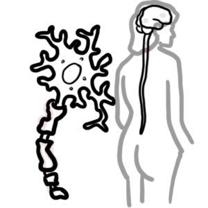
Pathophysiology
- Genetic mutations -> abnormal mRNA
- abnormal mRNA like these do not leave the nucleus
- The abnormal mRNA can cause these hairpin structures which disrupt existing proteins in the nucleus
- The abnormal mRNA causes existing proteins to lose function or gain function:
- Loss of function of the MBNL proteins
- Gain of function of CUGBP proteins
- These proteins are normally involved in splicing, mRNA transport, stability and decay
- These protein dysfunction are thought to be the pathological hallmark of Myotonic Dystrophy resulting in:
- Disruption of alternative splicing
- Disruption mRNA transport
- Disruption of mRNA decay
- Abnormal protein production and function causing complications of Myotonic dystrophy.
Classification
- DM1 is the most common
- Caused by mutation of the DMPK gene of chromosome 19 (Dystrophia Myotonica protein kinase gene)
- The mutation is a trinucleotide expansion of CTG on the DMPK gene
- DM2 less common
- Caused mutation of the CNBP gene in chromosome 3
- The mutation is a tetranucleotide expansion of CCTG on the CNBP gene.
- DM2 is associated with proximal muscle weakness
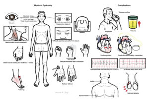
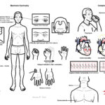
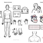
Clinical Manifestation
Myotonia is defines as continued contraction of the muscle after voluntary contraction ceases.

This image series is only available to members
Clinical features
- Delayed relaxation after contraction – hand shake slow to release
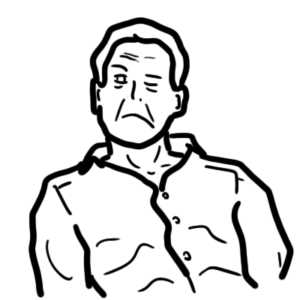
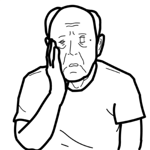
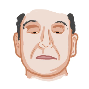
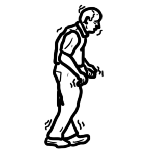
- Myotonic facies (expressionless, mouth open with a lean face)
- Facial muscle weakness
- Sternocledoid muscle weakness
- Difficulty opening eyes after firm closure
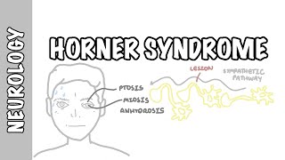
- Ptosis (often bilateral)
- Cataracts
- Balding
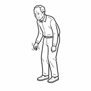
- Distal muscle wasting and weakness – hands
- Reduce reflexes
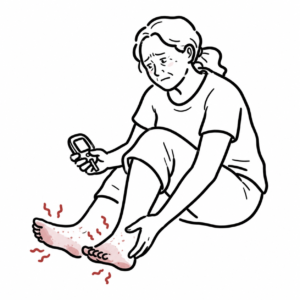
- Foot drop
Myotonic dystrophy type 2 has all the same features of the classic myotonic dystrophy with the difference being in proximal muscle wasting/weakness rather than distal.
Complications
What other disorders involve trinucleotide repeat?
Investigations and Diagnosis
Other investigations are mainly to look for complications of Myotonic Dystrophy
- Ophthalmology review
- Electrocardiogram
- 24 holter monitor
- Echocardiogram
- Sleep study
- Pulmonary function test
- HbA1c
- TFT
Diagnosis
- Genetic testing
- Nerve conduction study – myotonia
Treatment
Treatment again targets complications of Myotonic Dystrophy
- Pacemaker, ICD
- CPAP machine
- Incentive spirometry
- Insulin
- Thyroxine
- Speech pathology for dysphagia
- Physical and occupational therapy is recommended for strengthening weakened muscles, evaluation for orthotics, and durable medical equipment needs.
Complications
Endocrine complications
- Type II diabetes melitius
- Hypogonadism
- Nodular thyroid enlargement
Cardiovascular complications
- Resting ECG changes – everything prolonged (PR, QRS, QT)
- Arrhythmia
- Supraventricular tachycardia (most common)
- VT
- Conduction blocks
- Mitral valve prolapse
- Cardiomyopathy
Gastrointestinal complications
Respiratory complications
- Hypoventilation – poor sleep
- Respiratory failure follow anaesthesia
- Recurrent pneumonia


Discussion