

Liver Mass I Tumour

Author
- Armando Faigl MBBS
- Areej zeeshan
Overview
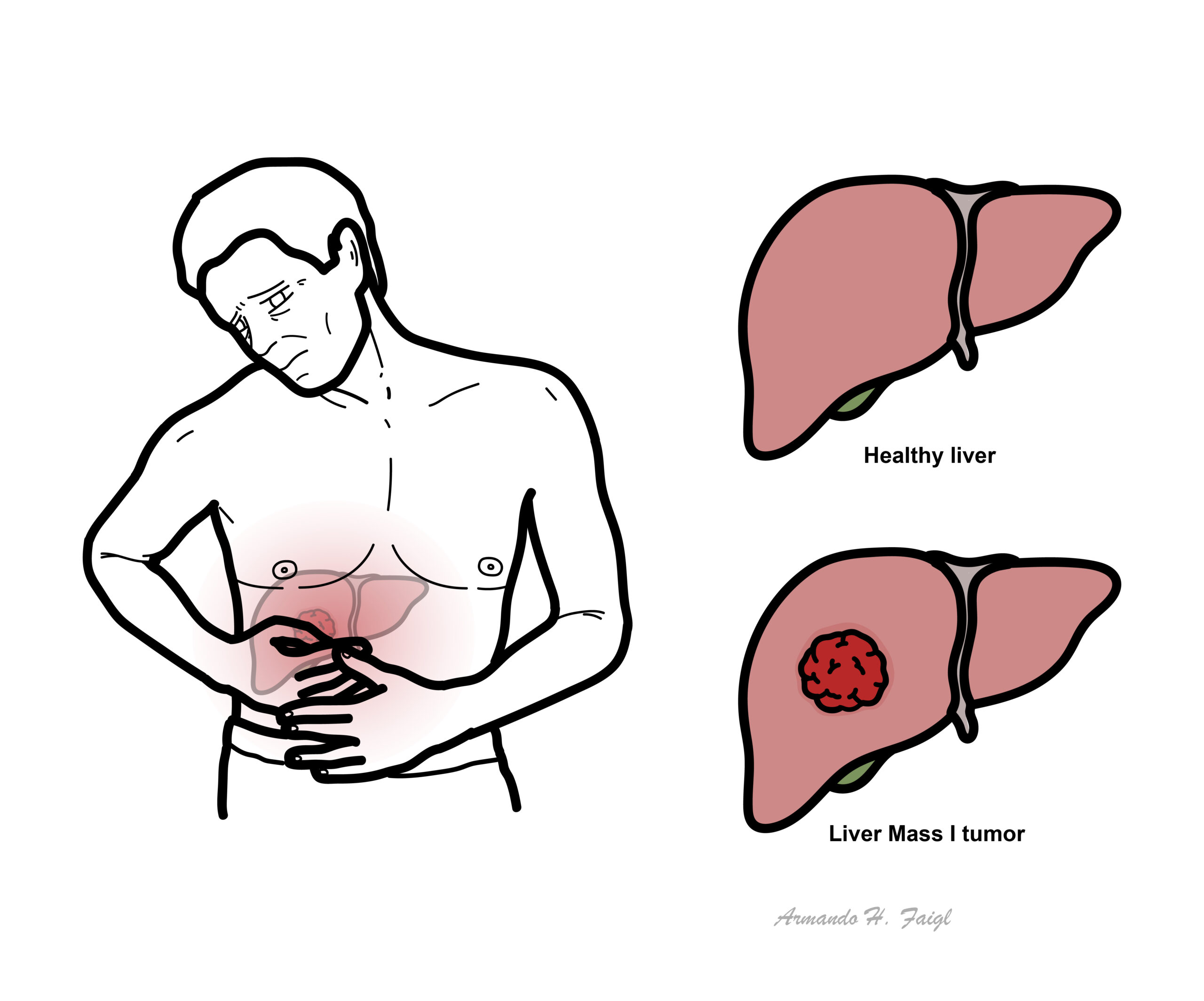
Liver tumors are divided into Benign and Malignant. Most liver tumors are benign, asymptomatic and found incidentally on unrelated imaging. Malignant liver tumours are associated with risk factors (Hep C, Cirrhosis) and present with a spectrum of clinical signs and symptoms such as right upper quadrant pain and liver failure.
Definition
Hepatic Adenoma: benign epithelial tumors of hepatocytes
Hepatic Carcinoma: primary malignant tumors of hepatocytes
Cirrhosis: morphological change in liver parenchyma as a result of chronic liver disease triggering an inflammatory response, characterized by fibrosis and regenerative nodules.
Classification
Benign
- Hepatocellular adenoma
- Focal nodular hyperplasia
- Hepatic hemangiomas
- Regenerative nodule
Malignant
Other
- Liver cysts – discussed separately
- Liver abscess – discussed separately
Clinical Manifestations

Hepatocellular adenoma
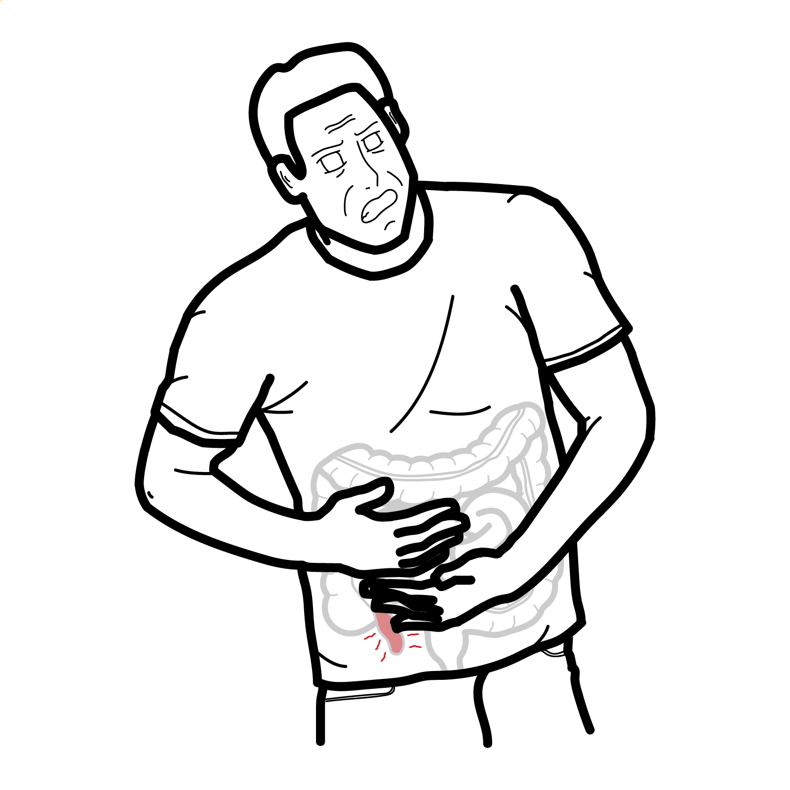
Is the most common liver tumor in young women who have a history of sex hormone use( oral contraceptives, anabolic steroids ) with the most common symptom being right abdominal pain.
Pain is caused by the mass pressing against the liver capsule or as a result of hemorrhagic necrosis as blood supply is compressed.
Hepatic adenomas have tendency to rupture which leads to life threatening intra abdominal bleeding.
It is important to distinguish it from other benign lesions as it has malignant potential. It is usually located in the right lobe of the liver and seen as round, well defined ,well circumscribed mass with yellow discoloration due to abundance of fat.
Treament
Usually as treated by surgical resection (tumors more than 5cm) but in cases where mass is too small sex hormone use is withdrawn.
Focal nodular hyperplasia
- Is the second most common benign liver lesion.
- Presents as a hyperplastic nodule which may be solitary or multiple which is well demarcated and poorly encapsulated. Has a central gray-white scar with radiating fibrous septa giving the center a stellate appearance.
- Occurs as a result of change in blood supply, such as congenital arteriovenous malformations which cause a regenerative hyperplastic response.This leads to hypoperfused parenchyma to produce septa while hyperperfused regions undergo hyperplasia.
- Since they are benign and do not have risk of haemorrhage or rupture they are treated conservatively.
Hepatic hemangiomas
- Are vascular liver lesions which are usually congenital, the most common being Cavernous hemangiomas.
- They are asymptomatic and seen as a sharply defined but unencapsulated mass filled with blood in vascular spaces lined by endothelial cells.
Biopsy is contraindicated as it may cause bleeding.
Regenerative nodules
Develop in response to liver injury and are comprised of a proliferation of hepatocytes and surrounding stroma. They are typically seen in the setting of cirrhosis
Malignant
Most malignant tumours of the liver are metastatic (from pancreas, colon, stomach, breast)
Hepatocellular carcinoma
Are the most common primary liver tumours with a background of chronic liver disease such as cirrhosis and hepatitis. Arises from liver parenchymal cells.
Risk factors
- Hepatitis B and C
- Aflatoxin (produced by aspergillus flavus)
Aspergillus is found in staple food crops of Asia and Africa such as corn ,rice and nuts.
- Alcohol
- Inherited disorders
- Alpha 1 antitrypsin deficiency
- hereditary hemochromatosis
- Wilsons disease
- Obesity
- Diabetes
- NAFLD
- Premalignant precursor lesions
- Hepatic adenoma
- High grade dysplastic nodules
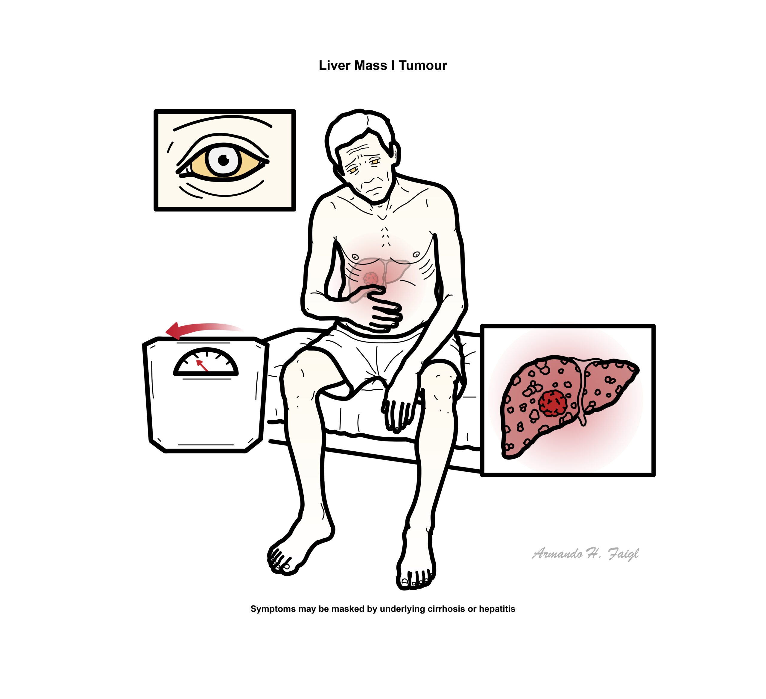
Clinical presentation is variable, it is masked by underlying cirrhosis or hepatitis in cases caused by these diseases. Commonly presents with rapid deterioration in pre-existing cirrhosis. Where aflatoxin may be the risk factor patients may present with abdominal fullness, upper abdominal pain, malaise, weight loss ,and jaundice.
- Benign liver tumors
- Cholangiosarcoma
- Hepatic angiosarcoma
- Metastatic liver from the GIT, lungs or breasts
Liver function tests maybe be elevated ,serum alpha-fetoprotein maybe elevated but is mainly associated with advanced HCC. Ultrasound is performed to confirm mass. Further imaging studies (CT,MRI) are performed to confirm the diagnosis.
Intrahepatic metastases by either vascular invasion or direct extension become more likely once tumors reach 3 cm in size.
- Depends on the stage of the disease
- Earlier stage smaller tumors can be removed surgically or via ablation. In advanced stages where the underlying cause may be cirrhosis liver transplant is usually considered.
- Radiofrequency ablation and chemoembolization is used to control local unresectable tumors.
- Metastasis
- Budd-Chiari syndrome
The 5-year survival of large tumors is dismal, and the majority of patients die within 2 years of diagnosis. Tumors that are resected at less than 3cm may increase survival to 3 years.
Targeted medications include atezolizumab/bevacizumab and sorafenib.
Cholangiocarcinoma
Are the second most common malignancy of the liver which affects the bile ducts. Arises in the extrahepatic biliary tree, but may be intrahepatic. Its classified according to its anatomical location
- Intrahepatic cholangiocarcinoma (10%)
- Extrahepatic cholangiocarcinoma (90%)
Risk factors
- Liver flukes
- Opisthorchis and Clonorchis species
- Chronic inflammatory disease
- Choledocholithiasis
- Hepatolithiasis
- Hepatitis B and C
- NAFLD
All these risk factors cause chronic inflammation and cholestasis, which are thought to promote mutations and epigenetic changes.
Clinical Manifestation
- Intrahepatic cholangiocarcinoma is usually asymptomatic and presents with non-specific symptoms, such as fever, malaise, and dull abdominal pain, in later stages of disease.
- Extrahepatic cholangiocarcinoma present with obstructive jaundice and related signs such as dark urine and pale stools.
Differential diagnosis
- Biliary cyst
- Hepatocellular carcinoma
- Hepatitis
- Cholecystitis
- Pancreatitis
Investigations
- Liver function tests showed increased bilirubin, alk phosphatase, INR, ALT and AST.
- Transabdominal ultrasound shows dilated bile ducts, intrahepatic cholangiocarcinoma usually spans the intrahepatic biliary tree and forms a large mass compressing the liver parenchyma.
- Extrahepatic tumors are generally small and firm nodules in the bile duct wall with ectasia, although the mass may or may not be seen on ultrasound.
- CT is done for staging while MRCP is performed for a definitive diagnosis.
Treatment
- In most cases by the time the tumor is detected surgical resection is not possible thus palliative treatment such as chemotherapy is chosen.
- Stenting of the bile duct can improve quality of life.
- When possible intrahepatic tumors are partially resected while extrahepatic tumors are completely resected but chances of recurrence are high.
Complications and prognosis
- Biliary obstruction
- Biliary cirrhosis
- Pancreatic duct obstruction
- Malignant ascites
Postoperative complications include:
- Liver failure
- Bile leak
- GI bleeding
5 year survival rate is 30% following surgery.


Members only discussions coming soon…