Breast Cancer

“A 35 Year old women presents with a 1cm hard lump in the upper quadrant of her right breast. She first noticed this when she was in the shower three months ago. She is worried that the lump might be malignant”
Breast cancer is the most common cancer in women worldwide. It accounts for about 25% of all female malignancies, with a higher proportion in developed country. Despite the increasing incidence of breast cancer, death rates are falling owing to earlier diagnosis, better surgical and radiotherapy techniques, and improved systemic therapies.
Mastalgia: Breast pain
Gynaecomastia: Enlarged breast tissue in male
Galactorrhea: Discharge of milk from the breast tissue. In Postnatal women is normal, but otherwise galactorrhoea can be caused by pituitary adenoma
The breast is a subcutaneous structure composed of 15 to 20 lobes of mammary gland tissue and fat and typically extends into the axilla as the axillary tail. The ligaments of Cooper (fibrous septa running from the subcutaneous tissue to the fascia of the chest wall) separates the breast lobules. The nipple is surrounded by the pigmented areola. Each lobule drains by its lactiferous duct on to the nipple.
Boundaries and Borders
Blood supply
Physiology – normal breast changes in women
| Risk Factors |
| Gender (99% female) |
| Age |
| Personal history |
| Family history |
| High breast density |
| Nulliparity, First pregnancy >30yo |
| Menarche <12yo, Menopause >55yo |
| Not breastfeeding long-term |
| Radiation exposure |
| Use of Hormone Replacement Therapy or oral contraceptives |
| Genetics (BRCA1 and BRCA2 gene mutation) |
| Alcohol use, obesity, sedentary lifestyle |
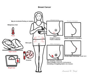
General presentation
nipple discharge is the commonest symptom of cancer after ‘lump’. Beware of neoplasia if discharge is blood-stained, persistent, and from a single duct.
Malignancy Triad: Sudden weight loss (anorexia), fever and night sweats.
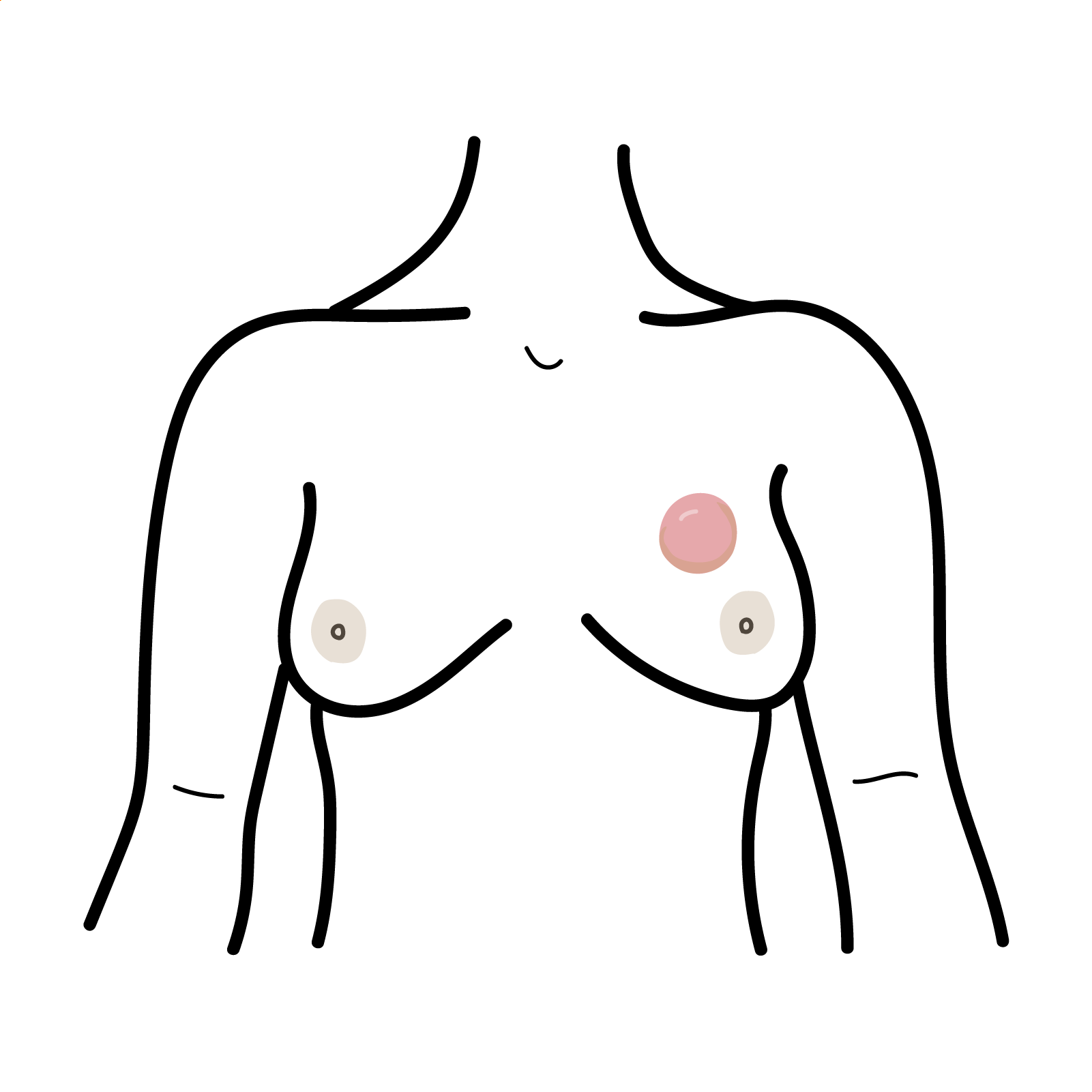
Examination
Breast lump
Nipple changes
Bowen’s disease is a common superficial cancer of the skin. It appears most commonly as a slow-growing, persistent red scaly patch on areas of skin exposed to the sun.
Diagnosis
More info on Breast Lumps
General
Mammographic abnormalities that warrant further investigation include: Radiological masses undetected on clinical examination Microcalcifications Stellate densities Architectural distortion Change from a previous mammogram.
Triple assessment
The predictive value for benign disease when all three components of the triple assessment are benign is 99%.
Staging – TNM classification
Diagnosis of breast cancer is based upon multidisciplinary team work using triple assessment of clinical examination, imaging (mammography and ultrasound), and needle biopsy.
Most carcinomas originate as in situ carcinoma before becoming invasive
Carcinoma in situ Carcinoma in situ (CIS) refers to the period during which normal epithelial cells undergo apparent malignant transformation but do not invade through the basement membrane. There are two forms:
| WHO histological classification of Breast Cancer |
| Epithelial tumoursInvasive Ductal carcinoma (most common)Lobular carcinomaInvasive Tubular carcinomaMedullary carcinomaColloid carcinomaPapillary carcinoma |
| Myoepithelial lesions (includes myoepitheliosis, adenomyoepithelioma, and malignant myoepithelioma) |
| Mesenchymal tumours (includes haemangiopericytoma, angiosarcoma, and leiomyosarcoma) |
| Fibroepithelial tumours (includes fibroadenoma, phyllodes tumour, and low-grade periductal stromal sarcoma) |
| Tumours of the nipple (includes Paget’s disease of the nipple) |
| Malignant lymphoma (includes diffuse large B-cell lymphoma, Burkitt’s lymphoma, and follicular lymphoma) |
| Metastatic tumours |
| Tumours of the male breast (includes invasive and in situ carcinoma) |
Medical Treatment
Aromatase antagonist inhibit the enzyme aromatase which normally converts testosterone to oestrogen. The majority of breast cancer is oestrogen receptor positive, meaning they grow in response to oestrogen, by inhibiting this cancer stops growing. Side effects (menopause): such as hot flashes, night sweats, and vaginal dryness, low libido. Serious side effect: osteoporosis (caution to use in frail women).
Tamoxifen is a nonsteroidal agent that binds to estrogen receptors (ER), inducing a conformational change in the receptor → blocking the effects of oestrogen on the cell. Side effects (menopause): hot flashes, vaginal dryness, low libido, mood swings, and nausea.
Surgical Treatment (usually in conjunction with chemotherapy +/- radiation)
If lymph node involvement is demonstrated following axillary lymph node sampling or sentinel node biopsy, axillary radiotherapy is given to help control axillary nodal metastasis, or an axillary clearance is performed. However, if an axillary clearance is performed as the primary intervention, radiotherapy for positive nodes is unnecessary‐all the diseased nodes have been removed. In addition, after clearance, radiotherapy is associated with a high incidence of lymphoedema of the arm because of the combined surgical and X‐ray damage to lymphatics.
Breast-conserving surgery should be followed by radiation therapy in women with early-stage invasive or locally advanced breast cancer.
Preoperative chemotherapy for locally advanced breast cancer increases the success of breast-conserving surgery.
Medication Complication
Radiotherapy Complication
Chemotherapy Complication
Surgery Complication
| PROGNOSITC FACTORS (other than TNM) | ||
| Biological factors | Favourable | Unfavourable |
| Histological type | Tubular, colloid, papillary | Scrirrhous |
| Grade | Low | High |
| Necrosis | Absent | Present |
| Lymphocytic infiltration | Present | Absent |
| Oestrogen status | Positive | Negative |
| Reactive lymph nodes | Present | Absent |
| Proliferative rate | Low S phase | Aneuploid |
| Chromosomal defects | – | Deletion/alteration 1, 3, 6, 7, 9. Shortening of allele on chromosome 11 |
| Proto-oncogenes | – | c‐erbB/c‐H‐ras |
| Growth Factors (GF) | – | Epidermal GF, Transforming GF, Platelet‐derived GF, Fibroblast GF, Insulin‐like GF |
The early detection methods are:
It is recommended that women aged 50–69 years attend the BreastScreen Australia Program every 2 years for screening mammograms.
Mammograms are performed earlier for women with family history of breast cancer.
<40yo screening is not recommended. However breast awareness is recommended for all ages.
Best Practice


Discussion