

Liver Abscess

Author
- Areej Zeeshan Ahmed
- Dr. Armando Faigl (editor)
Overview
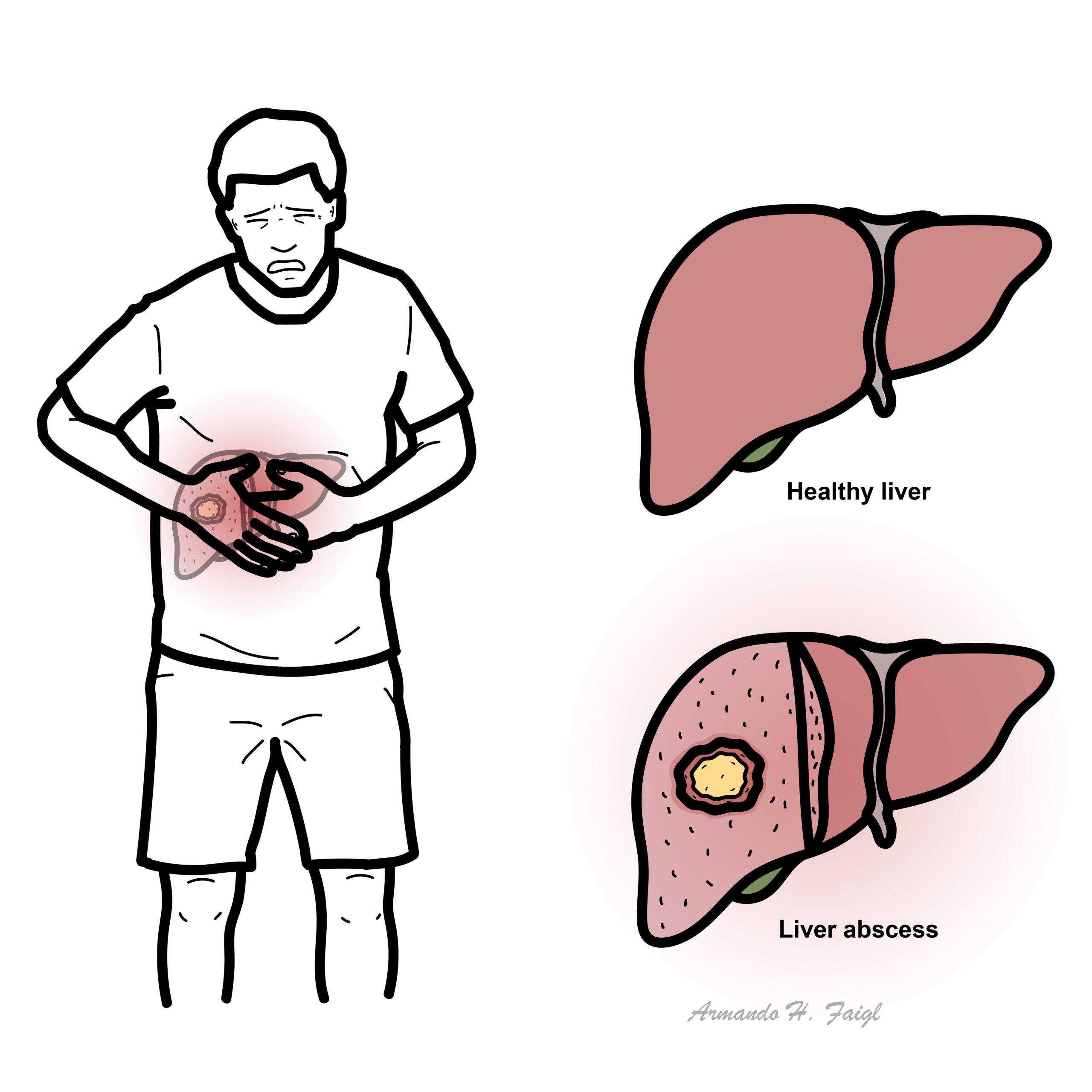
Collection of pus forming a mass in the liver as a result of infection. Mainly divided into two types
- Pyogenic
- Amoebic
Route of infection
- Biliary tract (most common)
- Gallstones
- Cholangitis
- Congenital anomaly causing to bile stasis
- Portal Vein
- Hepatic artery
- Trauma
- Penetrating
- Non-penetrating can cause bile leakage or haemorrhage and necrosis
Since the liver receives its blood circulation from the systemic and portal circulations, it is more susceptible to getting infections and abscesses from the blood.
Pyogenic Liver Abscess
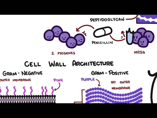
Can be solitary or multiple pus filled masses caused by bacteria and are generally polymicrobial involving gut-derived Gram-negative, gram positive and anaerobic organisms.
- E.coli
- Klebsiella
- Streptococcus
- Staphylococcus
Hemochromatosis is associated with Y. enterocolitica liver abscess. Reason being the bacteria thrives in iron rich environment.
Liver abscess can be one manifestation of the highly virulent klebsiella pneumoniae. Serotypes K1 and K2 is also associated with endopthlamitis.
- Biliary tract disease
- Diabetes mellitus
- Liver surgery
- Immunosuppression
- age over 50
- Malignancy
- Being male
- Proton pump inhibitors
- Biliary tract
- Obstruction of bile flow allows proliferation of bacteria thus increased susceptibility to infection.
- Portal Vein
- Peritonitis allows the bacteria to travel up the liver via the portal vein
- Trauma
- Directly introduces bacteria into the liver parenchyma when penetrating,when non penetrating necrosis or bile leakage create a suitable environment for bacterial growth
In patients who present with cholangitis , but who remain septic despite relief of biliary obstruction, it is vital to exclude liver abscess.
Clinical Manifestation
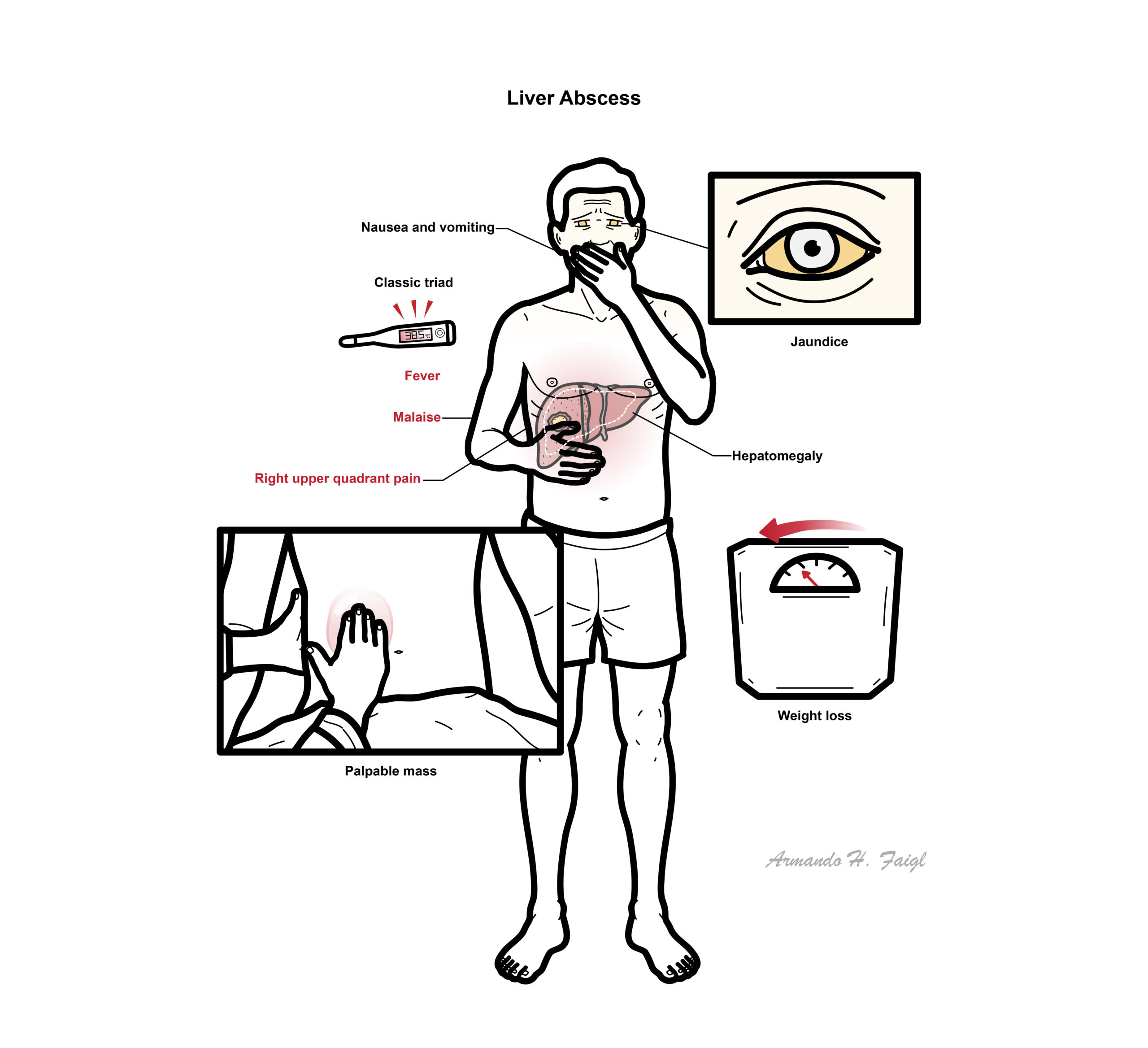
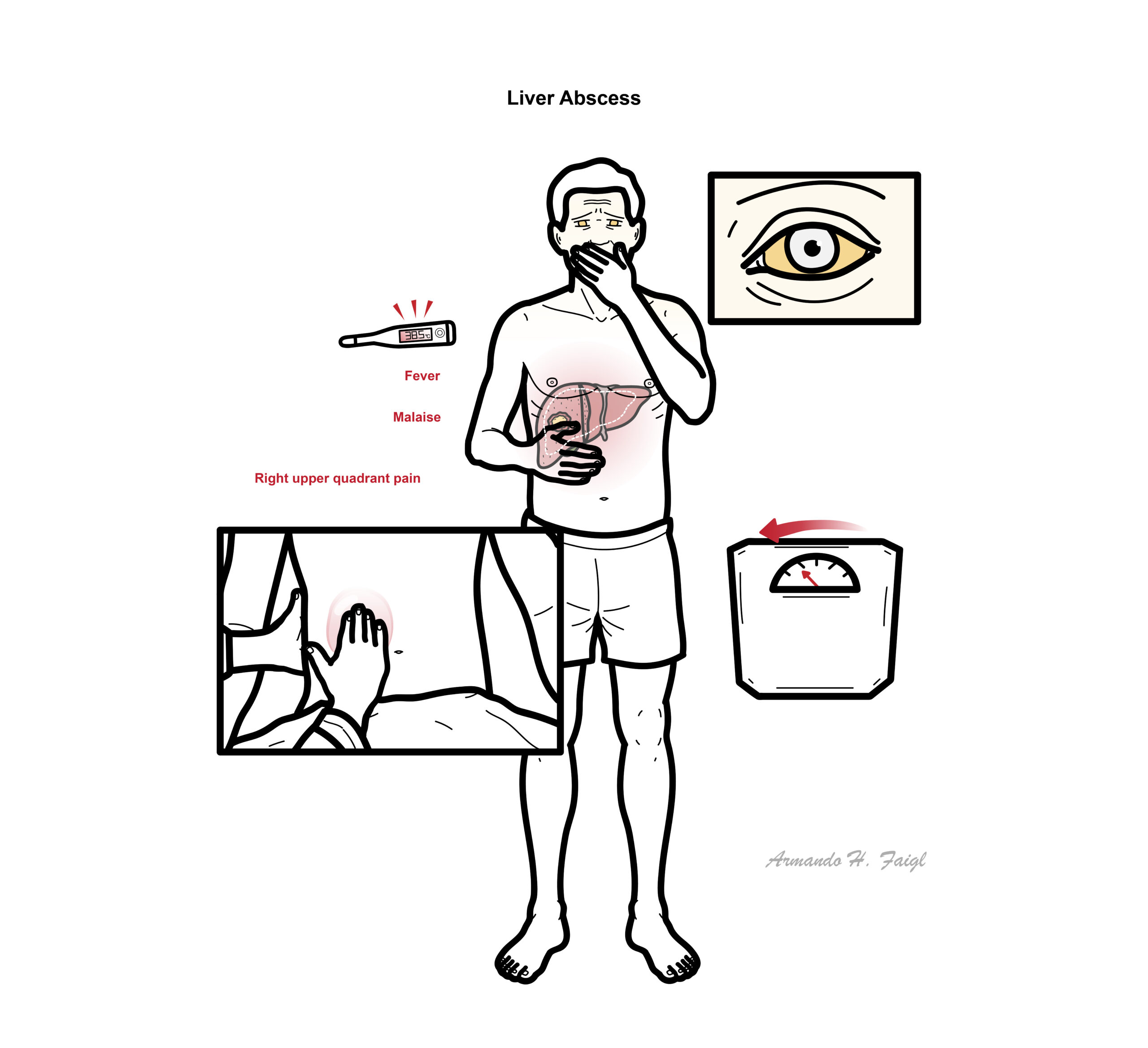
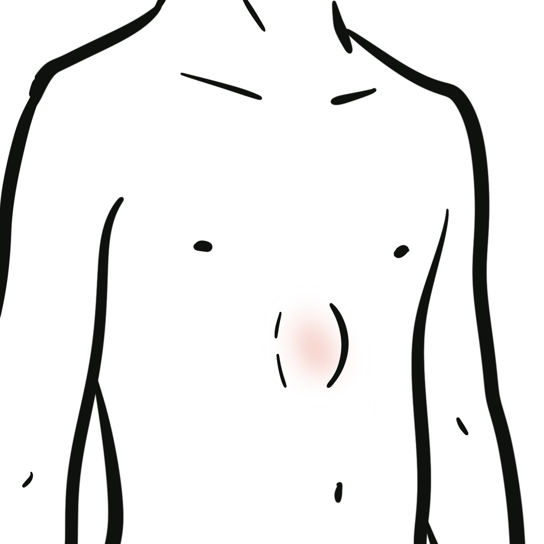
Patients complain of a classic triad of fever (with chills), right upper quadrant pain, malaise
Similar triad to ascending cholangitis – fever, RUQ pain and leukocytosis
Some may present more indolently (> 1 month) with fever of unknown origin (FUO) with non specific symptoms as nausea, vomiting and unexplained weight loss.On GPE jaundice with hepatomegaly and large mass are observed.
Examination may reveal tender hepatomegaly, and occasionally jaundice.
- Amoebic liver abscess
- Benign liver tumor
- Ascending cholangitis
- Liver cyst
- Viral hepatitis
- FBC – neutrophilia and leukocytosis
- LFT
- ↑ESR/CRP.
- Blood cultures – positive in >50% of patients with pyogenic liver abscesses
Elevated serum Alk phosphatase are the most common finding.
Serological test for amebiases should be done in all patients.
Secondly abdominal imaging studies are performed
- Liver ultrasound showing echoic lesion
- CT scan (most sensitive)
- hypodense lesion with “rim enhancement” with contrast
- liver lesions where gas within is highly suggestive of pyogenic abscess
The characteristic presentation is solitary or multiple lesions in the right liver lobe with a edematous surrounding parenchyma.
Abscess aspiration (if blood cultures negative and failure to respond to initial treatment).
Consider colonoscopy to exclude colon cancer in Streptococcus milleri abscess.
Treatment
Percutaneous CT or Ultrasound guided drainage for lesions more than 3-5 cm.
Prolonged Antibiotics (IV and oral) 4-6 weeks
- Antibiotics empirical pending results bacterial vs amoebic – Metronidazole + ceftriaxone or Tazocin
- Once organism identified tailor antibiotics
Surgical drainage
- Large (>5cm), multiple, loculated, or viscous abscesses
- Failure to respond to percutaneous drainage within 7 days
- Underlying disease requiring surgical management
Complications and Prognosis
Complications include:
- Abscess rupture
- Peritonitis if into abdomen
- Empyema if chest
- Retroperitoneal
- Metastatic complications (more common in Klebsiella pneumoniae)
- Sepsis
- Pneumonia
- Pleuropulmonary fistula
Prognosis for treated PLA is good but 100 percent of untreated PLAs succumb to death.
Increased morbidity is associated with increasing age, pyogenic abscess with sepsis, immunosuppression, bacteraemia, multiple comorbidities and abscesses.
Recurrence of a liver abscess is more common in patients with an underlying biliary tract abnormality than in those without biliary disease.
Amoebic Liver Abscess
Infection is caused by anaerobes ,the most common being Entamoeba histolytica, which is transmitted feco-orally.
Patients usually present with fever (no chills), right upper quadrant pain, chest pain and sometimes diarrhoea. However Amebiasis presents with a variety of clinical states
- Asymptomatic cyst passer – do not invade or cause issues
- Diarrhoea or dysentery – moderate disease
- Invasive intestinal amebiasis – dysentery, colitis, toxic megacolon and amebomas
- Invasive extraintestinal amebiasis – liver abscess, peritonitis pleuropulmonary abscess, cutaneous and genital amebic lesions.
The main route of infection for this protozoan is the portal vein.
E histolytica is endemic in Central and South America, Africa, and Asia thus is suspected in individuals who travel there.
- Stool culture can not differential pathogenic to non pathogenic Entamoeba bacteria
- To identify E. histolytica cysts – stool antigen or Stool PCR
- Abscess drainage – fluid culture, antigen and PCR
The abscess is filled with a reddish brown paste like fluid and is a solitary mass in the right lobe of the liver.
Treatment
- Metronidazole
- Following treatment the use of a luminal agent (e.g. paromomycin) is required to eliminate intraluminal cysts.
- Similar guidelines for drainage as of pyogenic abscess.
References
BMJ
AMBOSS
NCBI
Oxford Handbook of Gastroenterology


Members only discussions coming soon…