Hyperparathyroidism

This section will mainly focus on Primary Parathyroidism
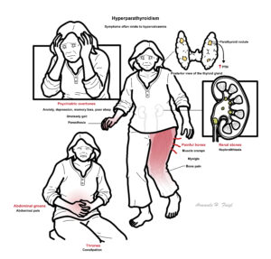
Primary Hyperparathyroidism (PHPT) is generalised disorder calcium, phosphate and bone metabolism due to increased secretion of parathyroid hormone normally leading to hypercalcaemia and hypophosphatemia. PHPT may present with recurrent nephrolithiasis, peptic ulcers, mental and muscle changes and less frequently bone resorption. Frequently, routine blood tests will pick up hypercalcaemia asymptomatically.
Hyperparathyroidism: Abnormally high concentration of parathyroid hormone in the blood, resulting in weakening of the bones through loss of calcium.
Primary Hyperparathyroidism: Excessive production of parathyroid hormone due to hyperactivity or growth of the parathyroid gland.
Secondary Hyperparathyroidism: Excessive production of parathyroid hormone in response to low serum calcium.
Tertiary Hyperparathyroidism: Excessive production of parathyroid hormone after a long period of secondary hyperparathyroidism.
Multiple Endocrine Neoplasia (MEN): hereditary group of disorders that affect the body’s network of hormone-producing glands. The main types are MEN1 and MEN2.
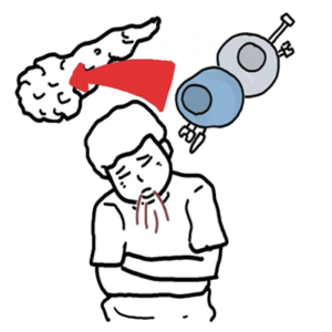
Hypoparathyroidism causes hypocalcaemia leading to muscle cramps and twitching and can lead to tetany.
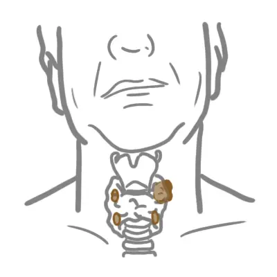
Anatomy of the Parathyroid Gland
The parathyroid glands are tan-coloured, bean-shaped structures, about the size of a grain of rice. There are 4 parathyroid gland which lie on the posterior surface of the thyroid gland.
Embryology Parathyroid development begins around the fifth or sixth week of life at the level of the pharynx, with all parathyroids migrating down into the neck.
Arterial Blood supply
Lymphatic drainage – along with those of the thyroid gland
Nerve supply
Pathology The parathyroid gland are made up of 4 main cells:
Parathyroid gland main purpose is to synthesise and secrete parathyroid hormone (PTH). The parathyroid hormone does this in response to blood calcium levels.
The normal range of serum calcium is 2.25-2.65mmol/L
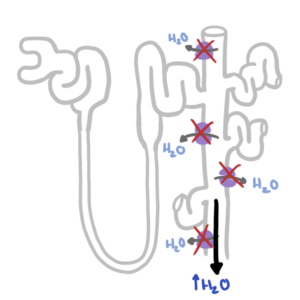
The skeleton contains 98 percent of total body calcium; the remaining 2 percent circulates throughout the body. One half of circulating calcium is free (ionized) calcium, the only form that has physiologic effects. The remainder is bound to albumin, globulin, and other inorganic molecules. Low albumin levels can affect the total serum calcium level. Measuring the corrected calcium is diagnostic for hypercalcaemia.
Primary hyperparathyroidism is caused by the inappropriate secretion of PTH, leading to hypercalcaemia. Causes:
Multiple Endocrine Neoplasia (MEN) are a group of familial endocrine diseases that affect multiple endocrine glands. There are two main types: MEN-1 and MEN-2.
Usually present asymptomatic 50%
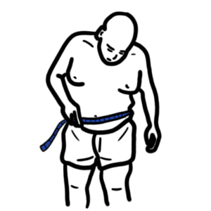
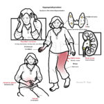
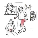
Stones, Bones, Thrones, Abdominal Groans, Psychiatric Overtones
Sudden = more likely malignancy. Chronic = more likely hyperparathyroidism. >3.7 really high = malignancy (parathyroid).
More info on Hypercalcaemia here
| DIFFERENT TYPES OF HYPERPARATHYROIDISM | ||
| Condition | Aetiology | Biochemical changes |
| Primary parathyroidism | Adenoma (80%) | ↑PTH,↑Ca, ↓PO4 |
| Secondary parathyroidism | Chronic Kidney Disease, Vitamin D deficiency | ↑PTH, ↓Ca, ↑PO4 |
| Tertiary parathyroidism | Long-term Secondary parathyroidism | ↑PTH, ↑Ca, ↑PO4 |
Secondary parathyroidism is due to Vitamin D deficiency which together with low Ca2+ stimulates the parathyroid gland (hyperplasia). With an increase in PTH phosphate levels drops but Ca2+ is still low because of no Vitamin D.
| PTH LEVEL | CAUSE |
| High PTH | Primary or tertiary hyperparathyroidismLithium-induced hyperparathyroidismFamilial Benign Hypocalciuric Hypercalcaemia |
| Low PTH | Malignancy (lung, breast, renal, ovarian, colonic, thyroid carcinoma, lymphoma, multiple myeloma)Elevated Vitamin D (Vitamin D intoxication, HIV, Sarcoidosis) Thyrotoxicosis Paget’s disease |
Once you have confirmed hyperparathyroidism (i.e. high PTH, high calcium, high urine calcium , low or normal phosphate, high ALP, more likely to be chronic, long standing, hypercalcaemia) then you can consider imaging:
The reality of treatment in primary hyperparathyroidism (90% adenoma) is surgical treatment – parathyroidectomy. If asymptomatic – can watch and wait – can give bisphosphonates for bone protection and increase oral fluids to reduce hypercalcaemia and prevent stones – review with serum calcium, PTH measurements and BMD every 6-12 months.
| Indications for Surgical Management |
| <50 |
| Clear cut related symptoms (peptic ulcer, stones, renal impairment, osteopenia) |
| Severe 3.7-4.5mmol serum calcium |
| Consistent follow-up is unlikely |
| Co-existing illness would complicate management |


Discussion