Systemic Sclerosis-Associated Interstitial Lung Disease 

Overview

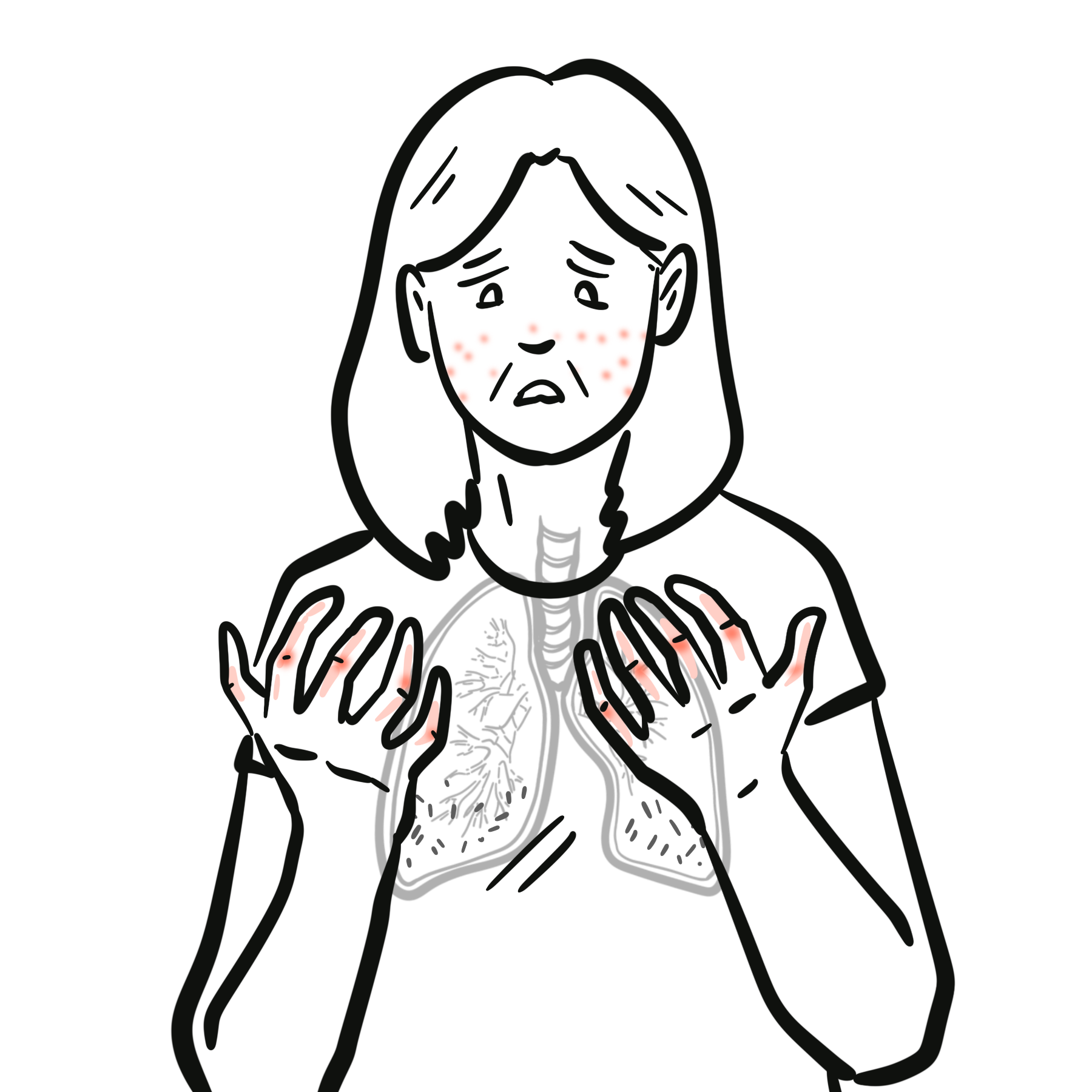
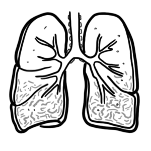
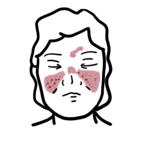
Systemic sclerosis–associated interstitial lung disease (SSc-ILD) is one of the most common and severe internal organ manifestations of systemic sclerosis (SSc), occurring in up to 50–60% of patients on HRCT, with clinically significant disease in ~25–30%. It is a major cause of SSc-related mortality. The most frequent histopathological pattern is nonspecific interstitial pneumonia (NSIP), followed by usual interstitial pneumonia (UIP). Risk factors include diffuse cutaneous SSc, anti-topoisomerase I (Scl-70) antibody positivity, male sex, African-American ethnicity, and early disease onset. The most important complication is progressive pulmonary fibrosis leading to respiratory failure and death.

Definition

Systemic sclerosis (SSc): Chronic autoimmune connective tissue disease characterized by vasculopathy, immune dysregulation, and fibrosis of skin and internal organs.
Interstitial lung disease (ILD): Group of disorders involving inflammation and/or fibrosis of the lung interstitium.
Nonspecific interstitial pneumonia (NSIP): ILD pattern with uniform interstitial inflammation and fibrosis, common in SSc.
Forced Vital Capacity (FVC): Spirometric measure of lung volume, used to monitor ILD progression.

Anatomy and Physiology

  • Alveolar-capillary unit: Site of gas exchange; consists of alveolar epithelium, interstitial space, and capillary endothelium.
  • Type I pneumocytes: Thin cells covering most alveolar surface, specialized for gas exchange.
  • Type II pneumocytes: Produce surfactant, repair alveolar epithelium after injury.
  • Interstitial space: Contains fibroblasts, extracellular matrix; normally thin to allow efficient diffusion.
  • Pulmonary microvasculature: Maintains low-resistance blood flow for oxygen uptake.

Aetiology and Risk Factors

Aetiology

  • Autoimmune-mediated endothelial injury and fibroblast activation
  • Genetic predisposition (e.g., HLA alleles, IRF5, STAT4 variants)
  • Environmental exposures (silica, organic solvents)
  • Microchimerism (fetal cell persistence in maternal circulation)

Risk Factors

  • Diffuse cutaneous SSc subtype
  • Anti-Scl-70 antibody positivity
  • Male sex
  • African-American ethnicity
  • Early disease onset (<5 years from SSc diagnosis)
  • High baseline CRP or ESR
  • Smoking

Remember

Anti-centromere antibody is associated with lower ILD risk but higher pulmonary hypertension risk.

Pathophysiology 

  • Endothelial injury → microvascular dysfunction, increased permeability
  • Immune activation → infiltration by T cells, B cells, macrophages
  • Cytokine release (TGF-β, IL-6, PDGF) → fibroblast activation
  • Fibroblast-to-myofibroblast transition → excessive collagen and ECM deposition
  • Interstitial thickening → impaired gas exchange, restrictive lung physiology
  • Progressive fibrosis → irreversible architectural distortion, honeycombing (UIP)

Early microvascular injury precedes fibrosis — screening should target early disease phase.

Clinical Manifestations

  • Progressive exertional dyspnea
  • Nonproductive cough
  • Bibasilar “Velcro” crackles
  • Fatigue, reduced exercise tolerance
  • Digital ulcers, Raynaud’s phenomenon (coexistent SSc features)
  • Signs of pulmonary hypertension in advanced disease

Remember

Anti-Scl‑70 positivity and diffuse cutaneous SSc are strong predictors of ILD development.

Diagnosis

Diagnostic criteria

  • Systemic Sclerosis diagnosis
  • HRCT showing ILD pattern
  • Exclusion of other ILD causes

Investigations

  • High resolution CT (HRCT)
    • NSIP (ground-glass opacities, subpleural sparing) 
    • UIP (honeycombing, reticulation)
  • Pulmonary Function Tests: restrictive pattern, ↓ FVC, ↓ DLCO
  • Serology: ANA, anti-Scl-70, anti-centromere, anti-RNA polymerase III
  • Echocardiography: screen for pulmonary hypertension
  • 6-minute walk test: functional assessment

Differential Diagnoses

ConditionDifferentiating Feature
Idiopathic NSIPNo systemic features, negative autoantibodies
IPF (UIP)Older age, no SSc features
Hypersensitivity pneumonitisExposure history, centrilobular nodules
Drug-induced ILDTemporal relation to drug exposure

Remember

HRCT is more sensitive than CXR; baseline and follow-up imaging are essential.

Classification

  • By HRCT/histopathology:
    • Nonspecific Interstitial Pneumonia (NSIP) – most common
    • Usual Interstitial Pneumonia (UIP)
    • Organizing Pneumonia (OP)
    • Lymphoid Interstitial Pneumonia (LIP)
  • By extent:
    • Limited (<20% lung involvement)
    • Extensive (>20% lung involvement)

Treatment

  • Immunosuppressants:
    • Glucocorticoids – not recommended given risk of renal crisis
    • Mycophenolate mofetil (first-line)
    • Cyclophosphamide OR Rituximab OR Tocilizumab
  • Anti-fibrotics:
    • Nintedanib (approved for SSc-ILD)
  • Supportive:
    • Oxygen therapy
    • Pulmonary rehabilitation
    • Vaccinations (influenza, pneumococcal)
  • Monitoring:
    • Serial PFTs every 3–6 months
    • HRCT as indicated
  • Refractory/severe disease
    • Autologous stem cell transplant (skin and lung)
    • CAR T-cell therapy

Remember

Nintedanib slows FVC decline in SSc-ILD regardless of background immunosuppression.

Complications and Prognosis

Complications

  • Progressive pulmonary fibrosis
  • Pulmonary hypertension
  • Respiratory failure
  • Increased infection risk (immunosuppression-related)

Prognosis

  • NSIP: better prognosis than UIP
  • Poor prognostic factors:
    • Male sex
    • African-American ethnicity
    • Extensive lung involvement (>20%)
    • Rapid FVC decline (>10% in 12 months)
    • UIP pattern

Early detection and aggressive management improve survival.

References

  1. Hoffmann-Vold AM et al. The identification and management of interstitial lung disease in systemic sclerosis: evidence-based European consensus statements. Lancet Rheumatol. 2020;2(2):e71–e83.
  2. Distler O et al. Nintedanib for systemic sclerosis–associated interstitial lung disease. N Engl J Med. 2019;380:2518–2528.
  3. Volkmann ER. Natural history of systemic sclerosis–related interstitial lung disease: how to identify a progressive fibrosing phenotype. J Scleroderma Relat Disord. 2020;5(2_suppl):31–40.
  4. Goh NS et al. Interstitial lung disease in systemic sclerosis: a simple staging system. Am J Respir Crit Care Med. 2008;177:1248–1254.
  5. Bruni C et al. Predicting ILD in systemic sclerosis: the ILD-RISC model. BMC Pulm Med. 2025;25:3722.
  6. Khanna D et al. 2013 ACR/EULAR classification criteria for systemic sclerosis. Arthritis Rheum. 2013;65:2737–2747.

Related Content

Discussion

0 Comments
Most Voted
Newest Oldest
Inline Feedbacks
View all comments

Table Of Contents

AH Community Platform is coming
Get access to member features
Early access launches soon
Armando hasudungan brain logo
Armando Hasudungan
By Visualising Medicine
© 2025 Visualising Medicine. All rights reserved.
Become a member to access note taking
Orangise your medical learning
This is just one of the many AH community member perks
Become a member to access quizzes
Strengthen your medical knowledge
This is just one of the many AH community member perks
0
Would love your thoughts, please comment.x
()
x