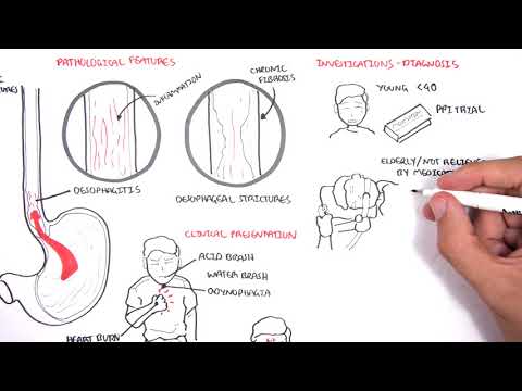
0:00 In this video, we're going to look at causes, different causes of upper GI 0:09 bleeding. 0:10 So essentially from your mouth, all the way down to the duodenum. 0:16 And bleeds around these area can cause the patient to vomit up blood, so hemato 0:22 emesis. 0:23 So now let's begin with, so now let us look at some common causes of upper GI 0:29 bleeding. 0:29 Starting with esophageitis, which is essentially inflammation of the esophagus. 0:34 So here we have the stomach, and here we have inflamed esophagus. 0:38 And there are different types of esophageitis. 0:40 We can have reflux esophageitis, gourd, infective esophageitis, chemical irrit 0:46 ants can cause 0:47 inflammation of the esophagus, as well as, there's esophageitis. 0:52 And essentially, again, it's this inflammation of the esophagus. 0:57 But while we're talking about esophagus, let's talk about Barrett's esophagus. 1:02 And Barrett's esophagus is a condition where cells lining the lower esophagus 1:08 change in 1:08 morphology. 1:09 So the esophagus can be divided into three parts. 1:12 The last part, the third part, the cells that line this area essentially change 1:19 in shape. 1:20 So here, in this area we normally have squamous epithelial cells, but these 1:26 change to Barrett's 1:28 metoplasia, which can subsequently lead to adenocosinoma of the esophagus. 1:36 So the relevance of Barrett's esophagus is that change in morphology can lead 1:42 to adenocosinoma. 1:44 And also pathological findings reveal that there are goblet cells present in 1:48 the area, 1:49 and where they shouldn't be. 1:52 The next condition we're going to talk about is esophageal varices. 1:56 So here we have stomach again, and here we have the veins around the stomach 2:01 and the 2:02 esophagus. 2:03 And esophageal varices develops as a result of portal hypertension, usually. 2:11 And esophageal varices is where we have dilated vessels, veins, of the esoph 2:18 agus. 2:19 And these dilated veins, when they dilate, they can rupture. 2:24 And this is very serious and dangerous. 2:28 Diagnosis of esophageal varices is using an endoscope. 2:35 So let's zoom into this area here of the esophagus and learn a bit more about 2:39 esophageal 2:39 varices in a diagram. 2:41 So here I'm drawing the esophagus, and these are the veins that drain the esoph 2:46 agus. 2:46 And the veins that drain the esophagus contain blood. 2:50 Now in esophageal varices, blood essentially pools in the area, causing these 2:57 veins to 2:58 grow, and they can subsequently rupture, causing serious problems. 3:02 So here we have varices, the pooling of the blood. 3:06 And the veins that actually drain the esophagus, there are two veins, and they 3:10 are nastomous 3:11 with each other. 3:12 These are from the top, the esophagus vein, and the left gastric vein. 3:17 And these veins, they drain later to the portal vein than to the liver. 3:22 But if you have portal hypertension, which we see in conditions such as liver 3:28 cirrhosis, 3:29 this blood will backflow to this area, thus leading to esophageal varices. 3:34 Here I'm drawing an endoscope, which is used to diagnose esophageal varices, 3:38 but it can 3:39 be dangerous. 3:41 And so here I'm drawing an image of what we would see if we were to look inside 3:46 the esophagus. 3:47 So we can see these prominent bulging veins full of blood, which can rupture 3:53 again, and 3:54 that is going to cause internal hemorrhage, which is very dangerous. 4:00 The next condition we're going to talk about is Mallory Y syndrome. 4:04 Mallory Y syndrome has to do with the connection between the esophagus and the 4:09 stomach. 4:10 And essentially it's where we have a tear. 4:12 So in Mallory Y syndrome, we have longitudinal tears near the cardio esophageal 4:16 junction. 4:17 These are superficial lacerations. 4:21 So if we were to zoom in, in the area, these are the Mallory Y's tears. 4:30 Mallory Y syndrome is associated with chronic vomiting, retching, coughing, and 4:39 straining. 4:41 The next cause of upper GI bleeds is we can have erosive gastritis. 4:46 Now erosive gastritis has to do with the stomach. 4:49 So here we're looking into the stomach and we have essentially erosions within 4:53 the layer 4:54 of the stomach. 4:55 So erosive gastritis is gastric mucosal erosion caused by damage to the mucosal 5:02 defense. 5:03 We can have bleeding with few or no symptoms. 5:08 We diagnose it using an endoscope. 5:10 Treatment includes proton pump inhibitors or H2 blockers, which essentially 5:15 work by 5:15 a stopping acid secretion. 5:19 Causes of erosive gastritis include use of insides, alcohol, and stress. 5:25 The next potential cause of upper GI bleeding are mass lesions, which, and what 5:29 I'm talking 5:30 about are polyps and essentially tumas, cancer. 5:34 So for example, we can have mass lesions, which are esophageal tumas, which can 5:40 be adenocarcinoma 5:42 or squamous cell carcinoma. 5:44 We talked about how Barrett's esophagus can lead to adenocarcinoma due to the 5:49 metaplasia 5:50 that occurs at the lower part of the esophagus. 5:54 Now we can have also growth within the stomach. 5:56 They can be benign tumas or malignant tumas. 5:59 Let's talk about benign tumas. 6:01 We can have few types. 6:02 They can be inflammation and hyperplastic polyps, fundic gland polyps, and gast 6:10 ric adenomas. 6:12 And tumas of the stomach. 6:14 There are a few types. 6:16 These can be gastric carcinoma, which is the most common. 6:21 Up to 95%. 6:22 We can have gastric lymphomas, roughly 5%, and then carcinoid tumas and stromal 6:27 cell 6:27 tumas, which are less common. 6:36 The next condition is dulophoys lesion, which is also known as a calabir 6:40 persistent artery. 6:41 And it's a rare but potentially life-threatening because it causes hemorrhage 6:48 of the GIT. 6:49 And as the name suggests, as the other name suggests, calabir persistent artery 6:53 , it has 6:53 to do with the artery that supplies the stomach. 6:57 So essentially, if we were to zoom into the stomach here and we were to look at 7:00 it through 7:01 an endoscope, we would see that it's essentially where we have a bulging of a 7:09 lesion. 7:10 And this lesion could subsequently rupture causing a hemorrhage because of the 7:16 blood that 7:16 supplies the area. 7:19 And this can cause severe internal bleeding, which can be fatal. 7:25 The next potential cause of upper GI bleed is known as angiodisplasia. 7:30 And it's a common vascular abnormality in the GI tract. 7:35 It's similar to telenctasia and it occurs all around the GIT, but mainly the 7:42 colon. 7:42 Anyway, the morphology of angiodisplasia is that we have elastic dilated thin 7:48 walled 7:48 vessels that are lined by endothelium alone. 7:53 And as I mentioned, it's most often seen in the colon, but we can also see it 7:57 in the stomach. 7:58 So if we were to look into the stomach here using an endoscope, we would see 8:04 potentially 8:05 angiodisplasia such as this. 8:15 The next condition is a very common cause of upper GI bleed, it's just ulcer 8:19 ation. 8:19 Ulceration is erosion of the mucus membrane and the risk factors and stuff are 8:25 similar 8:26 to gastritis, but it is technically a different condition. 8:31 Risk factors for ulcer formation in the stomach include excess hydrochloric 8:37 acid secretion, 8:38 which can be caused by a few things, helicobacter pylori infection, use of NSA 8:44 IDs and stress. 8:46 And ulcers can occur not only in the stomach, but also the duodenum. 8:50 So we can have duodenal ulcers and gastric ulcers. 8:54 So just looking at a duodenal ulcer here, it's more common than a gastric ulcer 9:00 by the 9:00 way. 9:02 So here I'm drawing the layers of the duodenum, here's a lumen, and here we 9:06 have an ulcer, 9:07 which is an erosion that goes down through the submucosa. 9:12 Gastric ulcer, similar thing, we can have complications with ulcers, they can 9:19 be bleeding, 9:20 you can have perforation as well as narrowing and obstruction. 9:25 Perforation means that the content can perforate into the peritoneum, which can 9:30 cause peritonitis 9:31 and severe inflammation and pain. 9:34 So those were some causes of upper GI bleeds. 9:37 I hope you enjoyed this video, thank you for watching.