

Syncope
Overview
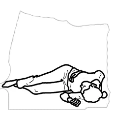
Syncope is a transient loss of consciousness (TLOC) due to global cerebral hypoperfusion, characterized by rapid onset, short duration, and spontaneous, complete recovery.
A sudden decrease in systemic blood pressure leading to a reduction in global cerebral blood flow for as little as 6–8 seconds (or a drop in Mean Arterial Pressure to < 30–40 mmHg) is sufficient to cause loss of consciousness.
Clinical Goal: Differentiate benign, self-limiting reflex/orthostatic causes from life-threatening cardiac etiologies (arrhythmias or structural heart disease) that carry a high risk of sudden cardiac death.
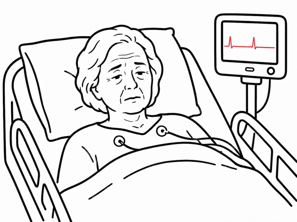
True syncope requires spontaneous, complete recovery without neurological deficit. If the patient has a prolonged post-ictal state, focal neurological signs, or requires pharmacological/electrical intervention to regain consciousness, consider Seizure, Stroke, or Cardiac Arrest rather than simple syncope.
Approach
Step 1: Primary Assessment & Red Flag Screening (San Francisco / Canadian Syncope Rules)
- Obtain immediate 12-Lead ECG, vital signs, point-of-care glucose, and bedside orthostatic blood pressures.
- High-Risk “Red Flag” Features (Mandatory Admission / Urgent Cardiac Workup):
- History: Syncope during exertion/supine position, preceding palpitations, sudden onset without prodrome, family history of premature sudden cardiac death (< 40 years).
- Comorbidities: Known structural heart disease, heart failure, ischemic heart disease, severe anemia.
- Exam: Abnormal vitals (SBP < 90 mmHg, sinus bradycardia < 40 bpm), new cardiac murmur (e.g., Aortic Stenosis/HCM).
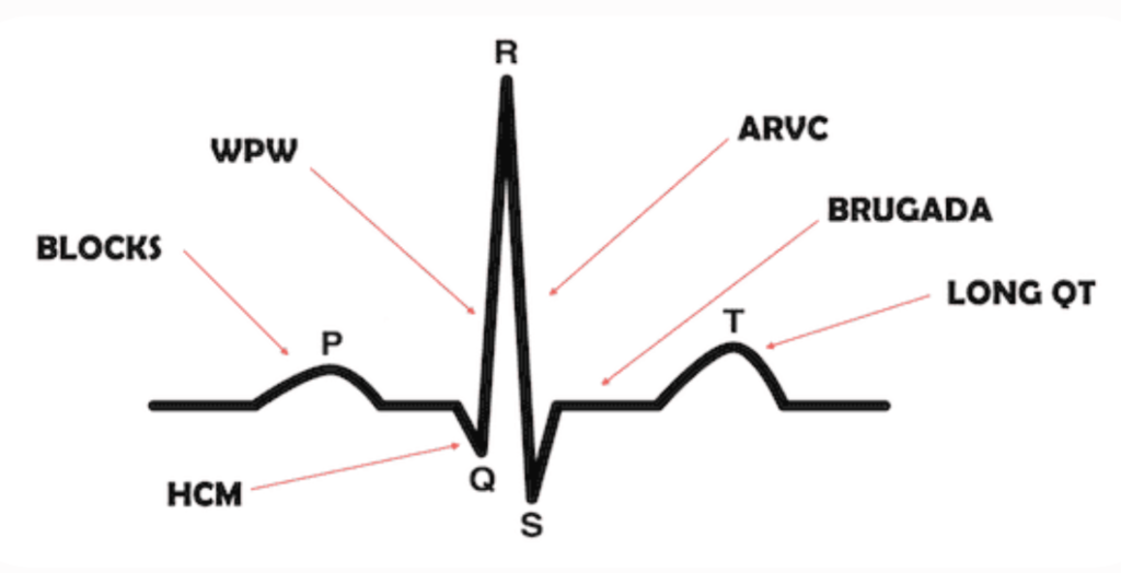
- ECG: Non-sinus rhythm, bifascicular block, QRS > 120 ms, pre-excitation (WPW), Long/Short QTc, Brugada pattern, pathological Q waves, ST-T segment changes.
Step 2: Diagnostic Branching Logic
- Cardiac Red Flags Present: -> Admit for continuous cardiac telemetry, echocardiogram, and urgent cardiology consultation.
- Clear Reflex Prodrome (Warmth, nausea, sweating, situational trigger) + Young/Normal Exam/Normal ECG: -> Reflex / Vasovagal Syncope (Reassure, outpatient discharge).
- Sustained Drop in SBP >= 20 mmHg upon Standing + Normal ECG: -> Orthostatic Hypotension (Review medications, hydration status).
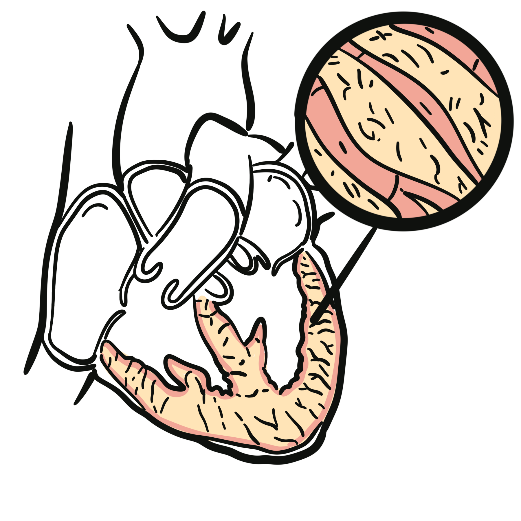
Syncope occurring during exertion (rather than immediately after exertion) is an alarming cardiac red flag indicating outflow tract obstruction (e.g., Hypertrophic Cardiomyopathy or Critical Aortic Stenosis) or a malignant ventricular arrhythmia until proven otherwise.
Differential Diagnosis
Reflex (Neurally Mediated) Syncope (~50% of cases – Benign):
- Vasovagal Syncope (VVS):
- Distinguishing features: Triggered by prolonged standing, pain, emotional distress, or venipuncture; classic prodrome (nausea, diaphoresis, pallor, tunnel vision).
- Situational Syncope:
- Distinguishing features: Occurs during or immediately after specific physiological triggers: micturition, deglutition, coughing, or defecation.
- Carotid Sinus Hypersensitivity:
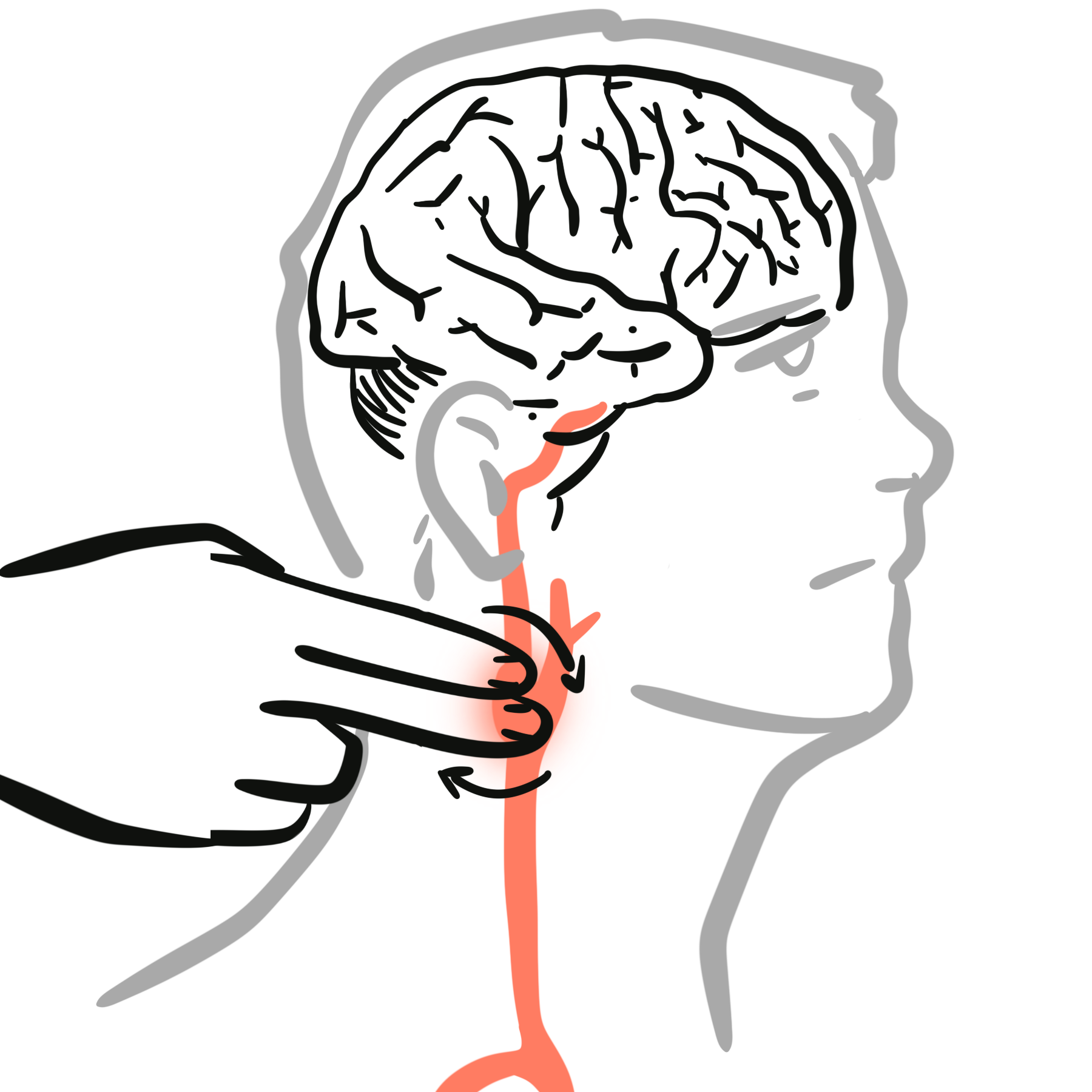
- Distinguishing features: Older individuals; triggered by neck turning, tight collars, or shaving; causes ventricular pauses > 3 seconds or SBP drop > 50 mmHg.
Orthostatic Hypotension (~15% of cases):
- Drug-Induced / Hypovolemic / Autonomic:
- Distinguishing features: Syncope upon standing; sustained SBP drop >= 20 mmHg or DBP drop >= 10 mmHg within 3 minutes; history of diuretics, vasodilators, dehydration, or Parkinson’s disease.
Cardiac Syncope (~15% of cases – High Mortality Risk):
- Arrhythmogenic (Electrical):
- Bradyarrhythmias: Sick Sinus Syndrome, High-degree AV Block (Mobitz II, 3rd Degree).
- Tachyarrhythmias: Ventricular Tachycardia (VT), Torsades de Pointes (Long QT), SVT.
- Inherited Channelopathies: Brugada Syndrome (coved ST elevation V1-V3), WPW Syndrome (delta waves, short PR), ARVC (epsilon waves).
- Structural / Mechanical:
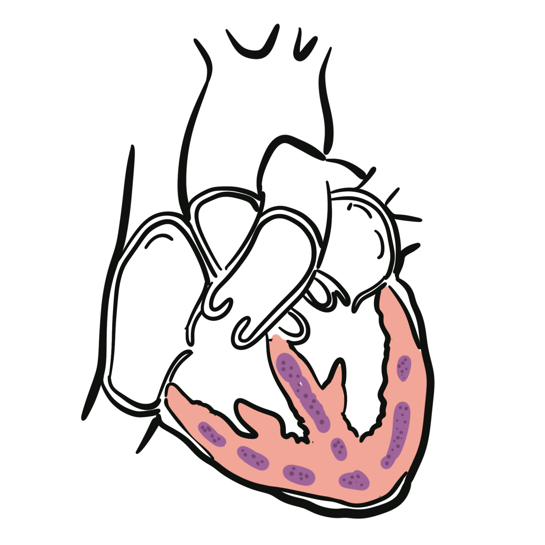
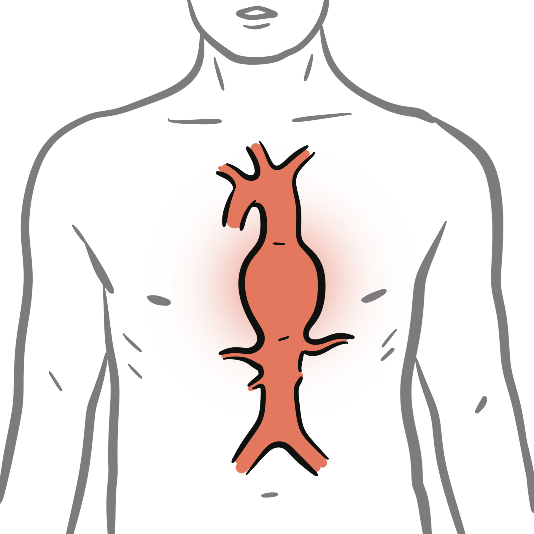
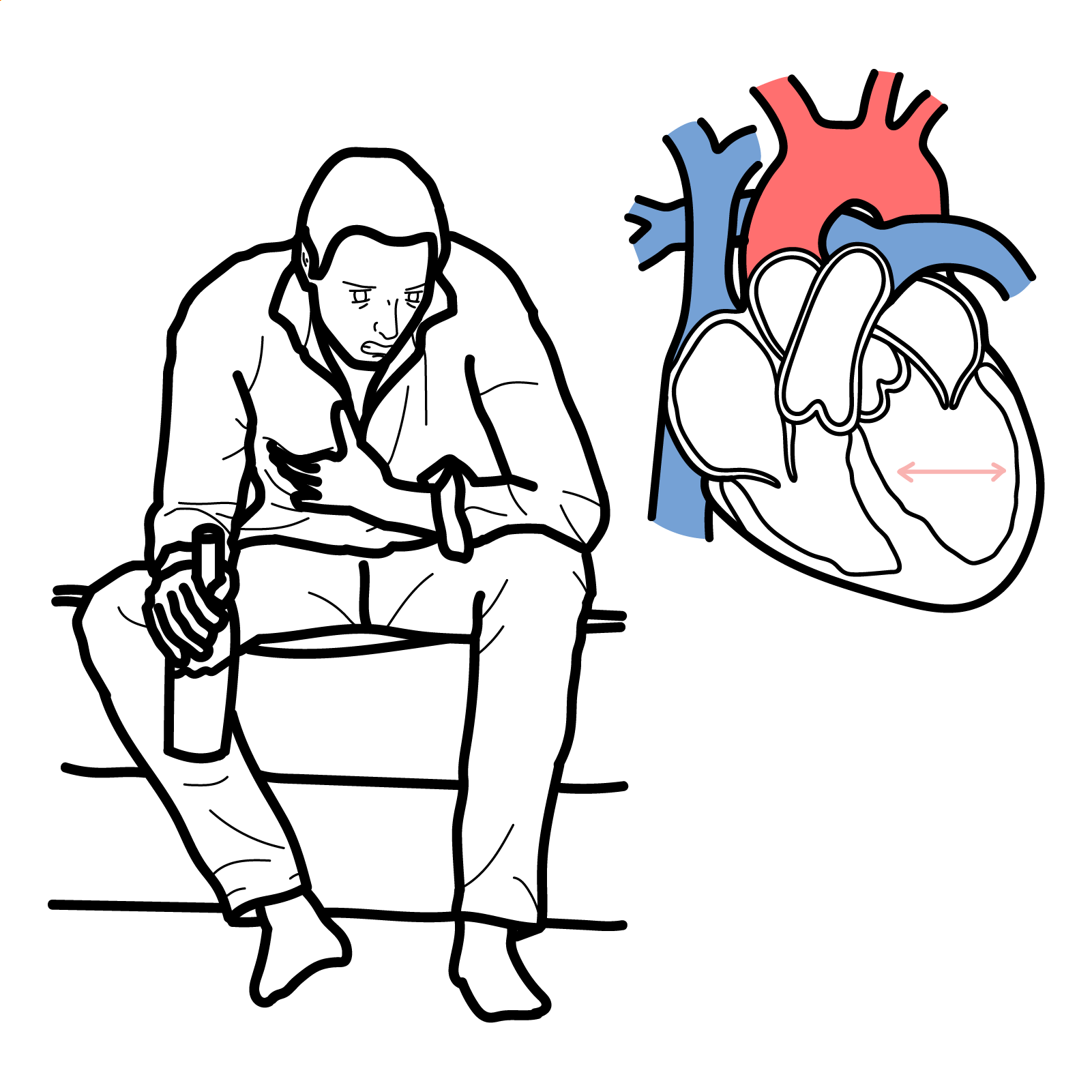
- Distinguishing features: Aortic Stenosis (crescendo-decrescendo systolic murmur), Hypertrophic Cardiomyopathy (systolic murmur increasing with Valsalva), Pulmonary Embolism, Acute Aortic Dissection, Atrial Myxoma.
Non-Syncopal Mimics:
- Seizure: Prolonged tonic-clonic activity, tongue biting (lateral), urinary incontinence, post-ictal confusion (> 15 min).
- Hypoglycemia: Diaphoresis, tremor, altered level of consciousness; confirmed by glucose < 3.0 mmol/L.
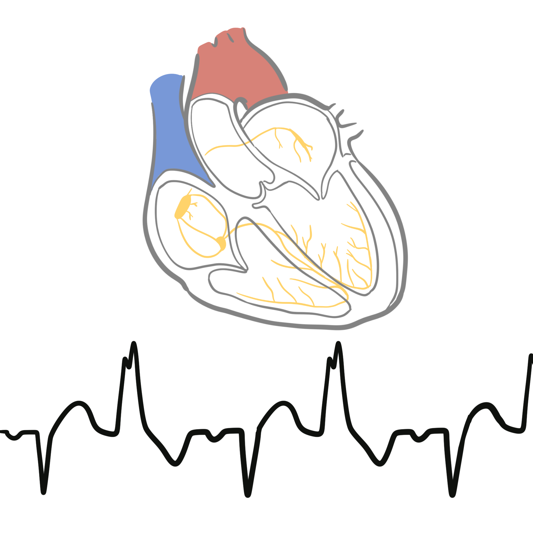
Brugada Syndrome is characterized by a type 1 coved-type ST-segment elevation >= 2 mm in right precordial leads (V1–V3) followed by a negative T wave. It predisposes young patients to polymorphic VT/VF and sudden cardiac death during sleep or fever.
Investigations
First-Line / Bedside & Emergency Diagnostics:
- 12-Lead ECG (MANDATORY in ALL Syncope): Look for PR interval, QRS duration, QTc duration, delta waves, Brugada pattern, ARVC epsilon waves, and ischemia.
- Active Stand Test (Bedside Orthostatics): Measure BP/HR lying supine for 5 minutes, then standing at 1 and 3 minutes.
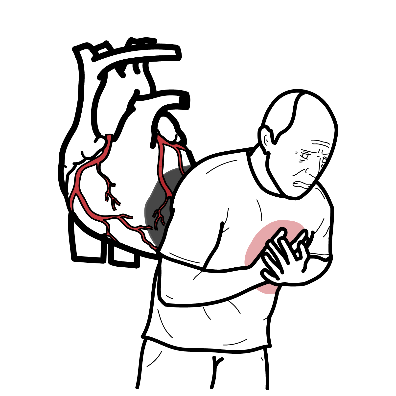
- Blood Tests: Full Blood Count (anemia), Electrolytes & Renal function, Beta-hCG (females of childbearing age), Troponin (if ischemic symptoms), D-Dimer (if PE suspected).
Targeted / Secondary Cardiac & Neurological Workup:
- Transthoracic Echocardiogram (TTE): Indicated in any patient with cardiac red flags, abnormal ECG, or suspected structural heart disease (evaluates EF, valvular lesions, HCM).
- Ambulatory ECG Monitoring:
- Holter Monitor (24–48h): Frequent symptoms (daily).
- Event / Patch Recorder (1–4 weeks): Intermittent weekly symptoms.
- Implantable Loop Recorder (ILR): Unexplained syncope with high suspicion of recurrent infrequent arrhythmia.
- Head-Up Tilt Table Testing (HUTT): Useful to confirm vasovagal syncope, psychogenic pseudosyncope, or delayed orthostatic hypotension in equivocal cases.
- CT Brain / EEG: Indicated ONLY if focal neurological deficits are present or if true seizure activity is suspected (NOT routinely recommended for straightforward syncope).
Routine CT Brain scan and EEG are low-yield and NOT indicated for uncomplicated syncope with a normal neurological exam. Direct resources toward an ECG, echocardiogram, and cardiac rhythm monitoring instead.
Critical Management
Immediate Resuscitation for High-Risk / Unstable Cardiac Syncope:
- Symptomatic Bradycardia / Complete Heart Block: Transcutaneous pacing / IV Atropine -> Emergency transvenous pacemaker placement.
- Ventricular Tachycardia / Torsades de Pointes: Synchronized Cardioversion (if unstable) or IV Amiodarone / Magnesium Sulfate (for Torsades).
- Structural Lesions (Severe AS, HCM): Avoid aggressive vasodilation or diuresis; urgent cardiothoracic surgery / cardiology consultation.
Reflex / Vasovagal Syncope (Long-Term Conservative Strategy):
- Patient Reassurance & Education: Explain benign nature of condition.
- Isometric Physical Counterpressure Maneuvers (PCM): Leg crossing, muscle tensing, arm-squeezing at onset of prodromal symptoms to raise MAP and prevent syncope.
- Fluid & Salt Intake: Increase fluid (2–3 L/day) and salt intake (unless contraindicated by hypertension or heart failure).
Physical counterpressure maneuvers (squatting, leg crossing, handgrip) at the onset of a vasovagal prodrome can rapidly increase venous return and cardiac output, successfully aborting an impending syncopal episode.
References
- European Society of Cardiology. 2018 guidelines for the diagnosis and management of syncope [Internet]. 2018 [cited 2026 Aug 20]. Available from: https://www.escardio.org/guidelines/clinical-practice-guidelines/all-esc-practice-guidelines/syncope/
- National Institute for Health and Care Excellence. Transient loss of consciousness (‘blackouts’) in over 16s (CG109) [Internet]. 2010 [updated 2023; cited 2026 Aug 20]. Available from: https://www.nice.org.uk/guidance/cg109


Members only discussions coming soon…