

Eye Trauma
Overview
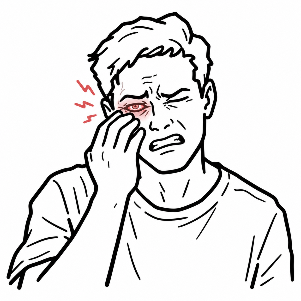
Ocular trauma encompasses mechanical, chemical, or thermal injury to the globe, ocular adnexa, or orbit ranging from superficial abrasions to sight-threatening globe rupture or optic neuropathy.
Classification:
- Open Globe: Full-thickness breach of cornea and/or sclera (Rupture from blunt trauma vs. Laceration from sharp penetration/intraocular foreign body [IOFB]).
- Closed Globe: Cornea/sclera remain intact (Contusion, lamellar laceration, superficial foreign body).
- Chemical Burns: True ophthalmic emergency requiring immediate copius irrigation prior to formal visual assessment.
In ocular trauma, always establish whether the globe is intact (closed) or breached (open). If an open globe is suspected, STOP physical examination immediately—do NOT palpate the eye, measure intraocular pressure (IOP), or instill eye drops. Place a rigid eye shield and consult ophthalmology urgently.
Approach
Step 1: Immediate Chemical Exposure Triage
- Alkali (Worst – Liquefactive Necrosis) / Acid (Coagulative Necrosis): Irrigate immediately with 1–2 L normal saline/Ringer’s lactate or continuous amphoteric solution before any exam. Target pH = 7.0–7.4 (check with litmus paper 5 min post-irrigation).
Step 2: Rule Out Open Globe Injury (OGI) Red Flags
- Key Physical Exam Signs:
- Teardrop/peaked pupil pointing toward the wound site.
- Extruding intraocular contents (uvea, vitreous, iris prolapse).
- Extravasation of aqueous humor (Positive Seidel Test under fluorescein).
- Subconjunctival hemorrhage (360 degrees or bullous).
- Asymmetric shallow or abnormally deep anterior chamber.
- Severe retrobulbar/orbital pain with acute reduction in visual acuity.
Step 3: Clinical Decision Branching
- Open Globe Suspected/Confirmed: -> Rigid Fox shield over orbit, keep patient NPO, IV antiemetics (prevent increased IOP from vomiting), systemic IV antibiotics, urgent CT Orbit (no MRI), immediate emergency ophthalmology consult.
- Closed Globe Injury: -> Proceed with complete slit lamp exam, fluorescein staining, eversion of lids, intraocular pressure measurement (Tonometer), and dilated fundus exam.
A teardrop-shaped pupil points directly toward the site of a full-thickness corneal or scleral laceration until proven otherwise.
Differential Diagnosis
Open Globe / Full-Thickness Injuries:
- Globe Rupture (Blunt Mechanism):
- Distinguishing features: History of high-energy blunt blow; severe vision loss, marked hypotony (IOP < 5 mmHg), 360-degree chemosis, occult scleral rupture behind insertion of rectus muscles.
- Penetrating / Perforating Laceration & IOFB:
- Distinguishing features: High-speed projectile history (metal-on-metal hammering, power tools); visible entry site, iris defect, traumatic cataract.
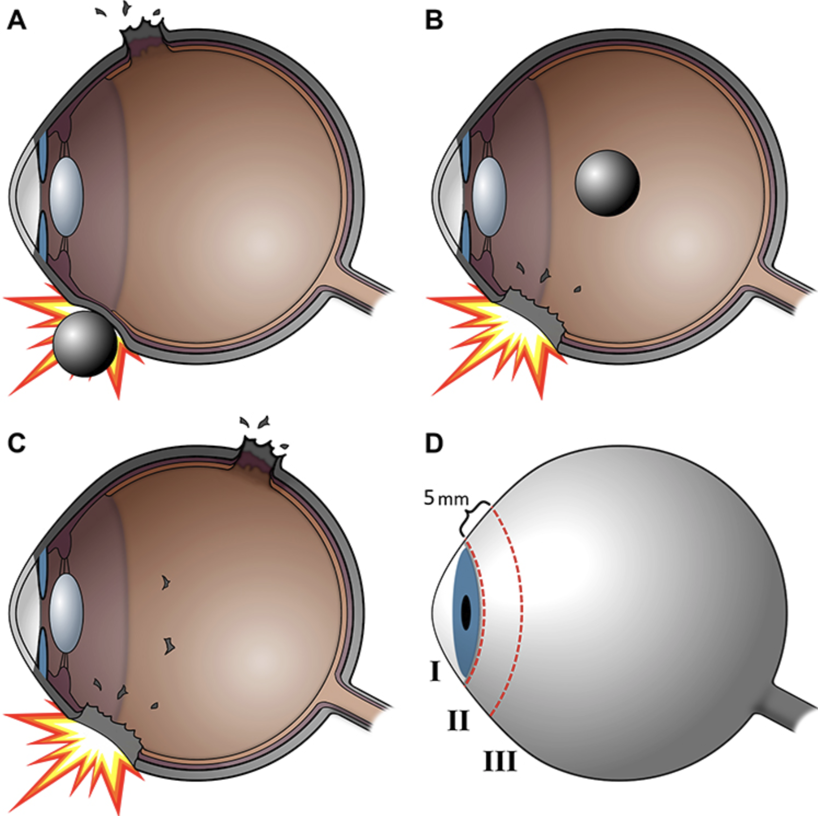
Open-globe injuries include rupture after blunt impact, a penetrating entry wound, and a perforating injury with entry and exit wounds.
What to notice
Detailed image description
A classification diagram showing how blunt impact can rupture the eyewall away from the point of contact, how a sharp object can produce one penetrating wound with a possible retained foreign body, and how a perforating injury creates both entry and exit wounds.
Closed Globe & Orbital Emergencies:
- Retrobulbar Hemorrhage / Compartment Syndrome:
- Distinguishing features: Rapidly progressive proptosis, severe pain, afferent pupillary defect (RAPD), elevated IOP (> 40 mmHg), restricted extraocular movements.
- Traumatic Hyphema:
- Distinguishing features: Blood layered in anterior chamber; risk of re-bleeding (days 2–5) and secondary glaucoma.
- Corneal Abrasion / Superficial Foreign Body:
- Distinguishing features: Intense foreign body sensation, photophobia, relief with topical anesthetic (tetracaine); focal epithelial defect uptake on fluorescein.
- Blowout Orbital Fracture:
- Distinguishing features: Enophthalmos, infraorbital nerve paresthesia (V2 distribution), diplopia on upward gaze due to inferior rectus entrapment.
- Traumatic Iritis / Traumatic Mydriasis:
- Distinguishing features: Post-blunt trauma anterior chamber cell/flare, ciliary flush, sluggish or irregular pupil without full-thickness laceration.
Retrobulbar hemorrhage with acute proptosis, RAPD, and elevated IOP is a surgical emergency requiring immediate bedside lateral canthotomy and cantholysis to decompress the orbit—do not delay for imaging or ophthalmology arrival.
Investigations
First-Line / Bedside Diagnostics:
- Visual Acuity Testing: Essential baseline metric for both eyes (Snellen chart or near-card; count fingers/light perception if severe).
- Penlight & Pupil Exam: Check for direct/consensual reflexes, shape (teardrop), and Relative Afferent Pupillary Defect (RAPD).
- Slit Lamp Examination (Only if Open Globe Ruled Out): Assess corneal integrity, anterior chamber depth/cells/blood (hyphema), and lens dislocation.
- Fluorescein Staining: Detect corneal defects or Seidel’s sign (streaming clearance of fluorescein by leaking aqueous humor under cobalt blue light).
- Tonometry: Measure IOP (Contraindicated if open globe is suspected).
Targeted / Diagnostic Imaging:
- Non-Contrast CT Orbits (Thin Cut 1 mm Axial/Coronal): Gold standard for detecting radiopaque IOFBs, occult scleral rupture, retrobulbar hematoma, and orbital wall fractures.
- Ultrasound B-Scan: Useful for evaluating posterior segment (retinal detachment, vitreous hemorrhage) only if globe is intact.
- MRI Orbits: STRICTLY CONTRAINDICATED if metallic IOFB is suspected (risk of ferromagnetic movement causing further ocular tissue destruction).
Never order an MRI in acute eye trauma until a metallic intraocular foreign body has been definitively ruled out by non-contrast CT.
Critical Management
Open Globe Injury (Immediate Pre-Operative Stabilization):
- Shielding: Place a rigid aluminum Fox shield resting on bony orbital margins (Do NOT place a pressure patch, gauze pad, or topical ointment).
- IOP Control: Administer IV Ondansetron/Metoclopramide to prevent retching/vomiting; provide aggressive analgesia.
- Infection Prophylaxis: IV systemic broad-spectrum antibiotics (e.g., Vancomycin + Ceftazidime or Fluoroquinolone) for endophthalmitis prevention; update Tetanus vaccination.
Retrobulbar Compartment Syndrome:
- Bedside Lateral Canthotomy and Cantholysis: Perform immediately at bedside if optic nerve is compromised (proptosis + elevated IOP + RAPD/vision loss).
Chemical Burns:
- Immediate Decontamination: Continuous irrigation with 1–2 L isotonic fluid until anterior chamber pH normalizes (7.0–7.4).
Never place a pressure patch or eye pad on a suspected open globe—any external pressure can cause irreversible extrusion of intraocular contents through the wound.
References
- American Academy of Ophthalmology. Open globe injury: assessment and preoperative management [Internet]. 2020 [cited 2026 Aug 20]. Available from: https://www.aao.org/eyenet/article/open-globe-injury
- Royal Children’s Hospital Melbourne. Clinical practice guidelines: penetrating eye injury [Internet]. [cited 2026 Aug 20]. Available from: https://www.rch.org.au/clinicalguide/guideline_index/Penetrating_eye_injury/



Members only discussions coming soon…