

Cardiac Tamponade
Overview
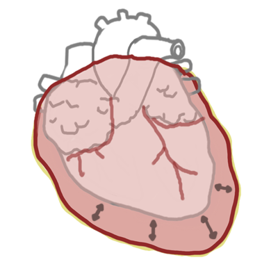
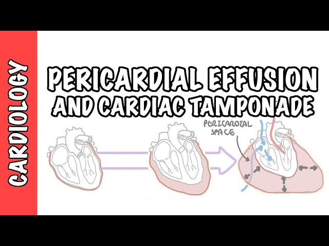
Cardiac tamponade is a life-threatening cardiovascular emergency caused by the accumulation of fluid, blood, pus, or gas within the pericardial sac. This accumulation causes a critical elevation in intrapericardial pressure that exceeds normal cardiac diastolic filling pressures, resulting in compression of the heart chambers, severely impaired diastolic filling, reduced stroke volume, and cardiac output failure.
The clinical presentation depends on both the volume of fluid and the rate of accumulation. A rapid accumulation of as little as 100 to 200 mL of fluid (e.g., trauma or aortic dissection) can cause acute catastrophic collapse, whereas slow accumulation of 1 to 2 liters (e.g., malignancy or tuberculosis) allows the pericardium to stretch before tamponade ensues. Immediate clinical recognition, fluid resuscitation to support preload, and urgent pericardial decompression are lifesaving.
Cardiac tamponade is primarily a hemodynamic and clinical diagnosis, not purely an echocardiographic one. If a patient is hypotensive, tachycardic, and has a pericardial effusion with signs of shock, proceed directly to emergency decompression—do not wait for formal lab reports or advanced imaging.
Definition
- Beck's Triad
- The classic triad of acute cardiac tamponade consisting of hypotension, jugular venous distention (JVD), and muffled (distant) heart sounds.
- Pulsus Paradoxus
- An exaggerated inspiratory decline in systemic systolic blood pressure exceeding 10 mmHg during normal quiet breathing.
- Ventricular Interdependence
- An exaggerated reciprocal coupling between the right and left ventricles within a fixed, non-distensible pericardial volume, where expansion of one ventricle directly compromises filling of the other.
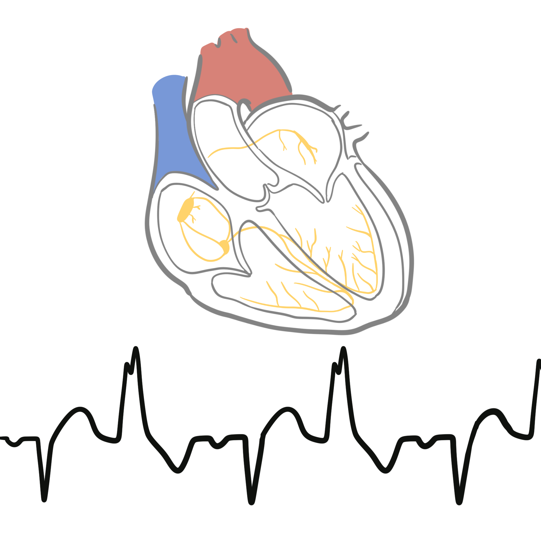
- Electrical Alternans
- Beat-to-beat alteration in QRS complex amplitude and axis on ECG, resulting from the physical "swinging" motion of the heart within a large pericardial fluid collection.
Why does Pulsus Paradoxus occur in Cardiac Tamponade?
During inspiration, negative intrathoracic pressure increases venous return to the right side of the heart, expanding the right ventricle (RV). Because the total space inside the rigid pericardium is fixed, the expanding RV pushes the interventricular septum toward the left side (ventricular interdependence). This bulging severely restricts left ventricular (LV) diastolic filling, reducing LV stroke volume and causing a transient drop in systemic systolic blood pressure (> 10 mmHg) during inspiration.
Low-pressure tamponade was first recognized in patients undergoing hemodialysis who developed tamponade despite normal or low central venous pressure. Intravenous fluid administration in these patients unmasks classic physical findings by restoring intravascular volume.
Aetiology & Risk Factors
| Category | Specific Causes | Clinical Features |
| Traumatic | Penetrating or blunt chest trauma, iatrogenic perforation (pacing wires, cardiac catheterization, central line insertion) | Rapid onset, hemopericardium, acute cardiovascular collapse. |
| Cardiovascular | Acute Type A Aortic Dissection (retrograde rupture), post-MI free wall rupture, post-pericardiotomy syndrome | Extremely high mortality; requires emergency surgical repair. |
| Malignancy | Metastatic lung cancer, breast cancer, lymphoma, melanoma, leukemia | Most common cause of subacute tamponade; large volume, recurrent effusions. |
| Infectious | Viral pericarditis (Coxsackie), Tuberculosis (dominant in endemic regions), Bacterial/purulent pericarditis | Often accompanied by fever, night sweats, or systemic signs of infection. |
| Systemic / Inflammatory | Uremia (end-stage renal disease), Systemic Lupus Erythematosus (SLE), Rheumatoid Arthritis, Hypothyroidism (myxedema) | Slow, insidious fluid accumulation; rich in protein or cholesterol. |
| Idiopathic | Non-specific pericarditis | Diagnosis of exclusion after ruling out infection and cancer. |
Pathophysiology
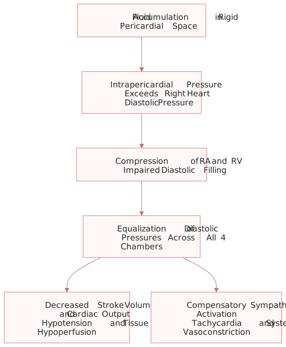
Cardiac tamponade pathophysiology: rising intrapericardial pressure compresses the right heart, limits diastolic filling and reduces cardiac output while triggering compensatory sympathetic activation.
What to notice
Detailed image description
Fluid accumulation within the rigid pericardial space raises intrapericardial pressure above right-sided diastolic pressure. Right atrial and right ventricular compression impairs diastolic filling, with equalization of diastolic pressures. The result is reduced stroke volume and cardiac output with hypotension and tissue hypoperfusion, accompanied by compensatory tachycardia and systemic vasoconstriction.
- Pressure Equalization: As fluid accumulates, intrapericardial pressure rises until it equals and exceeds normal right ventricular and right atrial diastolic pressures (typically 5 to 10 mmHg).
- Diastolic Collapse: Because right-sided chambers have thin walls and lower operating pressures, the right atrium collapses during late diastole/early systole, followed by right ventricular diastolic collapse.
- Diastolic Plateau: Diastolic pressures in the right atrium, right ventricle, pulmonary artery, and left ventricle equalize to a single uniform elevated pressure level.
- Hemodynamic Collapse: Reduced diastolic filling causes a dramatic reduction in end-diastolic volume, lowering stroke volume and cardiac output. Compensatory sympathetic activation causes sinus tachycardia and intense peripheral vasoconstriction to maintain blood pressure until final cardiovascular collapse.
Clinical Manifestations
- Dyspnea & Tachypnea: Most sensitive symptom (present in > 80% of patients).
- Chest Discomfort: Dull, aching chest pain or pleuritic pain that worsens when lying flat and improves when sitting forward.
- Weakness, lightheadedness, cold diaphoresis, syncope, and feeling of impending doom.
Clinical Examination
- Beck’s Triad:
- Hypotension with narrow pulse pressure.
- Distended neck veins (elevated JVP).
- Muffled / Distant heart sounds.
- Pulsus Paradoxus: Systolic blood pressure drops by > 10 mmHg during quiet inspiration.
- Jugular Venous Pulse (JVP) Contour: Elevated JVP with a preserved ‘x’ descent but a loss/absence of the ‘y’ descent (because passive early diastolic filling of the RV is completely blotted out by external compression).
- Tachycardia: Compensatory mechanism to maintain cardiac output ($CO = HR \times SV$).
- Peripheral Signs of Shock: Cold, clammy extremities, delayed capillary refill, oliguria, and altered mental status.
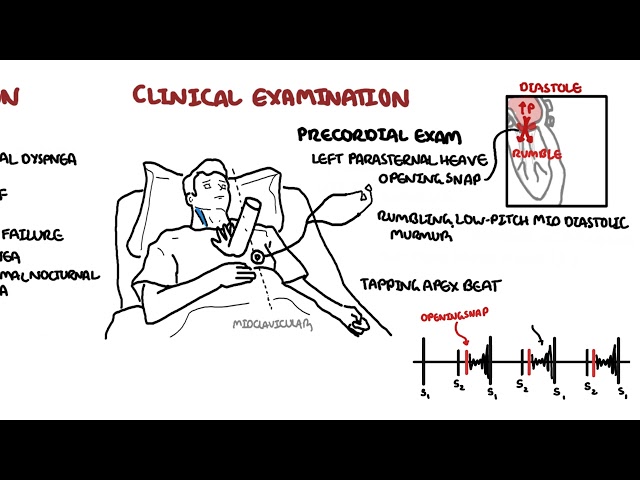
Beck’s Triad: hypotension (with pulsus paradoxus), increased JVP, muffled heart sound
Kussmaul’s Sign (an paradoxical rise in JVP during inspiration) is classic for Constrictive Pericarditis, whereas it is RARE in pure Cardiac Tamponade because passive RV filling is uniformly restricted throughout the entire cardiac cycle.
Diagnosis
Electrocardiogram (ECG)
- Sinus Tachycardia: Universal compensatory response.
- Low Voltage QRS: Defined as total QRS amplitude < 5 mm in limb leads or < 10 mm in precordial leads.
- Electrical Alternans: Beat-to-beat variation in QRS height and direction; pathognomonic when present, though seen in < 20% of cases.
Echocardiography (Gold Standard Imaging)
Echocardiography establishes both the presence of effusion and the hemodynamic severity of tamponade:
- Right Atrial (RA) Collapse: Inversion of the RA wall during late diastole/early systole (early, highly sensitive sign).
- Right Ventricular (RV) Collapse: Indentation of the RV anterior wall during early diastole (highly specific sign).
- Dilated Inferior Vena Cava (IVC): Plethoric IVC with < 50% reduction in diameter during inspiration.
- Doppler Flow Variations: Exaggerated respiratory variation in transmitral flow velocity (> 25% decrease on inspiration) and transtricuspid flow velocity (> 50% increase on inspiration).
Chest Radiography
- In acute tamponade, CXR is often completely normal because small fluid volumes (100–200 mL) do not enlarge the cardiac silhouette.
- In chronic tamponade, CXR shows a classic enlarged, globular, “water-bottle” heart silhouette with clear lung fields.
Invasive Hemodynamic Monitoring (Right Heart Catheterization)
- Demonstrates equalization of end-diastolic pressures across the right atrium, right ventricle, pulmonary capillary wedge pressure (PCWP), and left ventricle.
- JVP/RA pressure tracing shows a prominent x descent and an absent y descent.
Diagnostic Sensitivity vs Specificity on Echo
- RA Collapse: High sensitivity (occurs early when intrapericardial pressure temporarily exceeds RA pressure).
- RV Collapse: High specificity (occurs later when intrapericardial pressure exceeds higher-pressure RV diastolic relaxation).
Treatment
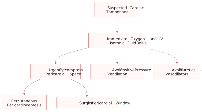
Emergency tamponade management: support preload, avoid interventions that reduce venous return, and proceed urgently to pericardial decompression.
What to notice
Detailed image description
Suspected cardiac tamponade requires immediate oxygen and IV isotonic fluid to support preload while urgent decompression is arranged. Positive-pressure ventilation, diuretics and vasodilators should be avoided where possible because they can further reduce venous return or preload. Definitive decompression is by percutaneous pericardiocentesis or a surgical pericardial window according to the clinical context.
Emergency Initial Stabilization
- Volume Expansion: Administer an immediate intravenous bolus of normal saline (500–1000 mL) to increase right-sided filling pressures and temporarily overcome external compression.
- CRITICAL CONTRAINDICATION – Positive Pressure Ventilation (PPV): Avoid intubation and mechanical ventilation prior to decompression whenever possible. Positive intrathoracic pressure severely decreases venous return, precipitating immediate complete cardiac arrest (Pulseless Electrical Activity – PEA).
- CONTRAINDICATED MEDICATIONS: Diuretics (e.g., Furosemide) and Vasodilators (e.g., Nitroglycerin) are strictly contraindicated because reducing intravascular volume or venodilating causes immediate cardiac collapse.
Definitive Decompression Procedures
- Echocardiogram-Guided Percutaneous Pericardiocentesis:
- First-Line Procedure: Insert a needle via the subxiphoid approach directed toward the left shoulder at a 30 to 45 degree angle under continuous echo guidance.
- Catheter Placement: Leave a multi-dusted pigtail catheter in place for continuous drainage until output drops to < 25–30 mL/day.
- Surgical Pericardiectomy / Pericardial Window:
- Indications: Traumatic hemopericardium, acute aortic dissection, purulent bacterial pericarditis, recurrent malignant effusions, or loculated regional tamponade.
Complications & Prognosis
Complications
- Pulseless Electrical Activity (PEA) Arrest: The primary fatal complication of unrecognized or delayed decompression.
- Low Cardiac Output Syndrome / Myocardial Stunning: Post-decompression pulmonary edema or left ventricular failure caused by a sudden surge in RV venous return overloading a chronically underfilled LV.
- Procedural Complications: Laceration of coronary arteries, RV wall puncture, pneumothorax, or liver laceration during pericardiocentesis.
Prognosis
Prognosis is primarily driven by the underlying etiology:
- Idiopathic / Viral / Traumatic (repaired): Excellent long-term survival following successful drainage (> 90%).
- Malignant / Aortic Dissection / MI Rupture: Poor prognosis; 1-year survival in malignant tamponade is < 25% despite drainage.
References
- Adler Y, Charron P, Imazio M, et al. 2015 ESC Guidelines for the diagnosis and management of pericardial diseases. Eur Heart J. 2015;36(42):2921-2964. doi:10.1093/eurheartj/ehv318
- Ristic AD, Imazio M, Adler Y, et al. Triage strategy for urgent management of cardiac tamponade: a proposal of the European Society of Cardiology Working Group on Myocardial and Pericardial Diseases. Eur Heart J. 2014;35(34):2279-2284. doi:10.1093/eurheartj/ehu217
- Spodick DH. Acute cardiac tamponade. N Engl J Med. 2003;349(7):684-690. doi:10.1056/NEJMra022630


Members only discussions coming soon…