Overview
Signs and symptoms of cerebellar disease presents on the same side (ipsilateral). Symptoms and signs consist of gait impairment, unclear (“scanning”) speech, visual blurring due to nystagmus, hand in-coordination, and tremor with movement.
| Remember |
| CAUSES OF CEREBELLAR DISEASE BASED ON ONSET | ||
| Acute | Subacute | Chronic |
| Drugs (phenytoin, lithium) | Alcohol | Chronic infection |
| Alcohol intoxication | Malnutrition (Vitamin B12 and B1 deficiency | Friederichs ataxia |
| Paraneoplastic syndrome | Hypothyroidism | |
| Hyponaturaemia | ||
| CAUSES OF CEREBALLAR DISEASE BASED ON LOCATION | |||
| Unilateral | Bilateral | Midline | Vermis |
| Space occupying lesion | Drugs (phenytoin) | Paraneoplastic Syndrome | Alcohol |
| Ischaemia | Alcohol | ||
| Multiple sclerosis | Friederichs ataxia | ||
| Trauma | Hypothyroidism | ||
| Large space occupying lesion | |||
| Multiple Sclerosis | |||
| Trauma | |||
| Frederichs ataxia is a autosomal recessive ataxia, comprising one-half of all hereditary ataxias. Two forms classic (frataxin) and association with vitamin E deficiency. |
Cerebellar Examination
General
- Titubation
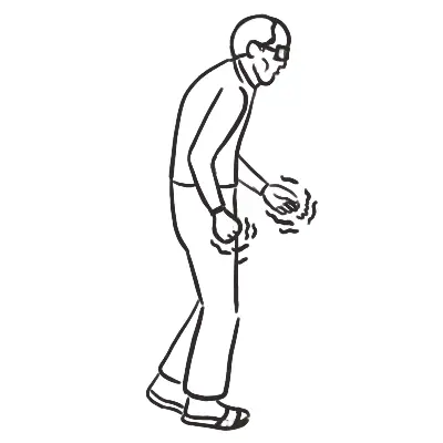
- Gait – ataxia, unable to tanden gait (heel to shin)
- Intention tremor
Head
- Auscultate over cerebellum for bruits
- Auscultate carotids – lateral medullary syndrome (Wallenburg syndrome)
- Cerebellopontine angle tumour – 5, 7, 8 cranial nerve affected
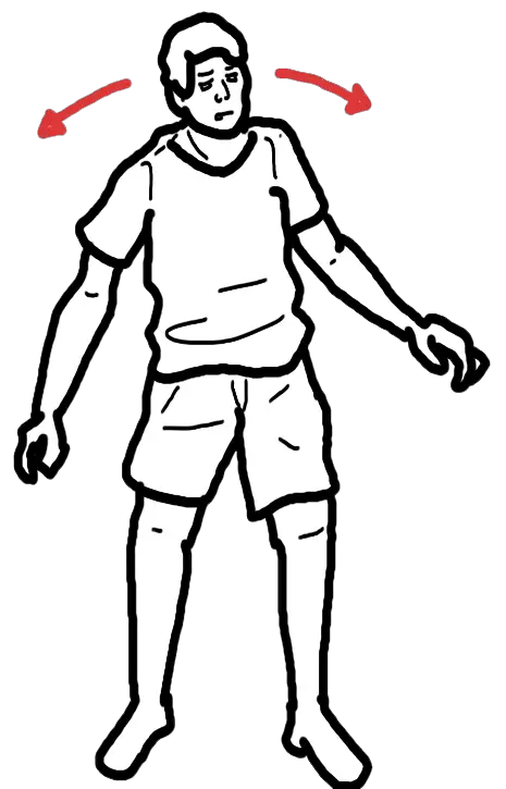
- Nystagmus
- Speech – explosive, jerky and loud with irregular seperation of syllables
Arms and legs
- Shake hands – tone
- Upward arm drift (due to hypotonia of the agonist muscles)
- Rebound – ask patient to raise arm quickly and stop (incoordination between antagonist and agonist muscles)
- Hypotonia – due to loss of facilitatory muscles
- Coordination
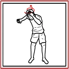
- Finger to nose – past pointing due to no connection between brainstem and cerebellum
- Heel-to-shin
- Dysdiadochokinesis – inability to perform rapid alternating movements
Special tests
- Trunkal ataxia (usually assocated with vermis pathology of the cerebellum)
- Reflex – pendular knee
| Side note There are many connections between the cerebellum and the parietal and frontal lobes of the brain and thus explains the clinical presentation that is associated with cerebellar disease. |
Pathways in the CNS
Pyramidal Pathways (through medulla)
- Anteriorcorticospinal tract
- Lateralcorticospinal tract
Extrapyramidal Pathways
- Rubrospinal tract
- Vestibulospinal tract
- Reticulospinal tract
- Tectospinal tract
- Olivospinal tract
Cerebellar Pathway
- Spinocerebellar tract
| CLINICAL DIFFERENCES BETWEEN THE CENTRAL NERVOUS SYSTEM PATHWAYS | ||||
| Clinical signs | Pyramidal | Extrapyramidal | Cerebellar | Functional |
| Power | Weak | No weakness | No weakness | Give-way weakness |
| Wasting | None (overtime no use maybe) | None | None | None |
| Tone | Spastic increase | Rigidity | Normal/reduced | Normal |
| Reflexes | Increased | Normal | Normal | Normal |
| Plantar response | Extensor | Flexor | Flexor | Normal |
| Coordination | Reduced by weakness | Normal but slow | Impaired | Normal (laborious) |
Discussion