

Dizziness and Vertigo
Overview
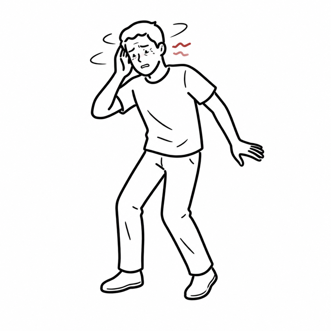
Dizziness is an umbrella term describing a non-specific symptom of spatial disorientation.
To narrow down the differential, dizziness must be classified into one of four distinct clinical categories:
- Vertigo (~50%): Illusion of motion (spinning, tilting, or swaying) caused by asymmetrical vestibular input (Peripheral vs. Central).
- Presyncope (~15%): Sensation of impending loss of consciousness (“feeling faint”) due to global cerebral hypoperfusion.
- Disequilibrium (~15%): Feeling off-balance or unsteady on one’s feet without a head sensation (proprioceptive, cerebellar, or motor deficit).
- Lightheadedness / Non-Specific (~10%): Vague, floating sensation often linked to psychiatric conditions (anxiety, hyperventilation) or medication side effects.
| Categories of dizziness | Pathophysiology | Aetiology |
| Vertigo (Spinning sensation) | Vestribular pathology | Vestibular neuritis Labyrinthitis Meniere’s disease |
| Presyncope (fainting sensation) | Reduced cerebral perfusion | Neurocardiogenic syncope Situational syncope Carotid sensitivity Postural hypotension |
| Dysequilibrium (imbalance sensation) | Gait disorder | Myelopathy Peripheral neuropathy Parkinson’s disease |
| Light-headedness (sensation of disconnection from the environment) | Psychological disorder | Anxiety Depression |
Focus on the timing and triggers rather than relying solely on the patient’s subjective description of “dizziness”. Ask: Is it continuous or episodic? If episodic, how long does it last, and what triggers it?
Vertigo is not only caused by pathology in the vestibular system but can also be caused by cerebellar and neurodegenerative disorders.
Approach
Step 1: Emergency Triage & Red Flag Screening (Rule Out Stroke / Central Cause)
- Screen for “DARS” Central Red Flags:
- Diplopia, Dysarthria, Dysphagia, Dysmetria / Ataxia.
- Acute focal neurological deficits or motor/sensory weakness.
- Rapid-onset, severe continuous vertigo with inability to sit or stand unassisted.
- Sudden-onset unilateral hearing loss or direction-changing / vertical nystagmus.
Step 2: Apply the HINTS Exam (Only in Acute Vestibular Syndrome – Continuous Vertigo + Nystagmus)
- HINTS Exam (Head Impulse, Nystagmus, Test of Skew):
- Head Impulse Test: Normal (no corrective saccade) -> CENTRAL (Stroke). Abnormal (corrective saccade) -> Peripheral (Neuritis).
- Nystagmus: Direction-changing or pure vertical/torsional -> CENTRAL. Unidirectional horizontal -> Peripheral.
- Test of Skew: Vertical ocular misalignment (skew deviation) -> CENTRAL. Normal (no vertical correction) -> Peripheral.
- (Mnemonic: INFARCT = Impulse Normal, Fast-phase Alternating [direction-changing nystagmus], Refixation Contralateral Test of Skew indicates Stroke).
Step 3: Branching Logic based on Timing & Triggers
- Acute Continuous Vertigo (Days): Vestibular Neuritis / Labyrinthitis vs. Posterior Circulation Stroke (Cerebellar / Brainstem Infarct).
- Episodic Positional Vertigo (Seconds – < 1 min): Benign Paroxysmal Positional Vertigo (BPPV – test with Dix-Hallpike Maneuver).
- Episodic Spontaneous Vertigo (Hours): Meniere’s Disease (with auditory symptoms) vs. Vestibular Migraine (with headache/phobia).
The HINTS exam is more sensitive than a diffusion-weighted MRI within the first 24 hours of symptom onset for detecting a posterior circulation (cerebellar/brainstem) ischemic stroke in patients with Acute Vestibular Syndrome, provided it is performed by a trained examiner.
Differential Diagnosis
Peripheral Vestibular Vertigo (Benign, Inner Ear):
- Benign Paroxysmal Positional Vertigo (BPPV):
- Distinguishing features: Brief episodes (< 1 min) of spinning triggered by head movement (rolling in bed, looking up); Dix-Hallpike test shows geotropic upbeat-torsional nystagmus with latency and fatigue; no hearing loss.
- Vestibular Neuritis / Labyrinthitis:
- Distinguishing features: Acute onset of continuous, severe vertigo lasting days; positive Head Impulse test; unidirectional horizontal nystagmus. Labyrinthitis presents with unilateral sensorineural hearing loss; Neuritis spares hearing.
- Meniere’s Disease:
- Distinguishing features: Episodic spontaneous vertigo lasting 20 minutes to 24 hours, accompanied by the classic triad: Low-frequency Sensorineural Hearing Loss, Tinnitus, and Aural Fullness in the affected ear.
Central Vestibular Vertigo (Life-Threatening, Brainstem/Cerebellum):
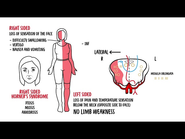
- Posterior Circulation Stroke / TIA (PICA / AICA / Basilar Infarct):
- Distinguishing features: Sudden onset continuous vertigo, severe gait ataxia (patient falls toward side of lesion), HINTS exam positive for central features, presence of cranial nerve deficits or cerebellar signs.
- Vestibular Migraine:
- Distinguishing features: Recurrent vertigo episodes (minutes to days) in patients with a history of migraine, accompanied by photophobia, phonophobia, or visual aura. Hearing is characteristically preserved.
- Acoustic Neuroma (Vestibular Schwannoma):
- Distinguishing features: Progressive unilateral sensorineural hearing loss, tinnitus, and mild imbalance/disequilibrium; rarely presents with acute vertigo.
Non-Vertiginous Causes of Dizziness:
- Orthostatic Hypotension / Cardiac Presyncope:
- Persistent Postural-Perceptual Dizziness (PPPD):
- Distinguishing features: Chronic non-spinning dizziness/unsteadiness present for >= 3 months; exacerbated by upright posture, motion, and complex visual environments.
BPPV is caused by canalithiasis (otoconia displaced into the posterior semicircular canal). It is treated mechanically with the Epley Maneuver, NOT with vestibular suppressant medications like Betahistine or Prochlorperazine.
Investigations
First-Line / Bedside Diagnostics:
- Bedside Neurological & Otological Exam: Evaluate cranial nerves, cerebellar signs (finger-to-nose, heel-to-shin), gait, and spontaneous/gaze-evoked nystagmus.
- Dix-Hallpike Maneuver: Diagnostic gold standard for Posterior Canal BPPV. (Positive if it reproduces vertigo and characteristic latency-delayed, fatigable geotropic torsional nystagmus).
- HINTS Exam (Head Impulse, Nystagmus, Test of Skew): Mandatory in all patients with acute continuous vertigo to rule out central stroke.
- 12-Lead ECG & Orthostatic Vitals: Mandatory if presyncope or cardiac etiology is suspected.
Targeted / Secondary Imaging & Audiology Strategy:
- MRI Brain with Diffusion-Weighted Imaging (DWI) + Internal Auditory Canals: Gold standard imaging modality to evaluate for posterior circulation stroke, cerebellar lesions, or vestibular schwannoma.
- CT Brain / CTA: Indicated if acute intracerebral/cerebellar hemorrhage or vascular dissection is suspected, but poor sensitivity (< 20-30%) for hyperacute ischemic stroke in the posterior fossa.
- Audiometry & Vestibular Function Testing: Pure-tone audiometry (confirms Meniere’s disease or acoustic neuroma); Videonystagmography (VNG) or Caloric testing for complex peripheral deficits.
A non-contrast head CT is completely unreliable for ruling out posterior fossa / cerebellar ischemic stroke. If a central cause is suspected, an MRI Brain (DWI) is mandatory.
Critical Management
- Benign Paroxysmal Positional Vertigo (BPPV):
- Particle Repositioning (Epley Maneuver): Immediate bedside treatment for posterior canal BPPV (success rate > 80-90% after 1-2 treatments).
- Acute Vestibular Neuritis:
- Short-Term Symptomatic Relief: Short-course Vestibular Suppressants (e.g., Prochlorperazine 5 mg PO/IV, Diazepam 2-5 mg PO, or Promethazine) for maximum 3-5 days ONLY.
- Corticosteroids: Oral Prednisolone (1 mg/kg for 5-7 days) started early may improve long-term vestibular recovery.
- Vestibular Rehabilitation Therapy (VRT): Crucial long-term physical therapy to promote central compensation; stop vestibular suppressants early to allow VRT to work.
- Acute Posterior Circulation Ischemic Stroke:
- Emergency Resuscitation: Immediate stroke code activation -> Thrombolysis (IV alteplase/tenecteplase) within 4.5 hours or Endovascular Thrombectomy (EVT) if large vessel occlusion present.
Long-term use of vestibular suppressants (Prochlorperazine, Benzodiazepines) is HARMFUL in peripheral vestibular neuritis—it delays central brain compensation and leads to chronic unsteadiness. Stop suppressants within 48-72 hours.
References
- Society for Academic Emergency Medicine. GRACE-3: acute dizziness and vertigo in the emergency department [Internet]. 2023 [cited 2026 Aug 20]. Available from: https://www.saem.org/publications/grace/grace-3
- American Academy of Otolaryngology–Head and Neck Surgery Foundation. Clinical practice guideline: benign paroxysmal positional vertigo (update) [Internet]. 2017 [cited 2026 Aug 20]. Available from: https://www.entnet.org/quality-practice/quality-products/clinical-practice-guidelines/bppv/
Members only discussions coming soon…