

Parkinson's Presentation
OVERVIEW
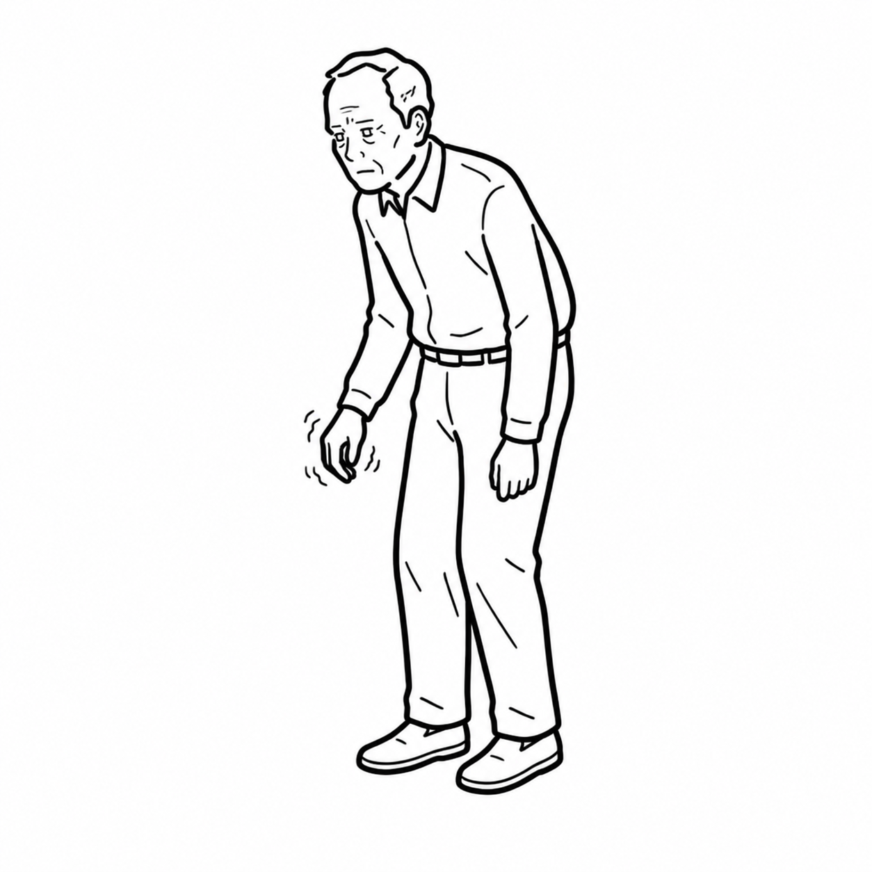
Parkinsonism is a clinical syndrome defined by bradykinesia together with rest tremor, rigidity, or both. Parkinson’s disease is the most common neurodegenerative cause of parkinsonism, but the syndrome may also result from medications, vascular disease, structural lesions or atypical neurodegenerative disorders.
Core Motor Features:
- Bradykinesia: Slowness of voluntary movement accompanied by progressive reduction in movement amplitude or speed during repetitive actions.
- Rigidity: Increased resistance to passive movement that is not dependent on movement velocity; tremor may produce cogwheel rigidity.
- Rest tremor: Typically a 4–6 Hz asymmetric “pill-rolling” tremor that is most apparent when the limb is relaxed and decreases during purposeful movement.
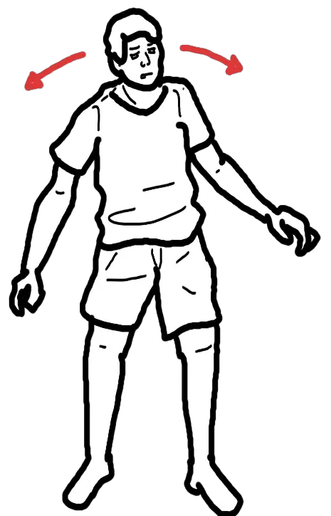
- Postural and gait impairment: Reduced arm swing, stooped posture, shuffling steps, freezing and impaired postural reflexes, usually appearing later in typical Parkinson’s disease.
Parkinson’s disease is diagnosed clinically. There is no single confirmatory blood test or routinely required scan. Early asymmetry, rest tremor, a clear and sustained response to dopaminergic therapy and levodopa-induced dyskinesia support the diagnosis. Early falls, rapid progression, severe autonomic failure, supranuclear gaze palsy, cerebellar signs or poor response to adequate levodopa suggest an alternative cause.
Tremor is not required for the diagnosis of parkinsonism. The essential motor feature is bradykinesia, demonstrated by progressive decrement in speed or amplitude during repetitive movement, together with rigidity or rest tremor.
APPROACH
Step 1: Confirm That Parkinsonism Is Present
Assess for true bradykinesia rather than subjective slowness alone:
- Ask the patient to perform repetitive finger tapping, hand opening and closing, pronation–supination, toe tapping and heel tapping.
- Look for progressive slowing, hesitations, interruptions or reduction in movement amplitude.
- Observe spontaneous movement, blinking, facial expression, speech volume and ease of rising from a chair.
- Test tone at the wrist, elbow and neck while the patient is relaxed.
- Use activation of the opposite limb if subtle rigidity is suspected.
- Observe for rest tremor while the patient is seated, walking and performing a distracting task.
- Examine posture, stride length, arm swing, turning, freezing and postural stability.
Step 2: Characterise the Pattern and Time Course
Establish:
- Onset: Gradual or abrupt; unilateral or bilateral.
- Progression: Slowly progressive, stepwise, fluctuating or rapidly deteriorating.
- Motor symptoms: Tremor, stiffness, slowness, reduced dexterity, micrographia, dragging one leg, difficulty turning in bed, freezing and falls.
- Bulbar symptoms: Hypophonia, dysarthria, drooling, dysphagia or choking.
- Non-motor symptoms: Anosmia, constipation, urinary symptoms, erectile dysfunction, orthostatic dizziness, sleep disturbance, depression, anxiety, hallucinations and cognitive change.
- Prodromal clues: Rapid eye movement sleep behaviour disorder, hyposmia, constipation and longstanding mood disturbance.
- Functional impact: Dressing, eating, handwriting, walking, driving, employment and medication self-management.
- Family history: Parkinson’s disease, tremor, dementia or early-onset movement disorder.
- Exposure history: Pesticides, manganese, carbon monoxide, recurrent head injury or illicit drugs contaminated with parkinsonian toxins.
Step 3: Review Medications and Secondary Causes
Identify dopamine-receptor blocking or dopamine-depleting agents:
- Antipsychotics, particularly potent first-generation agents.
- Metoclopramide and prochlorperazine.
- Some other antiemetics and vestibular suppressants.
- Sodium valproate.
- Tetrabenazine, deutetrabenazine or reserpine.
- Flunarizine or cinnarizine where used.
- Other recently introduced medicines temporally associated with the syndrome.
Drug-induced parkinsonism is often symmetric and may lack a classic rest tremor, although substantial overlap exists. Symptoms may persist for months after withdrawal, and dopamine-blocking medication can unmask underlying degenerative Parkinson’s disease.
Step 4: Look for Features Supporting Parkinson’s Disease
- Clear unilateral onset with persistent asymmetry.
- Rest tremor of a limb.
- Marked improvement with dopaminergic treatment.
- Development of levodopa-induced dyskinesia.
- Gradual progression over years.
- Hyposmia or cardiac sympathetic denervation on appropriate specialist testing.
Step 5: Screen for Atypical or Secondary Parkinsonism
Features that should prompt early specialist reassessment include:
- Rapid progression of gait impairment or disability.
- Recurrent falls within the first few years.
- Early severe autonomic failure, urinary retention or symptomatic orthostatic hypotension.
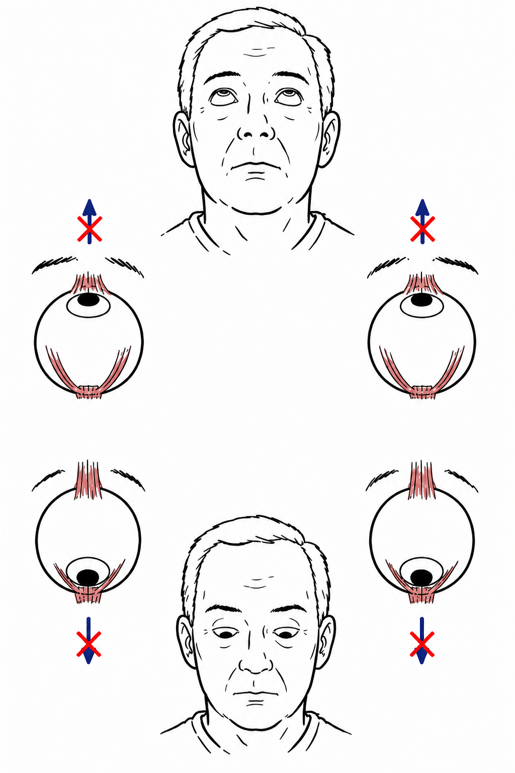
- Vertical supranuclear gaze palsy or slowing of vertical saccades.
- Cerebellar ataxia or unexplained pyramidal signs.
- Inspiratory stridor, early severe dysphagia, dysarthria or anterocollis.
- Disproportionate axial rigidity or symmetric onset.
- Cortical sensory loss, apraxia or alien-limb phenomena.
- Poor response to an adequate levodopa trial.
- Normal presynaptic dopaminergic functional imaging.
- Dementia or recurrent visual hallucinations before, or within one year of, the onset of parkinsonism.
Early recurrent falls, severe autonomic failure, vertical gaze palsy, cerebellar signs or a poor response to adequate levodopa are red flags against typical Parkinson’s disease and should trigger evaluation for atypical or secondary parkinsonism.
DIFFERENTIAL DIAGNOSIS
Idiopathic Parkinson’s Disease
- Gradual, asymmetric onset.
- Bradykinesia with rigidity or rest tremor.
- Reduced arm swing and dexterity commonly begin unilaterally.
- Clear and sustained response to levodopa is supportive.
- Postural instability generally develops later than bradykinesia and rigidity.
- Non-motor symptoms may predate the motor syndrome.
Tremor Disorders
- Essential tremor: Bilateral action or postural upper-limb tremor that may involve the head or voice. Bradykinesia with decrement and true rigidity are absent.
- Dystonic tremor: Irregular or jerky tremor associated with abnormal posturing and sometimes a “null point.”
- Enhanced physiological tremor: Fine, high-frequency postural tremor associated with anxiety, caffeine, hyperthyroidism, medications or metabolic disturbance.
Drug-Induced Parkinsonism
- Temporal relationship to a dopamine-blocking or dopamine-depleting medicine.
- Often bilateral and symmetric, although asymmetry can occur.
- Orofacial dyskinesia or akathisia may coexist.
- Improvement may take weeks to months after the causative medication is withdrawn.
- Persistent asymmetric symptoms may represent unmasked Parkinson’s disease.
Atypical Neurodegenerative Parkinsonism
- Multiple system atrophy: Early severe autonomic failure, urinary retention, erectile dysfunction or orthostatic hypotension; may include cerebellar ataxia, pyramidal signs, anterocollis or inspiratory stridor. Levodopa response is often limited or short-lived.
- Progressive supranuclear palsy: Early unexplained falls, axial rigidity and impaired vertical saccades or vertical supranuclear gaze palsy, with possible retrocollis, dysarthria and executive dysfunction.
- Corticobasal syndrome: Markedly asymmetric rigidity, apraxia, cortical sensory loss, dystonia, myoclonus or alien-limb phenomena.
- Dementia with Lewy bodies: Dementia, recurrent well-formed visual hallucinations, cognitive fluctuations and rapid eye movement sleep behaviour disorder. Dementia occurs before or within one year of the motor syndrome. Profound antipsychotic sensitivity may occur.
Secondary Parkinsonism
- Vascular parkinsonism: Predominantly lower-body gait disorder, short steps, freezing and postural instability, often with pyramidal signs, cognitive impairment and urinary symptoms.
- Normal-pressure hydrocephalus: Gait initiation failure with short, broad-based or “magnetic” steps, cognitive impairment and urinary urgency or incontinence.
- Structural brain lesion: Tumour, subdural collection, strategic infarction or another basal-ganglia lesion; consider with abrupt or rapid onset or focal neurological signs.
- Wilson disease: Consider in younger patients with parkinsonism, dystonia, tremor, psychiatric symptoms or unexplained liver disease.
- Toxin-related parkinsonism: Carbon monoxide, manganese, methanol or MPTP exposure.
- Post-encephalitic, infectious or traumatic parkinsonism: Suggested by the relevant history.
Parkinsonism Mimics
- Depression with psychomotor slowing.
- Frailty, arthritis or painful musculoskeletal disease causing reduced movement.
- Cervical myelopathy.
- Frontal gait disorder.
- Peripheral neuropathy or sensory ataxia.
- Functional movement disorder.
- Medication-related sedation.
- Hypothyroidism or another systemic metabolic disorder.
Essential tremor is primarily an action and postural tremor. Parkinsonian tremor is typically asymmetric and present at rest, but the decisive distinction is whether genuine bradykinesia with decrement and rigidity are present.
INVESTIGATIONS
Clinical Diagnosis
- Refer people with suspected Parkinson’s disease promptly and untreated to a clinician with expertise in its differential diagnosis.
- Perform a complete neurological examination rather than diagnosing Parkinson’s disease from tremor alone.
- Reassess the diagnosis over time because atypical features may emerge only with disease progression.
- Apply recognised clinical criteria, such as the Movement Disorder Society criteria: first establish parkinsonism, then assess absolute exclusion criteria, supportive features and red flags.

Medication and Functional Assessment
- Document all prescribed, non-prescribed and recently discontinued medicines.
- Establish the exact relationship between symptoms and medication exposure.
- Review the timing and clinical response to dopaminergic treatment.
- Assess falls, gait, swallowing, speech, cognition, mood, sleep, autonomic function and activities of daily living.
- Measure lying and standing blood pressure when orthostatic symptoms, falls or autonomic dysfunction are present.
Laboratory Investigations
Routine blood tests do not confirm Parkinson’s disease. Use targeted testing to exclude mimics or secondary causes:
- FBC, electrolytes, renal and liver function.
- Thyroid function.
- Vitamin B12 or folate where neuropathy, cognitive impairment or nutritional deficiency is possible.
- Glucose or HbA1c where metabolic disease contributes to gait impairment.
- Ceruloplasmin, serum copper, 24-hour urinary copper and slit-lamp examination when Wilson disease is clinically plausible.
- Targeted infectious, autoimmune or toxicological investigations according to the presentation.
Structural Neuroimaging
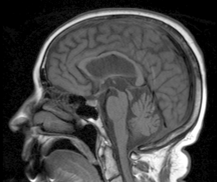
- Do not use structural MRI to diagnose typical Parkinson’s disease.
- Obtain brain MRI when the presentation is atypical; onset is abrupt, rapidly progressive or symmetric; focal, pyramidal or cerebellar signs are present; or vascular parkinsonism, normal-pressure hydrocephalus, tumour or another structural lesion is suspected.
- MRI may provide supportive patterns for atypical parkinsonian disorders but is not independently diagnostic.
Dopaminergic Functional Imaging
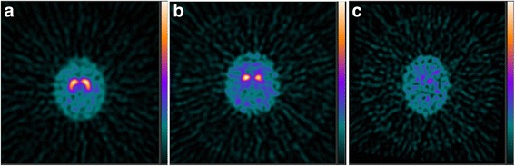
- Consider dopamine-transporter SPECT imaging when clinical examination cannot reliably distinguish essential tremor from degenerative parkinsonism.
- An abnormal scan supports presynaptic dopaminergic deficit but does not reliably distinguish Parkinson’s disease from multiple system atrophy, progressive supranuclear palsy or corticobasal degeneration.
- Normal presynaptic dopaminergic imaging argues strongly against degenerative Parkinson’s disease.
- Do not use functional dopaminergic imaging routinely when the clinical diagnosis is clear.
Additional Specialist Assessments
- Formal neuropsychological assessment: When the pattern or severity of cognitive impairment is uncertain.
- Speech and swallowing assessment: For hypophonia, dysarthria, coughing with meals, weight loss or aspiration risk.
- Autonomic testing: For severe or diagnostically important orthostatic hypotension.
- Sleep assessment: When rapid eye movement sleep behaviour disorder, excessive daytime sleepiness or sleep-disordered breathing is suspected.
- Genetic counselling and testing: Consider for very early onset, a strong family history or a phenotype suggesting a monogenic disorder.
Dopamine-transporter imaging can distinguish degenerative parkinsonism from conditions such as essential tremor, but it cannot reliably separate Parkinson’s disease from atypical degenerative parkinsonism. Parkinson’s disease remains a clinical diagnosis.
CRITICAL MANAGEMENT
Immediate Priorities After Diagnosis
- Explain the diagnosis, expected course and available treatment options in an individualised manner.
- Provide access to a Parkinson’s disease specialist and a continuing point of contact.
- Assess the impact of motor and non-motor symptoms on quality of life.
- Review driving, occupational safety, falls, swallowing and medication self-management.
- Involve physiotherapy, occupational therapy, speech pathology, nursing, pharmacy, dietetics and psychological services according to need.
- Encourage regular exercise incorporating aerobic activity, strength, balance and task-specific practice.
Initial Motor Treatment
- Levodopa: Offer when motor symptoms affect quality of life. It usually provides the greatest improvement in bradykinesia and rigidity. Use the lowest effective dose while monitoring nausea, orthostatic hypotension, hallucinations, wearing-off and dyskinesia.
- Dopamine agonists: May be considered in selected patients, particularly when symptoms do not yet substantially impair quality of life. Counsel about excessive daytime sleepiness, hallucinations, oedema and impulse-control disorders.
- Monoamine oxidase-B inhibitors: May provide modest symptomatic benefit in early disease or be used as adjunctive therapy.
- Treatment selection should account for age, cognition, comorbidity, occupation, falls risk, psychiatric history and patient preference.
Motor Fluctuations and Dyskinesia
- Confirm medication timing and adherence and review whether dietary protein or impaired gastric emptying is affecting levodopa absorption.
- Adjust levodopa dose size, timing or formulation under specialist guidance.
- Consider adjunctive catechol-O-methyltransferase inhibitors, monoamine oxidase-B inhibitors, dopamine agonists or amantadine according to the clinical problem.
- Consider device-assisted therapies for selected patients with disabling fluctuations despite optimised oral treatment: deep-brain stimulation, continuous subcutaneous apomorphine, intestinal levodopa infusion or other continuous dopaminergic infusion systems where available.
Non-Motor Management
- Orthostatic hypotension: Review causative medicines, hydration and salt intake where appropriate; use compression strategies and medication when necessary.
- Constipation: Address fluid, fibre, physical activity and appropriate laxative therapy.
- Depression and anxiety: Provide appropriate psychological and pharmacological treatment.
- Psychosis or hallucinations: Exclude delirium and infection, review Parkinson’s medicines sequentially with specialist input, and avoid inappropriate dopamine-blocking antipsychotics.
- Dementia: Consider a cholinesterase inhibitor when clinically appropriate.
- Sleep disturbance: Identify nocturnal immobility, rapid eye movement sleep behaviour disorder, restless legs syndrome, sleep apnoea and medication-related daytime somnolence.
- Dysphagia and weight loss: Arrange urgent speech-pathology and dietetic assessment and evaluate aspiration risk.
- Sialorrhoea: Optimise swallowing and consider specialist-directed pharmacological or botulinum-toxin treatment.
Acute Hospital and Perioperative Management
- Administer Parkinson’s medicines at the patient’s usual exact times rather than standard ward-round times.
- Never abruptly stop levodopa, dopamine agonists or other dopaminergic therapy.
- If oral administration is unsafe, obtain urgent pharmacy, neurology or Parkinson’s specialist advice, use an enteral route where possible and consider an appropriate temporary non-oral dopaminergic strategy.
- Avoid dopamine antagonists that can worsen parkinsonism, including metoclopramide, prochlorperazine, haloperidol and other potent dopamine-blocking antipsychotics.
- Look actively for infection, constipation, dehydration, pain, urinary retention and medication errors when mobility deteriorates suddenly.
Parkinsonism–Hyperpyrexia Syndrome
Suspect this neurological emergency when a patient develops severe akinesia or generalised rigidity, fever, reduced consciousness or confusion, autonomic instability, dysphagia, elevated creatine kinase, rhabdomyolysis or acute kidney injury. Common triggers include abrupt withdrawal or impaired absorption of dopaminergic medication, infection, surgery, dehydration and gastrointestinal dysfunction.
Management includes:
- Immediate hospitalisation and urgent neurological input.
- Airway, breathing and circulatory support.
- Treatment of hyperthermia, dehydration and electrolyte abnormalities.
- Investigation and treatment of infection or another precipitant.
- Prompt restoration of dopaminergic therapy through an appropriate route.
- Monitoring for aspiration, rhabdomyolysis, renal failure, thrombosis and respiratory complications.
Parkinson’s medication is time-critical. Abrupt withdrawal or missed doses—particularly of levodopa—can precipitate profound immobility, aspiration and potentially fatal parkinsonism–hyperpyrexia syndrome.
REFERENCES
- National Institute for Health and Care Excellence. Parkinson’s disease in adults. NICE guideline NG71 [Internet]. London: NICE; 2017, updated 2020. Available from: https://www.nice.org.uk/guidance/ng71
- Postuma RB, Berg D, Stern M, et al. MDS clinical diagnostic criteria for Parkinson’s disease. Mov Disord. 2015;30(12):1591–1601. doi:10.1002/mds.26424.
- Pringsheim T, Day GS, Smith DB, et al. Dopaminergic therapy for motor symptoms in early Parkinson disease practice guideline summary: a report of the AAN Guideline Subcommittee. Neurology. 2021;97(20):942–957. doi:10.1212/WNL.0000000000012868.
- Bloem BR, Okun MS, Klein C. Parkinson’s disease. Lancet. 2021;397(10291):2284–2303. doi:10.1016/S0140-6736(21)00218-X.
- National Institute for Health and Care Excellence. Parkinson’s disease. NICE quality standard QS164 [Internet]. London: NICE; 2018. Available from: https://www.nice.org.uk/guidance/qs164
- Seppi K, Ray Chaudhuri K, Coelho M, et al. Update on treatments for nonmotor symptoms of Parkinson’s disease—an evidence-based medicine review. Mov Disord. 2019;34(2):180–198. doi:10.1002/mds.27602.
- Parkinson’s UK. Emergency management of patients with Parkinson’s [Internet]. London: Parkinson’s UK; 2023. Available from: https://www.parkinsons.org.uk/professionals/resources/emergency-management-patients-parkinsons
- Greenland JC, Barker RA. The differential diagnosis of Parkinson’s disease. In: Stoker TB, Greenland JC, editors. Parkinson’s disease: pathogenesis and clinical aspects [Internet]. Brisbane: Codon Publications; 2018. Available from: https://www.ncbi.nlm.nih.gov/books/NBK536715/




Members only discussions coming soon…