

Chest Trauma
Overview
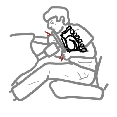
Chest trauma encompasses blunt or penetrating mechanical force applied to the thoracic cavity, causing injury to the chest wall, lungs, airways, heart, great vessels, or esophagus.
Thoracic trauma accounts for 20–25% of all trauma-related deaths. Over 85% of thoracic injuries can be definitively managed without open thoracotomy using basic resuscitation, chest drain insertion, and analgesia.
Core Physiology: Thoracic injuries rapidly impair ventilation, oxygenation, and systemic perfusion via direct lung contusion, space-occupying tension physiology, severe hemorrhage, or cardiac compromise.
The “Deadly Dozen” thoracic trauma injuries must be systematically evaluated during primary and secondary surveys. The immediate life threats—Tension Pneumothorax, Open Pneumothorax, Massive Hemothorax, Cardiac Tamponade, Flail Chest, and Tracheobronchial Rupture—must be diagnosed clinically and managed during the Primary Survey before waiting for imaging.
Approach
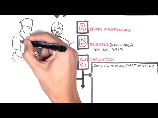
Step 1: Primary Survey & Immediate Resuscitation (ATLS – ABCDE)
- Airway: Assess patency, stridor, or tracheal deviation. Secure airway immediately if airway rupture or severe hypoxia is present.
- Breathing: Oxygenation/ventilation check. Inspect chest expansion, feel for subcutaneous emphysema/flail segments, auscultate breath sounds.
- Absent breath sounds + Hypotension + Tracheal shift ->> Tension Pneumothorax (Immediate decompression).
- Sucking chest wound ->> Open Pneumothorax (3-sided occlusive dressing).
- Paradoxical chest movement + Hypoxia ->> Flail Chest (Analgesia + positive pressure ventilation).
- Circulation: Check pulses, BP, JVP, heart sounds.
- Flat neck veins + Absent breath sounds + Dullness to percussion + Shock ->> Massive Hemothorax (Intercostal catheter + massive transfusion).
- Distended neck veins + Hypotension + Muffled heart sounds ->> Cardiac Tamponade (eFAST ->> Pericardial window / Resuscitative Thoracotomy).
- Disability & Exposure: GCS, expose chest fully, assess for exit/entry wounds.
Step 2: Emergency Resuscitative Interventions (Primary Survey)
- Tension Pneumothorax: Immediate Finger Thoracostomy (preferred in ventilated/adult trauma) or Needle Decompression (2nd intercostal space mid-clavicular line OR 4th/5th intercostal space anterior axillary line) followed by Intercostal Catheter (ICC).
- Massive Hemothorax: ICC placement (28–32 Fr); auto-transfusion if available.
- Indications for Emergency Department Thoracotomy (EDT): Penetrating thoracic trauma with witnessed loss of vital signs in ED/transport with initial signs of life.
Step 3: Clinical Decision Branching
- Hemodynamically Unstable: Immediate clinical intervention (Decompression, Chest Drain, Resuscitation) ->> eFAST at bedside.
- Hemodynamically Stable: Proceed to CXR, eFAST, and contrast CT Chest/Angiography.
A tension pneumothorax is a clinical diagnosis, NOT a radiologic one. Never delay emergency decompression to obtain a chest X-ray if clinical features (hypotension, unilateral absent breath sounds, tracheal deviation) are present.
Differential Diagnosis
Immediate Life-Threats (Primary Survey “Lethal Six”):
- Tension Pneumothorax:
- Distinguishing features: Severe respiratory distress, unilateral absent breath sounds, ipsilateral hyper-resonance, hypotension, JVP distension, contralateral tracheal shift.
- Massive Hemothorax (Blood accumulation >1500 mL or >200 mL/hr for 2–4 hours):
- Distinguishing features: Unilateral absent breath sounds, dullness to percussion, severe hemorrhagic shock, flat neck veins.
- Cardiac Tamponade:
- Distinguishing features: Beck’s Triad (Hypotension, Distended Neck Veins, Muffled Heart Sounds); Pulsus paradoxus; elevated JVP with loss of y descent.
- Open Pneumothorax (“Sucking Chest Wound”):
- Distinguishing features: Full-thickness chest wall defect (>2/3 tracheal diameter); bubbling/sucking wound with respiration.
- Flail Chest:
- Distinguishing features: >=2 contiguous ribs fractured in >=2 places; paradoxical inward movement of flail segment during inspiration; severe pain and underlying pulmonary contusion.
- Tracheobronchial Tree Rupture:
- Distinguishing features: Massive subcutaneous emphysema, persistent large air leak after chest drain insertion, pneumomediastinum, hemoptysis.
Potentially Lethal Injuries (Secondary Survey):
- Blunt Thoracic Aortic Injury (BTAI):
- Distinguishing features: High-deceleration injury mechanism; upper extremity hypertension with delayed/diminished femoral pulses; widened mediastinum on CXR.
- Pulmonary Contusion:
- Distinguishing features: Direct parenchymal damage leading to edema/hemorrhage; delayed hypoxia (developing over 24–48 hours) out of proportion to initial CXR.
- Blunt Cardiac Injury (Myocardial Contusion):
- Distinguishing features: Sternal fracture/anterior impact; unexplained sinus tachycardia, arrhythmias, elevated troponin, or new ST-T changes.
- Traumatic Diaphragmatic Rupture:
- Distinguishing features: Left-sided blunt impact (80%); bowel sounds in chest cavity, nasogastric tube curling up into thorax on CXR.
- Esophageal Rupture (Boerhaave / Traumatic):
- Distinguishing features: Pneumomediastinum, left pleural effusion, severe pain, subcutaneous emphysema (Mackler’s triad).
Differentiate Tension Pneumothorax from Massive Hemothorax by chest percussion: Tension Pneumothorax is hyper-resonant, whereas Massive Hemothorax is dull to percussion.
Investigations
First-Line / Bedside Diagnostics:
- Extended Focused Assessment with Sonography for Trauma (eFAST):
- Pneumothorax: Absence of lung sliding, absence of B-lines, presence of “Bar Code / Stratosphere sign” on M-mode, identification of “Lung Point”.
- Hemothorax / Tamponade: Pleural fluid collections, pericardial effusion/tamponade physiology.
- Portable Supine Chest X-Ray (CXR):
- Look for: Mediastinal widening (>8 cm), loss of aortic knob outline, apical cap, elevated hemidiaphragm, rib/sternal fractures, hemopneumothorax.
- Blood Tests & Gas: FBC, U&E, Coagulation panel, Group & Crossmatch (minimum 4–6 units), Troponin I/T, ABG (lactate, base deficit).
Targeted / Imaging Strategy:
- CT Angiography (CTA) Chest (Thin-slice contrast): Gold standard for stable patients. Evaluates aortic tear/dissection, occult pneumothorax, pulmonary contusions, rib/sternal fracture configurations, and active bleeding.
- Bronchoscopy / Endoscopy: Confirms suspected tracheobronchial or esophageal disruption.
- Echocardiography (TTE/TEE): Evaluates structural cardiac damage, valvular dysfunction, or pericardial effusion.
The eFAST exam can detect a pneumothorax faster and with higher sensitivity than a supine portable chest X-ray. Look specifically for the loss of “lung sliding” and the presence of a “lung point”.
Critical Management
Massive Hemothorax & Surgical Indications:
- Initial Drain Management: Insert 28–32 Fr chest tube at 4th/5th intercostal space anterior to mid-axillary line.
- Indications for Immediate Operative Thoracotomy:
- Immediate drainage of >1500 mL of blood upon chest tube placement.
- Ongoing drainage of >200 mL/hr for 2–4 consecutive hours.
- Persistent instability requiring ongoing massive blood transfusion.
Flail Chest & Pulmonary Contusion:
- Analgesia (Cornerstone): Multimodal analgesia (Regional anesthesia/epidural, IV patient-controlled analgesia) to prevent splinting, hypoventilation, and atelectasis.
- Fluid Management: Euvolemic fluid strategy; avoid fluid overload which exacerbates underlying pulmonary contusion.
Blunt Thoracic Aortic Injury (BTAI):
- Impulse Control: Target HR <60 bpm and SBP 100–120 mmHg (using short-acting beta-blockers e.g., IV Esmolol) to decrease aortic wall shear stress ($\mathrm{dP/dt}$) prior to definitive endovascular repair (TEVAR).
In flail chest and pulmonary contusion, effective regional analgesia (e.g., epidural or erector spinae plane block) is the most critical intervention to prevent respiratory failure and avoid mechanical ventilation.
References
- Coccolini F, et al. Thoracic trauma WSES-AAST guidelines. World J Emerg Surg. 2025. Available from: https://pmc.ncbi.nlm.nih.gov/articles/PMC12522690/
- Eastern Association for the Surgery of Trauma. Blunt thoracic trauma: pain management [Internet]. [cited 2026 Aug 20]. Available from: https://www.east.org/education-resources/practice-management-guidelines/details/blunt-thoracic-trauma-btt-pain-management-in

Members only discussions coming soon…